JCI-accredited · 45+ hospitals & clinics · 90+ countries served · 24/7 multilingual support
Nuclear Medicine Systems

Spect CT

Diagnosis
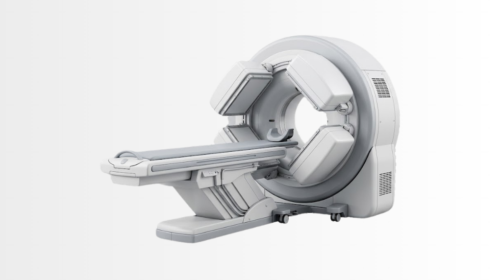
Advanced Spect CT imaging device at Acibadem Hospitals Group.

Quick answer

SPECT/CT is a hybrid nuclear medicine imaging technology that combines functional radiotracer imaging with detailed CT anatomy in a single examination. At Acibadem in Turkey, it is used to support evaluation of cardiac conditions, bone disorders and selected cancer-related findings, helping clinicians localize and interpret areas of abnormal activity.

SPECT/CT: Hybrid Imaging for Clearer Diagnosis and Personalised Care

SPECT/CT is an advanced nuclear medicine imaging technology that combines two complementary types of scans in one examination: single-photon emission computed tomography (SPECT) and computed tomography (CT).

In simple terms, SPECT shows how certain tissues and organs are functioning, while CT provides detailed pictures of the body’s anatomy. Together, these images can help specialists identify where an abnormal finding is located, assess its significance and plan the most appropriate next step.

At Acibadem International, SPECT/CT supports precise diagnostic assessment in cardiac, bone and selected tumour-related studies. It can help our Nuclear Medicine teams understand changes that may not be fully explained by structural imaging alone. By combining functional and anatomical information, the technology can support earlier detection, more confident interpretation and personalised treatment planning.

Every scan is considered individually. Your specialist will review your symptoms, medical history, previous imaging and laboratory findings to determine whether SPECT/CT is suitable for your care plan.

What Is SPECT/CT?

SPECT/CT is a hybrid imaging method used in nuclear medicine. “SPECT” stands for single-photon emission computed tomography. It uses a small amount of a radiotracer, a substance that can be detected by a specialised camera, to show activity within the body.

The CT component produces detailed cross-sectional images of bones, organs and other structures. When the SPECT and CT images are combined, clinicians can see both the functional activity of a tissue and its precise anatomical location.

For example, a SPECT scan may show an area of increased or reduced radiotracer uptake. The CT scan can help identify whether that activity is related to a bone, organ, lymph node, soft tissue area or another structure. This can improve diagnostic confidence and reduce uncertainty in selected clinical situations.

SPECT/CT does not replace every other imaging examination. Instead, it can complement tests such as ultrasound, MRI, PET/CT, conventional CT, X-ray and laboratory studies when additional functional information is needed.

How Does SPECT/CT Work?

A SPECT/CT examination usually begins with the administration of a radiotracer. Depending on the purpose of the scan, the radiotracer may be given through a small injection, swallowed or inhaled. The type of tracer and timing of the examination are selected according to the clinical question.

After the radiotracer is administered, there may be a waiting period while it travels through the body and accumulates in the target tissues. During this time, you may be asked to drink water, rest or follow other simple instructions from your care team.

When it is time for imaging, you will lie comfortably on a scanning table. A specialised gamma camera moves around your body to acquire the SPECT images. The CT scan is then performed as part of the same session. The equipment is open rather than tunnel-shaped, and the scan itself is generally painless.

Advanced software aligns the functional SPECT images with the anatomical CT images. Nuclear medicine physicians and radiology specialists then interpret the combined information in the context of your clinical history and other test results.

The full appointment length can vary depending on the type of scan, the radiotracer used and whether additional delayed imaging is needed. Your international patient coordinator and clinical team will provide personalised instructions before your appointment.

Clinical Applications of SPECT/CT

SPECT/CT may be used across a range of diagnostic pathways. It is particularly valuable when clinicians need to understand both tissue function and detailed anatomy. The technology can support the assessment of cardiac conditions, bone disorders and selected tumour-related findings.

Clinical applications How SPECT/CT may help Examples of conditions or clinical questions
Cardiac imaging Helps assess blood flow to the heart muscle and may support the evaluation of myocardial perfusion findings. Suspected coronary artery disease, chest pain assessment, known heart disease, evaluation of heart muscle blood supply.
Bone imaging Helps localise areas of bone activity and relate them to specific joints, vertebrae, fractures or other structures. Unexplained bone pain, stress injuries, degenerative joint changes, suspected infection, fracture assessment and selected metastatic bone evaluations.
Oncology and tumour-related studies May help clarify the anatomical location of radiotracer uptake and support staging, treatment planning or follow-up in selected cases. Selected tumour-related bone findings, evaluation of suspected spread to bone, follow-up of certain cancers and assessment of treatment response when clinically appropriate.
Parathyroid imaging Can support localisation of overactive parathyroid tissue before surgery in suitable patients. Primary hyperparathyroidism and selected cases requiring pre-operative localisation.
Thyroid and endocrine studies Can provide functional information alongside anatomical detail in selected thyroid and endocrine assessments. Selected thyroid nodules, differentiated thyroid cancer follow-up and other specialist endocrine indications.
Infection and inflammation assessment May help identify or localise areas of active inflammation or infection when conventional imaging does not provide a complete answer. Selected bone or joint infections, inflammatory processes and complex post-treatment findings.

The exact role of SPECT/CT depends on your individual diagnosis and the question your physician needs to answer. In some cases, another imaging method may be more appropriate. Your specialists will recommend the examination that best supports safe and effective care.

Why Hybrid Functional and Anatomical Imaging Matters

Traditional anatomical imaging can show the shape, size and location of structures in the body. Functional nuclear medicine imaging can show how tissues behave, such as how they take up a radiotracer or how blood reaches part of an organ. SPECT/CT brings these two perspectives together.

This combined approach can be particularly helpful when a finding is small, located in a complex area of the body or difficult to interpret on a single imaging method. A functional signal alone may not always identify the exact structure involved. CT correlation can help improve localisation and make the result more clinically meaningful.

For patients, this may mean fewer unanswered questions and a clearer route toward the next stage of care. For clinicians, it can provide valuable information for diagnosis, treatment selection, surgical planning or monitoring after treatment.

SPECT/CT is also designed to support targeted imaging. The scan protocol, radiotracer selection and CT settings are tailored to the medical indication wherever possible. This helps ensure that imaging is clinically useful while following radiation protection principles.

Patient Benefits at a Glance

Benefit What it can mean for patients
Functional and anatomical information in one examination Supports a more complete understanding of where an abnormality is located and how the tissue is functioning.
Improved diagnostic confidence Can help specialists interpret unclear findings and make more informed clinical decisions in selected cases.
Earlier clarification of certain conditions May identify functional changes before some structural changes become obvious on conventional imaging alone.
Support for personalised treatment planning Provides information that may guide decisions about medication, surgery, follow-up imaging or referral to another specialist.
Largely non-invasive experience The examination generally involves a radiotracer and imaging, without surgery or major invasive procedures.
Useful for complex clinical questions Can be valuable when symptoms, laboratory results and other imaging findings need further clarification.
Coordinated multidisciplinary review Results can be assessed alongside information from cardiology, oncology, orthopaedics, endocrinology, surgery and other relevant specialties.

What to Expect During a SPECT/CT Procedure

Before your examination, a specialist will review your medical history, current medications, allergies, pregnancy or breastfeeding status where relevant, and previous imaging reports. You may be asked to bring copies of earlier scans, laboratory results, pathology reports or referral documents.

Preparation instructions depend on the type of SPECT/CT study. Some examinations may require fasting, avoiding certain foods or medicines, drinking extra water or allowing time between the radiotracer administration and the scan. Your care team will provide clear instructions in advance.

Arrival and consultation

On the day of your appointment, the Nuclear Medicine team will confirm your identity, review the planned procedure and answer any questions. International patients can receive support with appointment coordination, medical document review and communication throughout the process.

Radiotracer administration

A radiotracer is selected specifically for the clinical purpose of the scan. In many studies, it is given through a small intravenous injection. The amount used is carefully calculated and kept as low as reasonably achievable while still allowing high-quality imaging.

You may not feel any different after receiving the radiotracer. Some patients are asked to wait before imaging begins so the tracer has time to reach the area being assessed.

The scan

During imaging, you will lie on a padded table and should remain as still as possible. The gamma camera will move around or near the area being scanned. It does not normally touch you, and the examination is not painful.

The CT portion is usually quick. Depending on the study, it may be used primarily for anatomical localisation and image correction, or it may provide more detailed diagnostic information. Your team will explain the protocol planned for your case.

After the examination

In most cases, patients can return to normal daily activities shortly after the scan. You may be advised to drink fluids and use the bathroom regularly to help your body eliminate the radiotracer. Specific post-scan instructions can vary, especially for certain nuclear medicine studies.

Your images are reviewed by experienced specialists. The findings are then shared with the referring physician or discussed with the relevant Acibadem medical team as part of your wider care plan.

Safety, Radiation Protection and Clinical Quality

SPECT/CT is a routinely used medical imaging method when clinically indicated. Like other imaging examinations involving radiation, it is performed only after the expected diagnostic benefit has been considered.

The radiotracer used for nuclear medicine imaging is generally administered in a small, controlled amount. The CT component also uses X-rays. Your team applies established radiation protection principles and seeks to use the lowest appropriate radiation exposure needed to obtain clinically useful images.

It is important to tell your physician if you are pregnant, think you may be pregnant or are breastfeeding. You should also inform the team about any recent nuclear medicine examinations, allergies, kidney concerns, diabetes, medications or other health conditions that may affect preparation or imaging.

Acibadem International hospitals are JCI-accredited, reflecting a strong commitment to internationally recognised quality and patient safety standards. SPECT/CT examinations are performed by trained Nuclear Medicine professionals, supported by physicians, radiology teams, nurses, medical physicists and other specialists where required.

Quality does not depend on technology alone. Accurate interpretation also requires clinical expertise, appropriate protocols and close communication between specialties. Your imaging results are assessed in the context of your complete medical picture, not as an isolated scan.

SPECT/CT for Cardiac Studies

In cardiac care, SPECT/CT can be used to assess blood flow to the heart muscle, often as part of myocardial perfusion imaging. This may help specialists investigate symptoms such as chest discomfort, shortness of breath on exertion or reduced exercise tolerance when coronary artery disease is a concern.

The scan can help identify areas of the heart muscle that may receive less blood flow during stress or at rest. CT information may also help improve image interpretation by accounting for tissue-related effects that can influence nuclear imaging.

Cardiac imaging decisions are always individual. Depending on your symptoms and clinical history, your cardiologist may recommend an exercise-based test, medication-based stress imaging, CT coronary angiography, echocardiography, invasive coronary angiography or another approach.

SPECT/CT for Bone Studies

Bone tissue is constantly renewing itself. Certain radiotracers can highlight areas where bone activity is increased or changed. SPECT/CT can then help localise this activity precisely, for example to a specific vertebra, joint, rib, foot bone or area around an orthopaedic implant.

This can be useful for investigating persistent pain, suspected stress injury, degenerative changes, fractures that are difficult to assess, selected inflammatory or infectious conditions and tumour-related bone findings. It may also help differentiate between possible causes of abnormal uptake when a conventional bone scan result is not specific enough.

In oncology, bone SPECT/CT may be used as part of an assessment for possible bone involvement in selected cancers. Results are considered together with medical history, tumour type, blood tests, MRI, CT, PET/CT and other relevant investigations.

SPECT/CT in Tumour-Related Assessment

Selected cancers and treatment pathways may benefit from SPECT/CT imaging. The technology can support the interpretation of radiotracer uptake, clarify the location of a finding and contribute to staging or follow-up decisions when appropriate.

Its role varies widely according to the type of tumour, the biological behaviour of the disease and the imaging tracer used. In some cases, PET/CT or MRI may be preferred. In others, SPECT/CT can provide valuable complementary information.

Our teams take a multidisciplinary approach to cancer care. When needed, Nuclear Medicine physicians work alongside medical oncologists, radiation oncologists, surgeons, pathologists, radiologists and other specialists to help ensure that imaging findings are integrated into a coordinated treatment plan.

Why SPECT/CT Matters for International Patients

Travelling abroad for diagnosis or treatment can feel complex, especially when several consultations, imaging appointments and specialist opinions are needed. Acibadem International supports international patients with coordinated care designed to make the process clearer and more manageable.

Before travel, you can share available medical documents for preliminary review. This may include previous imaging reports, laboratory results, pathology reports, discharge summaries and physician letters. A specialist can then assess the information and advise whether SPECT/CT or another diagnostic pathway may be appropriate.

Our international patient teams help coordinate appointments with the relevant departments, including Nuclear Medicine, cardiology, oncology, orthopaedics, endocrinology or surgery. Language support is available to help you understand preparation requirements, appointment timing, consent processes and follow-up recommendations.

Where possible, care planning is organised to reduce unnecessary delays during your visit. If further tests or consultations are recommended after SPECT/CT, your coordinator can assist with arranging the next steps. Your final plan remains personalised and is based on the findings of your medical assessment.

Frequently Asked Questions

Is SPECT/CT safe?

SPECT/CT is routinely used by trained Nuclear Medicine specialists when there is a clear clinical reason for the examination. It involves a controlled amount of radiotracer and CT-related radiation exposure. Your team will explain the expected benefits, possible considerations and any instructions specific to your case.

Is the scan painful?

The scan itself is generally painless. You may feel a brief pinch if the radiotracer is administered by injection. During imaging, you will need to lie still for a period of time, but the scanner does not usually cause discomfort.

How long will the appointment take?

The timing depends on the type of scan and radiotracer used. Some studies include a waiting period between the tracer administration and imaging, while others may require delayed images. Your team will provide an estimated schedule before your appointment.

Do I need to prepare for SPECT/CT?

Preparation varies by examination. You may need to fast, adjust some medications, drink water or avoid particular foods. Always follow the instructions provided by your specialist and let the team know about any medications or health conditions.

Will my insurance cover the examination?

Insurance coverage depends on your policy, insurer approval process and the medical indication. You can share your insurance details with the international patient team, who can help review the available options and explain the process before treatment.

How do I get started?

You can submit a consultation request and share your available medical reports. A specialist will review your case and advise whether SPECT/CT is appropriate within your diagnostic or treatment plan. The international patient team can then help coordinate your next steps, travel-related planning and language support.

Add Acıbadem on Google

Add us as a Preferred Source to see more of our trusted health content across Google Search, AI Overviews and Discover.

Share this page
Where it is used

Related Medical Units

Where to find it

Available at these Hospitals

Treatments

Related Treatments

Speak with our medical team

Find out how this technology could support your diagnosis or treatment.

We’re With You at Every Step

How can we help you today?

We value your privacy We use essential cookies to run this site and, with your consent, analytics cookies to understand how it is used and improve it. You can accept, reject, or choose what to allow. See our Cookie Policy.









