Fetal MRI: When It Is Recommended and What Parents Should Expect

Fetal MRI is usually recommended as a follow-up test when ultrasound findings need further evaluation. It does not use ionizing radiation and is generally considered safe when medically indicated during pregnancy.
Key Takeaways
- Fetal MRI is usually recommended as a follow-up test when ultrasound findings need further evaluation.
- It does not use ionizing radiation and is generally considered safe when medically indicated during pregnancy.
- The scan most often helps assess the baby’s brain, spine, chest, abdomen, placenta, or amniotic fluid-related concerns.
- Most fetal MRI exams do not require contrast dye or sedation for the pregnant patient.
- Results can help parents and doctors plan pregnancy monitoring, delivery, and newborn care.
Fetal MRI is a specialized imaging test used during pregnancy when doctors need more information than ultrasound can provide. It can help clarify how a baby is developing and support care planning before and after birth.
Overview: What fetal MRI is
Fetal MRI is a type of magnetic resonance imaging performed during pregnancy to create detailed pictures of the developing baby inside the womb. It is usually not the first imaging test in pregnancy. In most cases, it is recommended after an ultrasound shows a possible concern or cannot fully answer an important question.
The scan uses a strong magnetic field and radio waves to produce images. Unlike X-rays and CT scans, it does not use ionizing radiation. This is one reason it can be an appropriate tool in selected pregnancies when additional detail is needed.
Fetal MRI is especially helpful for looking at structures that can be difficult to assess on ultrasound alone, such as the brain, spinal cord, lungs, bowel, kidneys, or placenta. It may also be useful when the baby’s position, maternal body habitus, low amniotic fluid, or other factors limit ultrasound images.
For many families, the test is not about replacing ultrasound but about adding information. The goal is to help doctors understand what is happening as clearly as possible, so they can guide monitoring, counseling, delivery planning, and early newborn care.
When fetal MRI is recommended
Doctors may suggest fetal MRI when a routine or targeted ultrasound identifies something that needs closer evaluation. This does not always mean there is a serious problem. In some cases, the scan simply helps confirm that a finding is less concerning than first suspected.
One of the most common reasons for fetal MRI is to evaluate the baby’s central nervous system. It can provide more detail about the brain and spine when there are concerns about ventricles, brain structure, neural tube defects, or other developmental differences. MRI can also help assess possible chest masses, diaphragm problems, abdominal wall defects, bowel findings, kidney and urinary tract issues, or certain placental abnormalities.
Fetal MRI may also be considered when ultrasound images are limited. For example, image quality can be affected by the baby’s position, decreased amniotic fluid, multiple pregnancy, or maternal factors. In these situations, MRI can add clarity without exposing the baby to radiation.
Depending on the situation, the scan may be part of a broader fetal medicine assessment that includes detailed ultrasound, genetic counseling, blood tests, and consultations with specialists such as maternal-fetal medicine doctors, pediatric radiologists, neonatologists, or pediatric surgeons. When a concern involves the brain or spinal cord, further evaluation may overlap with care pathways used for brain conditions or other complex neurologic findings after birth.
What fetal MRI can show

Fetal MRI is valued for the extra anatomical detail it can provide. In the baby’s brain, it may help doctors look more closely at structures, development, fluid spaces, and areas that are difficult to define on ultrasound. In the spine, it can help clarify suspected structural abnormalities and how they relate to surrounding tissues.
In the chest, MRI may be used to assess lung development, chest masses, or a suspected diaphragmatic hernia. In the abdomen, it can help evaluate the bowel, liver, kidneys, bladder, and abdominal wall. It may also assist with understanding some placental findings or the effects of low amniotic fluid.
Even when MRI adds important information, it may not answer every question before birth. Some conditions become clearer as pregnancy progresses or after delivery, when direct examination and postnatal imaging are possible. Parents are often told that fetal imaging provides the best information available at that time, but a final diagnosis may still evolve.
The images can help specialists decide whether a pregnancy needs closer monitoring, whether delivery should take place in a center with neonatal or pediatric surgical support, and what kind of follow-up the baby may need after birth. In selected cases, this planning may include discussion of advanced imaging and MRI scan services after delivery if further assessment is needed.
Safety of fetal MRI during pregnancy
Many parents understandably ask whether fetal MRI is safe. Current medical practice considers fetal MRI generally safe when it is medically indicated and performed appropriately. The scan does not use ionizing radiation, which differs from imaging methods such as CT.
Most fetal MRI examinations are performed without contrast dye. Gadolinium contrast is usually avoided in pregnancy unless there is a very specific reason and the expected benefit clearly outweighs possible risks. In addition, sedation is typically not needed for the pregnant patient, and there is no need to sedate the baby.
The timing of the scan depends on the clinical question. Many fetal MRI exams are done in the second or third trimester, often after a detailed ultrasound. The referring team will weigh the value of earlier information against whether waiting a little longer may provide clearer images.
As with any medical test in pregnancy, the decision to perform MRI should be individualized. A qualified specialist can explain why it is being recommended, what information it may provide, and whether there are any limitations or alternatives in that specific case.
What parents should expect during the scan
Before the exam, parents usually receive instructions from the imaging center or fetal medicine team. These may include guidance about clothing, jewelry, and metal objects, since MRI uses a magnet. Patients are commonly asked to arrive early to complete a safety questionnaire about implants, devices, prior surgeries, or other factors that may affect MRI compatibility.
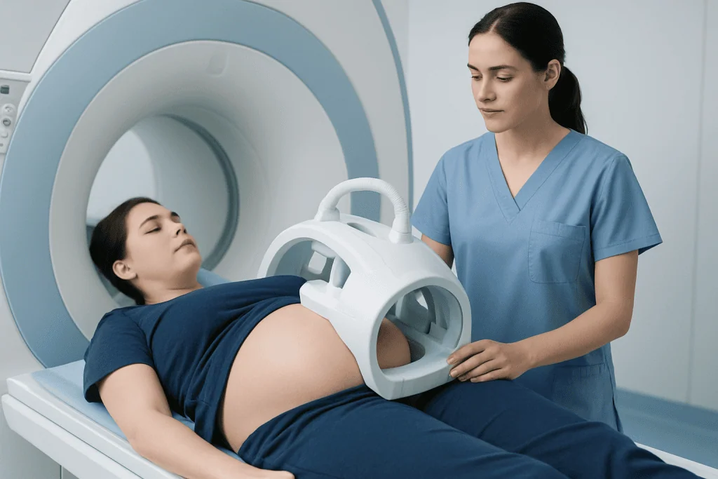
During the scan, the pregnant patient lies on the MRI table while the machine takes images. The team may adjust positioning for comfort and safety, especially later in pregnancy. The test is painless, but the machine can be noisy, so ear protection is usually provided. Some people feel mildly uncomfortable from lying still or from the enclosed space, and they should let the staff know if they feel anxious or unwell.
The scan length can vary depending on the reason for the exam and how much the baby moves. Fetal movement is normal, and radiology teams are used to working around it by taking multiple image sequences. In many cases, parents can return home shortly after the test and resume normal activities unless their doctor advises otherwise.
In centers with advanced prenatal imaging, fetal MRI is often coordinated with pregnancy follow-up and delivery planning so the findings can be reviewed in the context of the overall pregnancy. If a concern affects multiple organs or may require treatment soon after birth, other imaging tools such as ultrasound may continue to be used alongside MRI for ongoing assessment.
How results are interpreted and used
Fetal MRI images are usually reviewed by a radiologist with expertise in prenatal or pediatric imaging, often together with a maternal-fetal medicine specialist. The report is interpreted in the context of ultrasound findings, gestational age, family history, and any genetic or laboratory information available.
Results may do one of several things: confirm an ultrasound finding, provide more detail, change the suspected diagnosis, or show that a previously unclear area appears normal. This can be helpful emotionally as well as medically, because it gives parents a more informed basis for decision-making and counseling.
When a condition is identified or strongly suspected, the information may guide referral to additional specialists. Depending on the finding, the family may meet with neonatology, pediatric neurology, pediatric surgery, genetics, or cardiology teams. If the issue could affect the newborn’s airway, feeding, movement, or surgery needs, delivery planning may be adapted so the right team is available at birth.
It is also important to understand that MRI findings can have limits. Not every developmental issue can be detected prenatally, and some findings may look different later in pregnancy or after delivery. Parents should feel comfortable asking what is known, what remains uncertain, and what follow-up is recommended.
Emotional support, follow-up, and practical planning
Being referred for fetal MRI can be stressful, even when doctors say it is being done out of caution. Parents may experience worry during the waiting period before the scan or while expecting results. Clear communication from the care team can make a meaningful difference, and it is reasonable to ask for explanations in simple language.
Helpful questions may include what the ultrasound showed, what the MRI is expected to clarify, how soon results will be available, and whether the findings are likely to change care during pregnancy or after birth. Some families also benefit from bringing a support person to appointments or writing down questions in advance.
Follow-up after fetal MRI depends on the findings. Some pregnancies continue with routine obstetric care plus a few extra ultrasounds. Others may require monitoring in a fetal medicine clinic, consultations with surgeons or newborn specialists, or plans for delivery in a tertiary care hospital. If surgery or specialized treatment may be needed after birth, coordination across teams becomes especially important.
For international patients seeking coordinated evaluation, Acibadem International’s multidisciplinary specialists and JCI-accredited hospitals diagnose and treat complex fetal and newborn conditions, with care plans tailored to the pregnancy and anticipated neonatal needs.
When to speak with a doctor promptly
Parents should contact their obstetrician, midwife, or fetal medicine team if they have questions about why fetal MRI was recommended or if they do not understand the results. It is also important to ask for medical advice if there are new pregnancy concerns such as bleeding, leaking fluid, severe pain, contractions, fever, or a noticeable change in fetal movements, according to the stage of pregnancy and the advice already given by the care team.
If anxiety about the scan is significant, parents should mention this before the appointment. Imaging teams can often help with practical adjustments and reassurance. In some cases, discussing options in advance can make the experience much easier.
After the scan, families should keep all follow-up appointments, even if they feel well. The purpose of fetal MRI is not only to make a diagnosis but also to support safe pregnancy management and newborn planning. Timely review with the referring doctor ensures the results are placed in the right clinical context.
Whenever there is uncertainty, a qualified clinician is the best source of personalized advice. Prenatal imaging findings can be complex, and decisions are safest when they are based on the full medical picture rather than on general information alone.
Frequently asked questions
Is fetal MRI safe for the baby?
Fetal MRI is generally considered safe when it is medically indicated and performed appropriately during pregnancy. It does not use ionizing radiation, which is one reason it is used when more detail is needed than ultrasound can provide.
Why would a doctor order fetal MRI after an ultrasound?
Ultrasound is usually the first imaging test in pregnancy, but sometimes it cannot fully explain a finding or provide enough detail. Fetal MRI can give clearer information about certain parts of the baby’s body, especially the brain, spine, chest, and abdomen.
At what stage of pregnancy is fetal MRI usually done?
Many fetal MRI exams are performed in the second or third trimester, often after a detailed ultrasound. The exact timing depends on the clinical question and when the images are likely to be most useful.
Does fetal MRI require contrast dye or sedation?
Most fetal MRI exams are done without contrast dye and without sedation. Contrast is usually avoided in pregnancy unless there is a specific reason, and sedation is not commonly needed for the pregnant patient.
How long does a fetal MRI take?
The length of the exam can vary, but it often takes longer than a routine ultrasound because multiple image sequences are needed. Movement of the baby can also affect timing, although radiology teams are experienced in adapting the scan.
What happens if fetal MRI finds an abnormality?
If an abnormality is seen, the findings are usually reviewed with the referring doctor and, if needed, additional specialists. The results can help guide monitoring during pregnancy, delivery planning, and the type of care the baby may need after birth.
References
- American College of Obstetricians and Gynecologists
- Society for Maternal-Fetal Medicine
- Radiological Society of North America
- American College of Radiology
- National Institute of Child Health and Human Development
This article is for general information only and is not a substitute for professional medical advice. Please consult a qualified doctor about your individual situation.
Explore treatments in Turkey — costs, top hospitals & a free quote
JCI-accredited · board-certified surgeons · reply within 24h
Add us as a Preferred Source to see more of our trusted health content across Google Search, AI Overviews and Discover.