Mcl Tear: A Complete Medical Overview

An MCL tear affects the ligament on the inner side of the knee and often happens after a twist or a blow to the outside of the knee. Common symptoms include inner knee pain, swelling, tenderness, and a feeling that the knee is unstable.
Key Takeaways
- An MCL tear affects the ligament on the inner side of the knee and often happens after a twist or a blow to the outside of the knee.
- Common symptoms include inner knee pain, swelling, tenderness, and a feeling that the knee is unstable.
- Mild to moderate MCL tears usually heal without surgery through activity modification, bracing, and physiotherapy.
- Doctors diagnose an MCL tear with a physical exam and sometimes imaging, especially if another knee injury is suspected.
- Prompt medical evaluation is important if walking is difficult, the knee locks, or major swelling or instability develops.
An MCL tear is a stretch or tear of the medial collateral ligament, the band of tissue on the inner side of the knee that helps keep the joint stable. Many MCL injuries improve with rest, bracing, and rehabilitation, but recovery time and treatment depend on how severe the tear is and whether other knee structures are also injured.
Overview
An MCL tear is an injury to the medial collateral ligament, which runs along the inner side of the knee. This ligament helps prevent the knee from bending inward too far and supports stability during walking, turning, and sports. An MCL injury may range from a mild stretch to a partial or complete tear.
This type of knee ligament injury is common in athletes, but it can also happen during daily activities such as slipping, twisting awkwardly, or falling. A direct blow to the outer side of the knee can force the joint inward and strain the ligament. Because the knee contains several important ligaments and cartilage structures, doctors also look for related injuries when an MCL tear is suspected.
Many people recover well with non-surgical care. Rest, ice, a brace, and structured rehabilitation are often enough for healing, especially in lower-grade tears. Severe injuries, or those combined with damage to other parts of the knee, may need more specialized treatment.
Symptoms of an MCL Tear

Symptoms can begin suddenly after an injury or become more noticeable over the next several hours. The most common complaint is pain along the inner side of the knee. Some people feel a sharp pull at the moment of injury, while others mainly notice soreness, swelling, or difficulty moving the joint.
The severity of symptoms usually depends on how much of the ligament is damaged. A mild sprain may cause tenderness with movement but still allow walking. A more serious tear may make the knee feel unstable, as if it could give way during standing or turning.
- Inner knee pain or tenderness
- Swelling around the knee
- Stiffness or reduced range of motion
- Bruising in some cases
- A feeling of looseness or instability
- Discomfort when twisting, pivoting, or going up and down stairs
Because symptoms of knee injuries can overlap, an MCL tear may sometimes occur together with damage such as a meniscus injury or an anterior cruciate ligament problem. If symptoms include catching, locking, or a strong sense of instability, a broader knee assessment is often needed, including evaluation for ACL tear.
Causes and Risk Factors
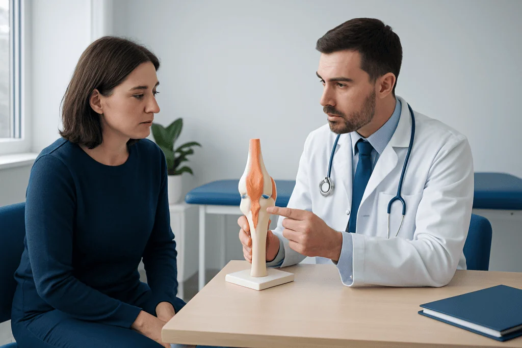
The MCL is commonly injured when the knee is forced inward while the foot remains planted. This can happen during contact sports such as football, soccer, basketball, or skiing, but it is not limited to athletes. Everyday movements, especially sudden twists or falls, can also strain the ligament.
Direct impact is a classic cause. For example, a blow to the outside of the knee during sport can overload the ligament on the inner side. Non-contact mechanisms are also possible, particularly when a person changes direction quickly or lands awkwardly from a jump.
Several factors can increase the risk of an MCL tear:
- Participation in sports that involve cutting, pivoting, or contact
- Poor conditioning or weak supporting muscles around the knee and hip
- Previous knee injuries
- Inadequate warm-up or training errors
- Uneven playing surfaces or inappropriate footwear
Doctors often describe MCL injuries by grade. Grade 1 means the ligament is overstretched but not torn significantly. Grade 2 indicates a partial tear with more noticeable laxity. Grade 3 refers to a complete tear, which causes greater instability and may occur alongside injuries such as meniscus tear.
How Doctors Diagnose It
Diagnosis begins with a medical history and physical examination. A doctor asks how the injury happened, where the pain is felt, whether swelling appeared quickly, and if the knee feels unstable. The pattern of injury can give important clues about which structures may be affected.
During the examination, the doctor checks tenderness along the inner side of the knee, range of motion, swelling, and joint stability. A valgus stress test, in which gentle pressure is applied to the knee, can help assess the MCL. The doctor may also examine other ligaments and the meniscus to look for combined injuries.
Imaging is not always necessary for a straightforward mild sprain, but it can be helpful when symptoms are severe or the diagnosis is unclear. X-rays may be used to rule out a fracture. Magnetic resonance imaging is especially useful for showing soft tissue injuries and can confirm the degree of ligament damage. In some cases, this may be arranged as part of a knee MRI scan to evaluate the MCL and nearby structures in detail.
Accurate diagnosis matters because treatment decisions depend not only on the grade of the MCL tear, but also on whether the person has associated cartilage, bone, or other ligament injuries.
Treatment Options
Treatment depends on the severity of the tear, the person’s activity level, and whether other parts of the knee are injured. Most isolated MCL tears, especially grade 1 and many grade 2 injuries, heal with conservative care. This usually includes reducing activities that worsen pain, applying ice, using compression, and elevating the leg during the early recovery period.
A knee brace may be recommended to support the joint while the ligament heals. Short-term use of crutches can help if walking is painful. Doctors may also suggest pain-relieving or anti-inflammatory medicines when appropriate, based on the individual’s health history.
Rehabilitation is a central part of recovery. Physiotherapy focuses on restoring knee motion, rebuilding strength in the quadriceps and hamstrings, improving balance, and gradually returning the person to normal activity. A structured physical therapy and rehabilitation program can also lower the risk of reinjury by improving movement control.
Surgery is less common for isolated MCL tears because the ligament often heals well on its own. However, an operation may be considered if the tear is complete, the knee remains unstable after rehabilitation, or there are additional injuries such as major ligament damage. In selected complex cases, care may involve evaluation by specialists in knee surgery. Treatment plans should always be individualized after a full orthopedic assessment.
Recovery, Prevention, and Self-care
Recovery time varies. Mild injuries may improve within a few weeks, while more significant tears can take longer and require a gradual return to sports or demanding work. Even when pain improves quickly, the ligament and surrounding muscles still need time to regain full function. Returning too soon can increase the chance of reinjury.
Self-care in the early phase often follows simple protective principles: rest from aggravating activity, ice for comfort, compression if advised, and elevation to help limit swelling. Gentle movement is usually introduced early to prevent stiffness, but exercises should follow medical guidance rather than pain alone.
Prevention focuses on supporting the knee before injury occurs and after recovery:
- Warm up properly before exercise or sport
- Strengthen the thigh, hip, and core muscles
- Practice balance and agility training
- Use proper technique for jumping, landing, and cutting movements
- Wear suitable footwear for the activity and surface
- Follow a gradual return-to-sport plan after injury
People with recurrent knee instability or repeated sports injuries may benefit from a more detailed evaluation. Near the end of the care pathway, some patients choose specialist assessment at centers such as Acibadem International, where multidisciplinary teams in JCI-accredited hospitals diagnose and treat knee ligament injuries for international patients.
When to Seek Medical Care
Any significant knee injury deserves attention if pain is persistent or normal walking is difficult. While some mild sprains improve with simple measures, it is important to seek medical care when symptoms suggest a more serious injury or when recovery is not progressing as expected.
Medical review is especially important if the knee feels unstable, swelling is marked, or the person heard or felt a pop at the time of injury. Prompt assessment can help identify whether the MCL is the only structure involved or whether another ligament, cartilage, or bone injury is present.
- Severe pain or rapid swelling
- Inability to bear weight or walk normally
- The knee gives way, buckles, or feels loose
- Locking, catching, or inability to fully bend or straighten the knee
- Numbness, major bruising, or visible deformity
- Symptoms that do not improve after several days of self-care
Urgent evaluation is particularly important after high-impact trauma, sports injuries with immediate instability, or if symptoms are worsening rather than gradually improving. A qualified doctor can confirm the diagnosis and advise the safest recovery plan.
Frequently asked questions
What is an MCL tear?
An MCL tear is an injury to the medial collateral ligament, which is located on the inner side of the knee. This ligament helps stabilize the knee and prevent it from bending inward too far. The injury can be mild, partial, or complete.
Can an MCL tear heal without surgery?
Yes, many MCL tears heal without surgery, especially grade 1 and grade 2 injuries. Treatment often includes rest, bracing, and rehabilitation exercises to restore strength and motion. Surgery is usually reserved for severe tears or injuries combined with damage to other knee structures.
How long does it take to recover from an MCL tear?
Recovery depends on the severity of the injury and the person’s overall condition. Mild sprains may improve within a few weeks, while more serious tears can take several weeks to months. A doctor or physiotherapist can give a more accurate timeline based on the injury grade and progress.
Is walking with an MCL tear safe?
Some people with mild injuries can still walk, although it may be uncomfortable. If the knee is unstable, swollen, or very painful, walking without support may worsen symptoms or increase the risk of further injury. Medical advice is best if weight-bearing is difficult.
What does MCL tear pain feel like?
Pain is usually felt on the inner side of the knee. It may feel sharp at the moment of injury and then become sore, tender, or stiff afterward. Some people also notice swelling and a sensation that the knee may give way.
How is an MCL tear different from an ACL tear?
An MCL tear affects the ligament on the inner side of the knee, while an ACL tear affects a ligament deeper inside the knee joint. MCL injuries are often linked to pain and tenderness along the inside of the knee, whereas ACL injuries more commonly cause a feeling of instability with pivoting. However, both can occur together, so a proper examination is important.
References
- American Academy of Orthopaedic Surgeons
- National Institute of Arthritis and Musculoskeletal and Skin Diseases
- American Orthopaedic Society for Sports Medicine
- MedlinePlus
- Mayo Clinic
This article is for general information only and is not a substitute for professional medical advice. Please consult a qualified doctor about your individual situation.
Joint and spine care in Turkey — expert assessment & treatment
JCI-accredited · board-certified surgeons · reply within 24h
Add us as a Preferred Source to see more of our trusted health content across Google Search, AI Overviews and Discover.