Understanding Chordoma: A Complete Patient Guide

Chordoma is a rare tumor that most often affects the skull base, sacrum, or mobile spine. Symptoms depend on location and may include headache, vision changes, pain, weakness, or bowel and bladder problems.
Key Takeaways
- Chordoma is a rare tumor that most often affects the skull base, sacrum, or mobile spine.
- Symptoms depend on location and may include headache, vision changes, pain, weakness, or bowel and bladder problems.
- Diagnosis usually involves MRI or CT imaging followed by biopsy reviewed by expert pathologists.
- Treatment often combines surgery and carefully planned radiation therapy.
- Because chordoma can return after treatment, regular long-term monitoring is a key part of care.
Chordoma is a rare tumor that develops from notochord-related cells, usually in the skull base or spine. It often grows slowly, but because it can press on important nerves, the brainstem, or the spinal cord, early specialist assessment and long-term follow-up are important.
Overview: what chordoma is and why it needs specialist care
Chordoma is a rare tumor that arises from remnants of the embryonic notochord, a structure involved in early spinal development. In adults, it most commonly appears at the base of the skull, in the sacrum near the tailbone, or along the mobile spine. Although it usually grows slowly, it can still cause serious problems because it develops close to critical nerves, the brainstem, and the spinal cord.
Many people first learn about chordoma after symptoms have been present for some time. Its slow growth can make it harder to recognize early, and symptoms may resemble those of other spine or nerve conditions. What makes chordoma distinct is not only where it forms, but also the need for careful planning before treatment starts.
Chordoma is generally managed by a multidisciplinary team that may include neurosurgeons, orthopedic spine surgeons, radiation oncologists, radiologists, pathologists, and rehabilitation specialists. This team-based approach helps guide diagnosis, treatment sequencing, and long-term monitoring, which are all important because these tumors can be challenging to remove completely and may recur.
Where chordoma occurs and how location affects symptoms
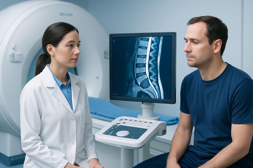
The exact symptoms of chordoma depend largely on its location. Skull base chordomas form near the clivus, a bone deep behind the nose and below the brain. Because this area is close to the brainstem and cranial nerves, even a slow-growing mass can affect eye movement, facial sensation, swallowing, hearing, or balance.
Sacral chordomas develop lower in the spine, near the pelvis and tailbone. These tumors may cause deep, persistent lower back pain, pain when sitting, numbness in the legs or groin, and changes in bowel or bladder function. Some people notice a sense of pelvic pressure or a lump in the lower back area.
Chordomas of the cervical, thoracic, or lumbar spine can lead to neck or back pain, weakness, numbness, or walking difficulty if the tumor presses on the spinal cord or nearby nerves. Depending on symptoms and imaging findings, doctors may also consider other conditions such as spinal tumors or brain tumors before confirming the diagnosis.
Symptoms and warning signs

Chordoma symptoms often begin gradually. In many cases, pain is the earliest sign, especially with tumors in the sacrum or spine. The pain may be dull, persistent, and not clearly linked to an injury. It can slowly worsen over time or become more noticeable at night or while sitting.
When chordoma affects the skull base, symptoms may include headaches, double vision, blurred vision, facial numbness, difficulty swallowing, changes in speech, or hearing problems. Some people also report dizziness or unsteadiness. These symptoms happen because the tumor may press on nearby cranial nerves or structures at the base of the brain.
Symptoms from spinal or sacral chordoma may include:
- Ongoing neck, back, or tailbone pain
- Numbness or tingling in the arms, legs, or groin
- Muscle weakness
- Difficulty walking or balance problems
- Changes in bladder or bowel control
- Pain that radiates into the hips or legs
These symptoms do not always mean chordoma, but they should be assessed if they are persistent, progressive, or associated with neurological changes. Early evaluation can help identify whether the cause is a tumor, a structural spine disorder, or another neurological condition.
Causes, risk factors, and who gets chordoma
Chordoma develops from remnants of notochord tissue left behind after fetal development. In most cases, there is no clear lifestyle-related cause, and people should not assume they did something to bring it on. It is generally considered a sporadic tumor, meaning it arises without an obvious preventable trigger.
Researchers have identified certain molecular features in chordoma cells, including expression of the brachyury protein, which can help support diagnosis. Rare familial cases have been described, but these are uncommon. For most patients, there is no strong inherited pattern and no established screening test for the general population.
Chordoma is more often diagnosed in adults than in children, although it can occur at any age. Risk is not strongly tied to common environmental exposures in the way some other cancers are. Because the condition is rare, diagnosis and treatment are usually best handled in centers familiar with complex skull base and spine tumors.
How chordoma is diagnosed
Diagnosis usually begins with a detailed medical history and neurological examination. Doctors ask about pain, weakness, sensory symptoms, changes in vision or swallowing, and bladder or bowel function. The pattern of symptoms helps determine which areas need imaging and how quickly evaluation should proceed.
MRI is often the main imaging test because it shows the relationship between the tumor and nearby nerves, the brainstem, or the spinal cord. CT scans may also be used to assess bone involvement, especially in the skull base and sacrum. Imaging helps define the tumor’s extent and supports planning for biopsy and treatment.
A biopsy is usually needed to confirm chordoma. This means removing a small tissue sample so a pathologist can examine the cells. Biopsy planning is important because the route used to obtain tissue can affect later surgery. Once confirmed, patients may be referred for advanced treatment planning that can include neurosurgery for skull base or spinal lesions and discussion of whether radiotherapy will be part of care.
After diagnosis, additional imaging may be recommended to look for spread or to map the tumor more precisely before treatment. Although chordoma is often locally aggressive rather than widely metastatic at presentation, staging and careful pre-treatment review are still important.
Treatment options and long-term follow-up
Treatment for chordoma is highly individualized. The main goals are to remove or control the tumor, protect neurological function, and reduce the risk of recurrence. Because chordoma often grows in sensitive areas, treatment planning requires balancing tumor control with preservation of surrounding structures.
Surgery is often the first major treatment when it is feasible and safe. The ideal operation aims to remove as much of the tumor as possible while minimizing harm to nearby nerves, blood vessels, the brainstem, or the spinal cord. The exact surgical approach depends on tumor location; for some patients, this may involve advanced brain and nerve surgery or complex spine-based procedures.
Radiation therapy is commonly used after surgery, or sometimes when complete surgical removal is not possible. Highly targeted methods can help deliver treatment to the tumor while limiting exposure to surrounding tissue. Standard chemotherapy has a limited role in most conventional chordomas, though systemic therapies may be considered in selected cases, recurrent disease, or within clinical trials.
Long-term follow-up is essential because chordoma can come back even after apparently successful treatment. Follow-up usually includes regular MRI scans and clinical review over many years. Rehabilitation, pain management, mobility support, and continence care may also be important parts of recovery depending on the tumor’s location and the treatment performed.
Living with chordoma: recovery, self-care, and emotional support
Recovery from chordoma treatment can take time, especially after major skull base or spinal surgery. Some people need physical therapy to rebuild strength, balance, or walking ability. Others may benefit from occupational therapy, speech and swallowing support, or assistance managing changes in bowel or bladder function.
Self-care focuses on practical recovery rather than home treatment for the tumor itself. Taking medicines as prescribed, attending rehabilitation sessions, going to follow-up imaging appointments, and reporting new neurological symptoms promptly can all help. Good nutrition, gradual activity as advised by the care team, and support for sleep and mental wellbeing may also improve recovery.
Because chordoma is rare, patients and families may feel isolated or overwhelmed. Clear communication with the care team can help them understand the goals of treatment, what follow-up will involve, and which symptoms to watch for. Near the end of the care pathway, some patients seek treatment coordination through centers such as Acibadem International, where multidisciplinary specialists in JCI-accredited hospitals diagnose and treat complex skull base and spine tumors for international patients.
When to seek medical care
Medical care should be sought if back, neck, or tailbone pain is persistent, unexplained, or steadily worsening, especially when it is combined with numbness, weakness, or difficulty walking. Symptoms affecting vision, swallowing, speech, hearing, or facial sensation also deserve prompt assessment, particularly if they are new or progressive.
Urgent evaluation is important if there is sudden weakness, loss of balance, severe headache with neurological symptoms, or any change in bladder or bowel control. These signs can indicate pressure on important nerves, the spinal cord, or structures at the base of the brain and should not be ignored.
Even when symptoms turn out to have another cause, early assessment helps guide the next steps. A qualified doctor can decide whether imaging, referral to a neurologist, neurosurgeon, orthopedic spine specialist, or cancer specialist is needed.
Frequently asked questions
Is chordoma cancer?
Chordoma is generally considered a malignant bone tumor, even though it often grows slowly. It can invade nearby bone and soft tissue, and it may return after treatment, which is why specialist management and follow-up are important.
Where does chordoma usually develop?
Chordoma most often develops at the skull base, in the sacrum near the tailbone, or along the spine. The exact location strongly influences symptoms, treatment options, and the type of specialist involved in care.
Can chordoma be cured?
Some patients can achieve long periods without active disease, especially when the tumor can be removed effectively and treated with well-planned radiation. However, chordoma can recur, so doctors usually recommend long-term imaging and specialist follow-up.
How is chordoma different from other spinal tumors?
Chordoma arises from remnants of the notochord and tends to occur in characteristic midline locations such as the clivus or sacrum. Other spinal tumors may come from nerve tissue, meninges, bone, or metastatic cancer, so imaging and biopsy are needed to make the correct diagnosis.
Is a biopsy always needed for chordoma?
In most cases, yes, a biopsy is needed to confirm the diagnosis before treatment planning. The biopsy should be carefully planned by an experienced team, because the technique and entry path can affect later surgery.
Does chordoma spread to other parts of the body?
Chordoma is often most concerning for its local growth near critical structures, but it can spread in some cases. Doctors may order additional imaging during staging or follow-up to look for recurrence or spread.
References
- National Cancer Institute
- National Organization for Rare Disorders
- American Cancer Society
- National Comprehensive Cancer Network
- World Health Organization
This article is for general information only and is not a substitute for professional medical advice. Please consult a qualified doctor about your individual situation.
Joint and spine care in Turkey — expert assessment & treatment
JCI-accredited · board-certified surgeons · reply within 24h
Add us as a Preferred Source to see more of our trusted health content across Google Search, AI Overviews and Discover.