Nuclear Medicine — Explained by Medical Evidence, Not Myths
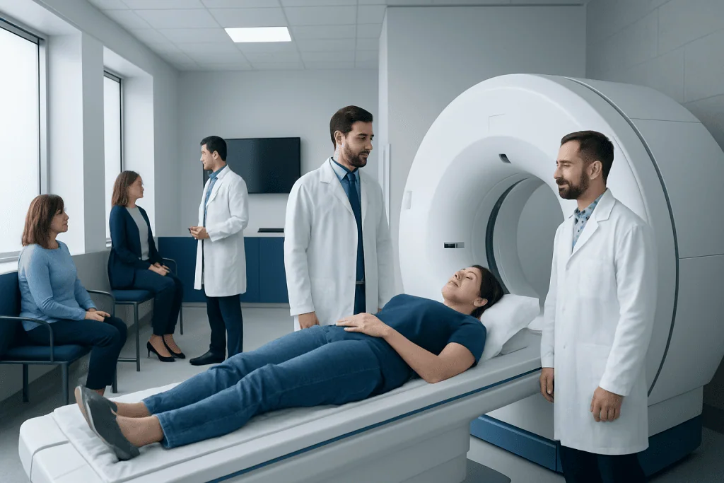
Nuclear medicine can reveal organ function and disease activity before structural changes appear on other scans. It includes both diagnostic tests, such as PET and bone scans, and targeted treatments, such as radioactive iodine therapy.
Key Takeaways
- Nuclear medicine can reveal organ function and disease activity before structural changes appear on other scans.
- It includes both diagnostic tests, such as PET and bone scans, and targeted treatments, such as radioactive iodine therapy.
- The radioactive materials used are carefully selected, controlled, and generally leave the body over time.
- Preparation depends on the test and may include fasting, pausing certain medicines, or avoiding close contact for a short period after treatment.
- Results are interpreted by nuclear medicine specialists together with a patient’s symptoms, lab tests, and other imaging findings.
Nuclear medicine is a medical specialty that uses small amounts of radioactive material to diagnose, monitor, and sometimes treat disease. Unlike many standard imaging tests, it helps show how organs and tissues function, not just how they look.
What nuclear medicine is
Nuclear medicine is a branch of medicine that uses tiny amounts of radioactive substances, called radiotracers or radiopharmaceuticals, to diagnose and sometimes treat disease. These materials are designed to travel to specific organs, bones, or types of cells. A special camera then detects the tracer and creates images that show how the body is working.
This is an important difference from many standard imaging tests. An X-ray, CT scan, or MRI mainly shows anatomy and structure. Nuclear medicine adds functional information, such as blood flow, metabolism, bone activity, or how well the thyroid, kidneys, lungs, or heart are working. In many situations, that functional detail helps doctors identify problems earlier or understand them more clearly.
Nuclear medicine is not one single test. It includes a range of scans and therapies. Common examples are bone scans, thyroid scans, renal scans, lung ventilation-perfusion scans, and PET scans. It also includes certain treatments that deliver radiation directly to diseased tissue, helping limit effects on healthy areas.
How it works and why it is different
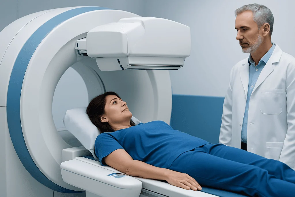
In a nuclear medicine test, the tracer may be injected into a vein, swallowed, or inhaled, depending on the organ being examined. After administration, there is often a waiting period while the tracer circulates and collects in the target tissue. A gamma camera, SPECT scanner, or PET scanner then records signals from the tracer and turns them into images.
Because the tracer participates in normal body processes, doctors can see biological activity rather than only shape. For example, a bone scan can highlight areas of active bone repair, a heart perfusion study can show reduced blood flow to heart muscle, and a PET scan can reveal increased metabolic activity in certain tumors or inflamed tissues.
This functional approach is why nuclear medicine often complements other tests rather than replacing them. A person may have ultrasound, CT, MRI, or blood tests alongside nuclear imaging. When these results are reviewed together, diagnosis is usually more accurate and treatment planning becomes more precise.
Hybrid technologies have made this even more useful. PET/CT and SPECT/CT combine functional and anatomical imaging in the same session. That allows doctors to match areas of abnormal tracer uptake with exact body structures, which can be especially helpful in oncology, cardiology, neurology, and some bone conditions.
Common uses of nuclear medicine
Nuclear medicine is used across many medical specialties. In cancer care, it can help detect disease, stage it, assess whether treatment is working, and monitor for recurrence. A PET scan may be used when doctors need more information about a suspicious area or when evaluating known cancer. In some patients, it works alongside cancer treatment planning and follow-up.
In heart care, nuclear cardiology studies can assess blood flow to the heart muscle during rest and stress. These tests can help evaluate chest pain, possible coronary artery disease, or reduced blood supply after a heart event. Similar techniques can also measure pumping function in certain settings.
In endocrine medicine, nuclear medicine plays an important role in thyroid diagnosis and therapy. Thyroid uptake scans can help explain overactivity, nodules, or other abnormalities. Radioactive iodine may also be used in selected cases of thyroid disease and after some forms of thyroid cancer.
Other examples include bone scans for unexplained bone pain or suspected spread of cancer to bone, kidney scans to assess function and drainage, and lung scans when pulmonary embolism is a concern. Some brain imaging applications are used in specific neurological situations, such as differentiating certain causes of cognitive or movement symptoms.
Safety, radiation exposure, and common myths
One of the most common concerns about nuclear medicine is radiation exposure. The amount used in diagnostic tests is usually small and chosen carefully to answer a specific clinical question. The overall benefit of making an accurate diagnosis often outweighs the risk, especially when the test is ordered for a clear medical reason.
It is also a myth that patients remain dangerously radioactive after every scan. Most diagnostic tracers lose activity quickly and leave the body through urine, stool, or natural decay. Patients are often advised to drink fluids and follow simple instructions after the test. More detailed precautions are usually needed only after certain therapeutic procedures, not routine scans.
Another misunderstanding is that nuclear medicine is only for cancer. In reality, it is also widely used for heart disease, thyroid disorders, bone problems, kidney function, infection evaluation, and some lung and brain conditions. It is better understood as a method of looking at body function than as a test for one disease only.
Pregnancy and breastfeeding require special consideration. A patient should tell the care team if she is pregnant, may be pregnant, or is breastfeeding. In those situations, the test may be postponed, modified, or replaced with another option depending on urgency and clinical need.
What to expect before, during, and after a test
Preparation depends on the type of nuclear medicine study. Some tests require fasting for several hours, while others do not. Patients may be asked to avoid caffeine, pause selected medications, empty the bladder before imaging, or arrive early for tracer administration. The imaging team provides instructions tailored to the specific examination.
During the scan, the patient usually lies still on a table while the camera moves around the body or the table passes through the scanner. The scan itself is typically painless. The injection, if needed, feels similar to a routine blood test or IV line. Some studies are completed within an hour, while others involve waiting periods or delayed images later the same day or even the next day.
After most diagnostic tests, normal activities can be resumed unless the doctor advises otherwise. Drinking extra water may be recommended to help clear the tracer. Results are reviewed by a nuclear medicine physician or radiologist, then shared with the referring doctor, who explains what they mean in the context of the patient’s symptoms and other findings.
Therapeutic nuclear medicine is different from diagnostic imaging and may involve more detailed counseling. Patients receiving radioactive iodine or other targeted therapy may be given temporary safety instructions about contact with others, hygiene, and travel. Following those instructions carefully helps protect family members and the public.
How doctors use the results
Nuclear medicine results are rarely interpreted in isolation. Doctors compare the scan with a patient’s medical history, physical examination, blood tests, pathology reports, and other imaging. This helps avoid overinterpretation, because tracer uptake can sometimes reflect inflammation, healing, or normal variation rather than a serious disease.
In many cases, nuclear medicine helps answer one of three practical questions: Is there disease present? How active is it? Is treatment working? That is why these tests are useful not only for diagnosis, but also for follow-up. For example, they can help assess whether a tumor has responded to therapy or whether heart muscle is receiving enough blood flow.
Sometimes a scan leads to further testing rather than an immediate diagnosis. A suspicious result may need confirmation with biopsy, endoscopy, ultrasound, CT, or MRI. In other situations, a normal nuclear medicine study can provide reassurance and help avoid more invasive procedures.
Because the technology is specialized, interpretation is best done by experienced teams. In centers where oncologists, cardiologists, endocrinologists, surgeons, and imaging specialists work together, results can be integrated into a clearer care plan. This may be especially helpful for complex conditions such as lung cancer or advanced thyroid disorders.
Treatment uses of nuclear medicine
Nuclear medicine is not limited to diagnosis. Some radiopharmaceuticals are used therapeutically, delivering radiation directly to overactive tissue or cancer cells. This targeted approach aims to treat the problem area while reducing exposure to the rest of the body.
A well-known example is radioactive iodine treatment for selected thyroid conditions, including hyperthyroidism and some forms of thyroid cancer care after surgery. Other therapies are used in certain cancers, including treatments that target bone metastases or specific tumor receptors. Whether a patient is suitable depends on the exact diagnosis, stage of disease, and overall health.
These treatments are planned carefully. Before therapy, doctors review prior scans, pathology, medications, kidney function, and pregnancy status where relevant. The patient also receives detailed instructions about preparation and aftercare, including temporary radiation safety measures when needed.
For patients needing coordinated evaluation and treatment, multidisciplinary services may include radiation oncology and surgical or medical specialties alongside nuclear medicine. Near the end of the care pathway, centers such as Acibadem International may support international patients through diagnosis and treatment at JCI-accredited hospitals with multidisciplinary specialist teams.
When to seek medical care
A person should seek medical care if a doctor recommends nuclear medicine to investigate symptoms that remain unexplained, such as persistent bone pain, ongoing thyroid symptoms, chest discomfort, unexplained weight loss, or concerns raised on blood tests or other scans. The test itself is not a treatment for symptoms, but it may help clarify the cause and guide the next step.
Medical advice is also important if a patient is pregnant, trying to conceive, or breastfeeding and has been scheduled for a scan or therapy. The care team can explain whether the test should proceed, be adapted, or be replaced. Patients should also report allergies, kidney problems, recent imaging tests, and all medicines and supplements they take.
After a nuclear medicine procedure, a patient should contact the healthcare team if instructions are unclear, side effects seem unusual, or symptoms worsen rather than improve. Most diagnostic scans do not cause significant after-effects, but ongoing symptoms always deserve medical review. For emergencies such as severe chest pain, sudden shortness of breath, or neurological symptoms, urgent care is needed immediately.
Frequently asked questions
Is nuclear medicine the same as a CT scan or MRI?
No. CT and MRI mainly show body structure, while nuclear medicine shows how tissues and organs function. In some cases, these methods are combined, such as PET/CT or SPECT/CT, to provide both functional and anatomical information.
Is nuclear medicine safe?
For most patients, nuclear medicine is considered safe when it is used appropriately and supervised by trained professionals. The radioactive dose is typically small for diagnostic tests, and the care team weighs the benefit of the information gained against the potential risk.
How long does a nuclear medicine scan take?
It depends on the type of study. Some scans are completed within about an hour, while others include a waiting period after the tracer is given and may take several hours or require delayed images. The imaging team usually explains the timeline in advance.
Will I feel anything from the radioactive tracer?
Most people do not feel the tracer working in the body. If it is injected, the main sensation is usually just the brief needle stick. The scan itself is generally painless, although lying still can be uncomfortable for some patients.
Do I need to avoid other people after a nuclear medicine test?
After most routine diagnostic scans, special restrictions are minimal or not needed. After certain treatments, such as radioactive iodine therapy, patients may need temporary precautions to limit close contact. The care team gives specific instructions based on the exact radiopharmaceutical used.
Can nuclear medicine detect cancer?
It can help detect, stage, and monitor many cancers, but it is not used alone in every case. Findings are interpreted alongside symptoms, lab results, biopsy results, and other imaging. An abnormal scan does not always mean cancer, because inflammation and healing can also affect tracer uptake.
This article is for general information only and is not a substitute for professional medical advice. Please consult a qualified doctor about your individual situation.
Explore treatments in Turkey — costs, top hospitals & a free quote
JCI-accredited · board-certified surgeons · reply within 24h
Add us as a Preferred Source to see more of our trusted health content across Google Search, AI Overviews and Discover.

More from the Health Library

Tachypnea: An Evidence-Based Guide for Patients

Impulse Control: An Evidence-Based Guide for Patients

Herbal: An Evidence-Based Guide for Patients

Imdur: A Complete Medical Overview

Cat Nap: A Complete Medical Overview