Brain Lesions: What Patients Need to Know

A brain lesion is a descriptive term for an abnormal area in the brain, not a single disease. Some brain lesions cause no symptoms and are found incidentally, while others may affect movement, speech, vision, balance, or thinking.
Key Takeaways
- A brain lesion is a descriptive term for an abnormal area in the brain, not a single disease.
- Some brain lesions cause no symptoms and are found incidentally, while others may affect movement, speech, vision, balance, or thinking.
- Common causes include stroke, infection, inflammation, injury, vascular abnormalities, and tumors.
- MRI is often the most useful imaging test to help characterize a brain lesion.
- Treatment may range from monitoring to medicines, rehabilitation, or surgery depending on the cause.
- New or sudden neurological symptoms need prompt medical assessment.
Brain lesions are areas of abnormal or damaged tissue in the brain seen on imaging such as MRI or CT. They are not a diagnosis by themselves, and the next steps depend on the lesion’s cause, size, location, symptoms, and whether it is stable or changing.
Overview: what brain lesions mean
Brain lesions are areas of tissue in the brain that look abnormal because they have been injured, inflamed, infected, deprived of blood flow, or changed by another underlying process. In practical terms, the phrase usually describes a finding on a brain scan rather than a final diagnosis. A lesion may be very small or larger, single or multiple, and may appear in different parts of the brain.
Some brain lesions are discovered while a person is being evaluated for symptoms such as headaches, seizures, weakness, or memory changes. Others are found by chance during imaging done for an unrelated reason. A lesion can sometimes remain stable for years and never cause meaningful problems, while in other situations it reflects a condition that needs treatment.
Because the brain controls many body functions, symptoms vary widely depending on where the lesion is located. The same imaging term can refer to very different causes, so doctors look at the person’s medical history, neurological examination, blood tests, and scan features together. That fuller picture helps determine whether a lesion is harmless, active, or urgent.
In some cases, a brain lesion may be related to a brain tumor or another structural condition, but many lesions are not cancer. This is why patient-friendly, step-by-step evaluation is important: the key question is not only whether a lesion exists, but what type it is and whether it is changing.
Symptoms and how they can vary
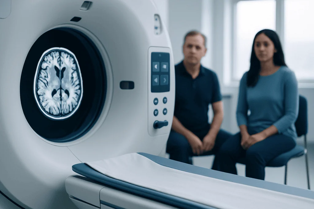
Brain lesions do not always cause symptoms. When they do, the pattern often depends on the lesion’s size, speed of development, and exact location. A small lesion in a sensitive area can cause noticeable symptoms, while a larger lesion elsewhere may remain silent for some time.
Possible symptoms can include persistent headaches, nausea, seizures, weakness or numbness on one side of the body, balance problems, dizziness, vision changes, speech difficulty, memory problems, personality or behavior changes, and confusion. Some people notice reduced coordination or trouble with everyday tasks. In children, symptoms may also include developmental changes or new difficulties at school.
Symptoms may begin suddenly, as can happen with bleeding or a stroke, or they may develop gradually over weeks or months. Sudden symptoms usually need urgent assessment because they can indicate a time-sensitive problem. Gradual symptoms also deserve medical review, especially if they are worsening.
- Headache that is new, persistent, or different from usual
- Seizures or fainting episodes
- Weakness, numbness, or facial droop
- Problems with vision, speech, memory, or concentration
- Unsteady walking or poor coordination
- Changes in mood, behavior, or alertness
Common causes and risk factors

Brain lesions can result from many different conditions. Reduced blood flow from an ischemic stroke, bleeding in or around the brain, infections such as abscesses, autoimmune inflammation, trauma, and problems affecting blood vessels are all possible causes. Demyelinating diseases that affect the brain’s protective nerve covering, such as multiple sclerosis, can also produce lesions that appear on MRI.
Other causes include benign or malignant tumors, congenital abnormalities, radiation effects, and older areas of scarring from a prior injury or infection. Sometimes age-related small vessel disease leads to tiny white matter lesions, especially in people with high blood pressure, diabetes, or vascular risk factors. These lesions may be found during evaluation for headaches, dizziness, or memory concerns.
Risk factors depend on the underlying cause. For example, stroke-related lesions are more likely in people with high blood pressure, smoking history, high cholesterol, diabetes, obesity, atrial fibrillation, or heart disease. Infection-related lesions may be more likely in people with weakened immune systems. Traumatic lesions can follow falls, sports injuries, or accidents.
Not every lesion can be linked to a clear risk factor right away. This can feel uncertain for patients, but it is common. Doctors often need imaging details, follow-up scans, or occasionally tissue sampling to tell whether a lesion represents inflammation, infection, vascular change, or a tumor.
How doctors diagnose brain lesions
Diagnosis usually begins with a careful history and neurological examination. The doctor asks when symptoms started, whether they are getting worse, and whether there are related issues such as fever, cancer history, recent infection, immune problems, trauma, or vascular risk factors. This information helps place the imaging finding in context.
Imaging is central to diagnosis. CT scans are often used urgently because they are quick and good at detecting bleeding, swelling, or major structural changes. MRI usually provides more detailed information about the lesion’s location, shape, and tissue characteristics. In many cases, doctors rely on advanced MRI scanning to distinguish among inflammation, stroke, tumor, infection, and old scarring.
Additional tests may include blood work, vascular imaging, electroencephalography if seizures are suspected, or lumbar puncture in selected cases. Sometimes repeat imaging after a period of time is the safest and most useful next step, especially if the lesion is small and the patient is stable. Growth, change in appearance, or new symptoms can provide important clues.
If a lesion appears suspicious for a tumor or remains uncertain after noninvasive testing, a neurosurgical opinion may be recommended. In selected situations, doctors may advise a biopsy or a procedure linked to neurosurgery to establish a definite diagnosis and guide treatment planning.
Treatment options depend on the cause
There is no single treatment for brain lesions because treatment targets the underlying condition. Some lesions only need observation with repeat MRI scans, especially if they are small, stable, and not causing symptoms. Others require medicines such as antibiotics for infection, anti-inflammatory treatment for autoimmune disease, antiseizure medication, or therapies to reduce stroke risk.
If a lesion causes significant pressure, bleeding, blockage of fluid flow, or concern for tumor, procedural treatment may be needed. Surgery may be used to remove a mass, drain an abscess, stop bleeding, or obtain tissue for diagnosis. When cancer is confirmed, management may include a combination of surgery, radiotherapy, and systemic treatment based on the exact tumor type.
For some vascular lesions or problems with brain blood vessels, minimally invasive procedures may be considered. Depending on the diagnosis, doctors may discuss options in interventional neuroradiology to treat certain abnormalities through image-guided techniques rather than open surgery.
Recovery often includes more than treating the lesion itself. Rehabilitation can help people regain strength, speech, balance, memory, or independence after a neurological injury. Even when a lesion cannot be removed, careful symptom management and supportive therapy can make a meaningful difference in daily life.
Living with a brain lesion: monitoring and self-care
For many patients, the main question after diagnosis is what daily life will look like. The answer depends on the cause and whether the lesion is active or stable. Some people simply need periodic follow-up scans and routine neurological review. Others may need ongoing treatment, rehabilitation, or monitoring for seizures and other complications.
Practical self-care focuses on protecting brain and vascular health. This includes taking prescribed medicines regularly, attending follow-up appointments, controlling blood pressure, blood sugar, and cholesterol, not smoking, limiting alcohol, sleeping well, and staying physically active within medical advice. If seizures are possible or have occurred, patients should follow safety guidance about driving, swimming, heights, and operating machinery.
It can also help to track symptoms over time. A simple record of headaches, weakness, speech changes, seizure-like episodes, or memory concerns can help the care team recognize patterns. Family members may notice subtle changes in mood, attention, or functioning that are worth mentioning during appointments.
Emotional support matters too. Uncertainty about a scan result can be stressful even when the lesion turns out to be benign or stable. Clear communication with the treating team, written questions before appointments, and asking when repeat imaging is needed can help patients feel more informed and reassured.
When to seek medical care
Immediate medical attention is important for sudden neurological symptoms. These include sudden weakness or numbness, facial droop, trouble speaking, sudden severe headache, new seizures, loss of consciousness, confusion, or sudden vision loss. These symptoms can signal stroke, bleeding, or another urgent problem and should not be ignored.
Medical review is also appropriate for symptoms that are less sudden but persistent or progressive. Ongoing headaches, worsening balance, repeated vomiting, new memory problems, personality changes, or steadily increasing weakness should be assessed by a doctor. The same is true when a known brain lesion is being monitored and symptoms change.
If imaging has already shown a lesion, patients should ask what the lesion most likely represents, whether more tests are needed, and when the next scan should be done. If the situation is complex, care from a multidisciplinary team can be helpful. Acibadem International’s multidisciplinary specialists and JCI-accredited hospitals diagnose and treat brain lesions for international patients, including imaging evaluation and advanced neurological care.
Prompt assessment does not always mean the cause is serious, but it helps ensure that urgent conditions are recognized early and treatable causes are not missed. When symptoms involve the brain or nervous system, timely medical guidance is the safest approach.
Frequently asked questions
Are brain lesions always cancer?
No. Brain lesions are not the same as cancer. A lesion is a general term for an abnormal area seen on imaging, and it may be caused by stroke, inflammation, infection, injury, vascular changes, or a tumor.
Can brain lesions be harmless?
Yes, some brain lesions are incidental findings and remain stable without causing symptoms. Even so, a doctor should interpret the imaging result because the significance depends on the lesion’s location, appearance, and the person’s medical history.
What is the best scan for brain lesions?
MRI is often the most informative scan for characterizing brain lesions because it shows brain tissue in greater detail than CT. CT is still very useful, especially in emergencies such as suspected bleeding or major swelling.
Do brain lesions go away?
Some do, depending on the cause. Inflammatory or infection-related lesions may improve with treatment, while old scars or stroke-related changes may remain visible even after symptoms improve.
Can brain lesions cause headaches?
They can, but not every headache is caused by a brain lesion. Headaches are common and often have other explanations, so doctors look at the whole pattern of symptoms, the examination, and imaging findings together.
When is surgery needed for a brain lesion?
Surgery may be considered when a lesion is causing pressure, bleeding, blockage, seizures that are hard to control, or concern for a tumor or abscess. In some cases, surgery is done to remove the lesion, and in others it is done to obtain tissue for diagnosis.
This article is for general information only and is not a substitute for professional medical advice. Please consult a qualified doctor about your individual situation.
Explore treatments in Turkey — costs, top hospitals & a free quote
JCI-accredited · board-certified surgeons · reply within 24h
Add us as a Preferred Source to see more of our trusted health content across Google Search, AI Overviews and Discover.
Check your numbers in seconds
BMI, calories, due date, blood pressure and 30+ more clinical calculators — free, instant, doctor-reviewed ranges.

More from the Health Library

Meconium: A Complete Medical Overview

B12 Deficiency: An Evidence-Based Guide for Patients

Strawberry Tongue: A Complete Medical Overview

Nose Leaking Clear Fluid: A Complete Medical Overview

Bac Chart: A Complete Medical Overview