Bibasilar Atelectasis: A Complete Medical Overview

Bibasilar atelectasis affects the bases of both lungs and is often seen on chest imaging. It may happen after surgery, with shallow breathing, mucus plugging, infection, or pressure on the lungs.
Key Takeaways
- Bibasilar atelectasis affects the bases of both lungs and is often seen on chest imaging.
- It may happen after surgery, with shallow breathing, mucus plugging, infection, or pressure on the lungs.
- Symptoms range from none at all to cough, chest discomfort, and shortness of breath.
- Treatment depends on the cause and may include deep-breathing exercises, airway clearance, oxygen, or treatment of an underlying lung condition.
- Prompt medical evaluation is important if breathing trouble is new, severe, or accompanied by fever, chest pain, or low oxygen levels.
Bibasilar atelectasis means part of both lower lungs is not fully expanding, usually because the tiny air sacs have partially collapsed or filled with mucus. It is a finding with many possible causes rather than a disease by itself, and treatment focuses on improving lung expansion and addressing the reason it developed.
Overview
Bibasilar atelectasis is partial collapse or incomplete expansion of the lower portions of both lungs. The term bibasilar refers to the bases of the lungs, and atelectasis means that some of the tiny air sacs, called alveoli, are not opening normally. This can reduce the amount of oxygen moving into the blood, although the effect may be mild or significant depending on how much lung is involved.
It is important to understand that bibasilar atelectasis is usually a radiology finding, not a final diagnosis on its own. A chest X-ray or CT scan may describe it when the lower lungs appear underinflated, compressed, or blocked by mucus. Doctors then look for the underlying reason, such as recent surgery, infection, pain causing shallow breathing, or another chest condition.
Many people recover well once the cause is treated and normal breathing patterns return. In other cases, especially in older adults or people with chronic lung disease, it may need closer monitoring to prevent complications such as low oxygen levels or pneumonia.
How the Lower Lungs Become Affected
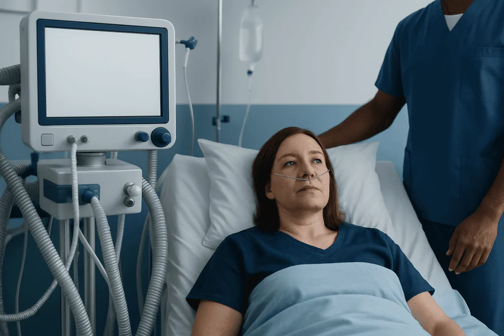
The lungs are made of millions of alveoli that open with each breath. When breathing is shallow, airflow is blocked, or pressure from outside the lung prevents expansion, some alveoli may collapse. The lower lobes are especially prone to this because gravity, body position, and reduced movement after illness or surgery can all make ventilation at the lung bases less effective.
Bibasilar atelectasis can occur in two broad ways. In obstructive atelectasis, a mucus plug, inhaled material, or less commonly a mass blocks the airway leading to part of the lung. In non-obstructive atelectasis, the problem comes from poor expansion rather than a blockage, such as after anesthesia, with pain from rib fractures, or when fluid around the lungs compresses them.
Because the lower lung regions are often where fluid, secretions, and pressure effects collect, these areas can show changes first on imaging. That is why bibasilar atelectasis is commonly mentioned in hospitalized patients, after surgery, or during evaluation for other respiratory concerns.
Symptoms and Possible Signs
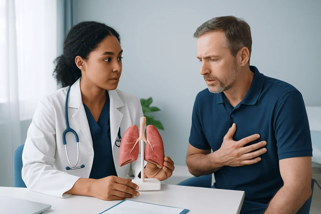
Bibasilar atelectasis may cause no obvious symptoms, especially when it is small and discovered incidentally on imaging. When symptoms do occur, the most common are shortness of breath, faster breathing, a dry or productive cough, and reduced exercise tolerance. Some people describe a sense that they cannot take a full deep breath.
Other possible signs include chest discomfort, fatigue, and lower oxygen levels. After surgery, a person may feel reluctant to breathe deeply because of pain, which can worsen the problem. On examination, a clinician may hear reduced breath sounds at the lung bases or notice signs of breathing effort.
Symptoms depend less on the phrase “bibasilar atelectasis” itself and more on how much lung is affected and what is causing it. For example, if it is related to infection, symptoms may overlap with pneumonia, including fever and mucus production. If it is due to fluid around the lungs, symptoms may come on gradually with increasing breathlessness.
Causes and Risk Factors
One of the most common settings for bibasilar atelectasis is the period after surgery, especially abdominal, chest, or upper abdominal procedures. Anesthesia, pain, reduced coughing, and lying in bed can all lead to shallow breathing and retained secretions. This postoperative form is often temporary and improves with breathing exercises and mobilization.
Other causes include respiratory infections, mucus plugging, asthma flare-ups, chronic obstructive lung disease, aspiration, chest trauma, and pressure from outside the lungs. Pleural effusion, a build-up of fluid around the lungs, can compress the lower lobes and create bibasilar changes on imaging. In some cases, obesity, prolonged bed rest, neuromuscular weakness, or sedation may contribute by limiting deep breathing.
Doctors also consider whether another condition is present that affects lung expansion or airflow. Relevant related problems may include chronic obstructive pulmonary disease or pleural effusion. Less commonly, a tumor, enlarged lymph node, or airway narrowing can lead to localized collapse by blocking an airway, which is why persistent or unexplained atelectasis may need further evaluation.
- Recent surgery or anesthesia
- Pain that limits deep breathing
- Mucus retention or ineffective cough
- Lung infection or inflammation
- Fluid around the lungs or external compression
- Smoking, chronic lung disease, or prolonged immobility
How Bibasilar Atelectasis Is Diagnosed
Diagnosis usually begins with symptoms, medical history, and a physical examination. The clinician asks about recent surgery, fever, cough, chest pain, smoking, known lung disease, aspiration risk, or worsening shortness of breath. Oxygen saturation may be checked to see whether the collapse is affecting breathing efficiency.
Imaging is central to diagnosis. A chest X-ray often identifies bibasilar atelectatic changes, while a CT scan can provide more detail if the cause is unclear or if a blockage, infection, or other structural problem is suspected. In some patients, doctors may also order blood tests, arterial blood gas testing, or sputum studies if infection or significant respiratory compromise is a concern.
If an airway blockage is possible, bronchoscopy may be used to look inside the airways and sometimes remove mucus plugs. Pulmonary function testing is not always needed immediately, but it may help in ongoing assessment of chronic respiratory problems. In many hospitals, imaging and specialist assessment work together with diagnostic imaging to clarify whether the finding is mild, temporary, or part of a broader lung condition.
Treatment Options and Recovery
Treatment for bibasilar atelectasis aims to reopen the affected air sacs and correct the underlying cause. For many patients, simple supportive measures are very effective. These may include deep-breathing exercises, coughing, early walking, position changes, and use of an incentive spirometer after surgery. Good pain control is also important so a person can breathe more fully.
When secretions are involved, chest physiotherapy, hydration, humidified air, and airway clearance techniques may help. If low oxygen levels are present, supplemental oxygen can be used while the lungs recover. If there is an associated infection, the doctor may treat the specific cause, and if an obstructing mucus plug is suspected, bronchoscopy may be considered to clear the airway. Some patients benefit from specialist care in chest diseases services when symptoms are persistent or the cause is complex.
If external compression is the problem, treatment targets that issue directly. For example, large pleural effusions may need drainage, and tumors or other structural causes may require further testing and tailored management. Surgery is not a routine treatment for bibasilar atelectasis itself, but it may be needed for the underlying condition in selected cases.
Recovery time varies. Mild postoperative atelectasis may improve within days, while atelectasis linked to chronic lung disease, recurrent aspiration, or significant infection can take longer. Follow-up imaging may be recommended if symptoms continue or if the initial finding was extensive.
Prevention and Self-care
Prevention focuses on keeping the lungs well expanded and the airways clear. After surgery or during recovery from illness, regular deep breaths, coughing as instructed, sitting upright, and walking as soon as it is safe can reduce the risk of bibasilar atelectasis. If an incentive spirometer is provided, using it correctly and consistently can be helpful.
People with chronic lung conditions should follow their treatment plan and seek help early when symptoms worsen. Stopping smoking, staying hydrated, and managing mucus-producing illnesses promptly can support better lung function. For those with swallowing difficulty or aspiration risk, careful eating habits and specialist assessment may reduce repeated lung problems.
At home, self-care should not replace medical review when shortness of breath is significant. Still, recovery often improves with rest balanced by movement, attention to breathing exercises, and treatment of any related condition. In international care settings, Acibadem International’s multidisciplinary specialists and JCI-accredited hospitals evaluate respiratory findings and provide personalized treatment plans for patients who need further assessment.
When to Seek Medical Care
Medical care should be sought promptly if shortness of breath is new, worsening, or severe. Immediate evaluation is especially important if symptoms are accompanied by chest pain, bluish lips or fingertips, confusion, fainting, high fever, or very low oxygen readings. These signs can point to significant breathing impairment or another urgent condition.
A doctor should also be consulted if cough, fever, or fatigue continue after surgery or a recent respiratory illness, or if imaging has shown bibasilar atelectasis without a clear explanation. Persistent collapse may need more detailed evaluation to rule out mucus plugging, infection, fluid around the lungs, or airway obstruction.
People with chronic respiratory disease, weakened immunity, or older age should not ignore subtle changes in breathing. Early medical review can help prevent complications and guide appropriate treatment, including further work-up through services such as pulmonary rehabilitation when reduced lung function is affecting recovery.
Frequently asked questions
Is bibasilar atelectasis serious?
It can be mild and temporary, especially after surgery, but its importance depends on how much of the lungs are affected and what caused it. Small areas may cause few symptoms, while larger areas or underlying infection can lead to more noticeable breathing problems.
Can bibasilar atelectasis go away on its own?
Mild cases sometimes improve quickly with deep breathing, movement, coughing, and recovery from the trigger that caused it. However, it should not be assumed to resolve without evaluation if symptoms are significant or persistent.
What does bibasilar atelectasis look like on an X-ray?
On a chest X-ray, it often appears as areas of increased density or underexpansion near the lower parts of both lungs. The report may describe linear, plate-like, or patchy bibasal opacities, but the exact wording varies.
Is bibasilar atelectasis the same as pneumonia?
No. Atelectasis means part of the lung is not fully inflated, while pneumonia is an infection of the lung tissue. The two can look similar on imaging and may occur together, so doctors use symptoms, examination, and sometimes further tests to tell them apart.
How is bibasilar atelectasis treated after surgery?
Postoperative treatment usually focuses on deep-breathing exercises, incentive spirometry, coughing, pain control, and early walking. These measures help reopen the lower lungs and reduce mucus build-up.
Can bibasilar atelectasis cause low oxygen levels?
Yes, it can, because collapsed or poorly expanded air sacs do not exchange oxygen as effectively. The degree of oxygen drop depends on the extent of the atelectasis and whether other lung or heart conditions are present.
References
- American Thoracic Society
- National Heart, Lung, and Blood Institute
- MedlinePlus
- Merck Manual Consumer Version
- Radiological Society of North America
This article is for general information only and is not a substitute for professional medical advice. Please consult a qualified doctor about your individual situation.
Explore treatments in Turkey — costs, top hospitals & a free quote
JCI-accredited · board-certified surgeons · reply within 24h
Add us as a Preferred Source to see more of our trusted health content across Google Search, AI Overviews and Discover.

More from the Health Library

Hydrogen Peroxide Ear Wax — Explained by Medical Evidence, Not Myths

Alfuzosin — Explained by Medical Evidence, Not Myths

Seropositive: An Evidence-Based Guide for Patients

Night Blindness: What Patients Need to Know

Inderal for Anxiety: What Patients Need to Know