

Cardiac CT Scan — Explained by Medical Evidence, Not Myths
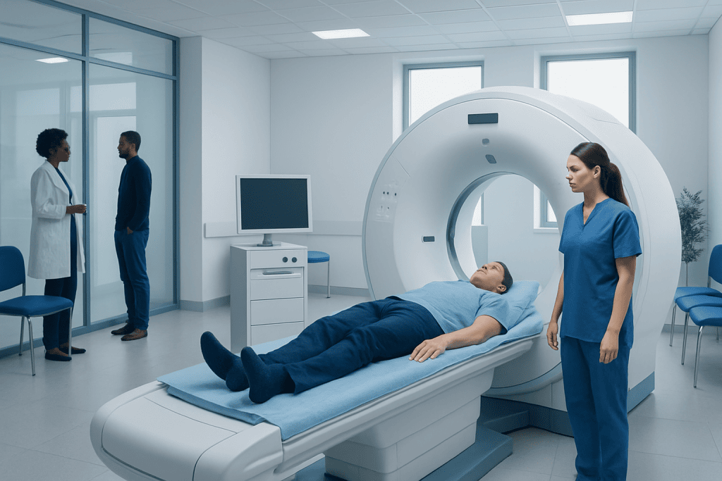
A cardiac CT scan provides detailed images of the heart and coronary arteries without surgery. It is commonly used to assess calcium buildup, possible artery narrowing, and some structural heart concerns.
Key Takeaways
- A cardiac CT scan provides detailed images of the heart and coronary arteries without surgery.
- It is commonly used to assess calcium buildup, possible artery narrowing, and some structural heart concerns.
- The test may involve contrast dye and sometimes heart-rate control medicines, depending on the type of scan.
- Results must be interpreted in the context of symptoms, risk factors, and other heart tests.
- Cardiac CT is useful, but it is not the right test for every person or every heart problem.
A cardiac CT scan is a noninvasive imaging test that uses X-rays and computer processing to create detailed pictures of the heart and blood vessels. It can help evaluate coronary artery disease, calcium buildup, heart structure, and certain causes of chest symptoms when used for the right medical reasons.
What a Cardiac CT Scan Is
A cardiac CT scan is a specialized computed tomography test that creates detailed cross-sectional images of the heart. In simple terms, it is an advanced X-ray technique that takes many pictures from different angles and combines them into highly detailed views of the heart muscle, chambers, valves, nearby vessels, and especially the coronary arteries that supply blood to the heart.
This test is often used to answer specific questions rather than as a general screening tool for everyone. Depending on the reason for testing, a clinician may order a coronary artery calcium scan to measure calcified plaque, or a coronary CT angiography to look more closely at the coronary arteries for narrowing or blockage. These are related tests, but they are not identical and are used for different clinical decisions.
One reason cardiac CT has become so valuable is that it can provide anatomical detail quickly and noninvasively. That means it may help clarify whether symptoms such as chest discomfort could be related to coronary artery disease or whether heart anatomy appears normal. It can also support planning for further care if another treatment or procedure is being considered.
Why Doctors Order a Cardiac CT Scan
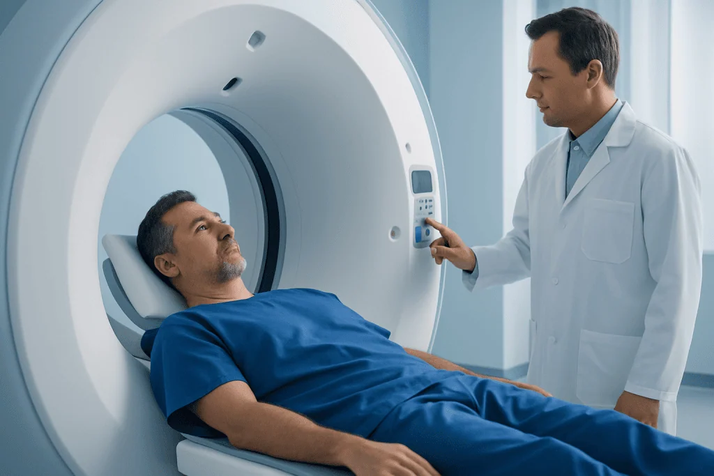
Doctors usually request a cardiac CT scan when they need a clearer picture of heart structure or coronary artery anatomy. It may be considered in people with chest pain or shortness of breath when the likelihood of coronary artery disease is uncertain, or when other tests have not given a clear answer. It can also help evaluate congenital heart anatomy, the aorta around the heart, or some abnormalities of the heart muscle and surrounding structures.
A coronary calcium scan is often used to refine cardiovascular risk in selected adults who do not have symptoms but may have risk factors such as high blood pressure, diabetes, smoking, high cholesterol, or a strong family history of heart disease. A higher calcium score suggests a greater burden of atherosclerotic plaque and may help guide prevention discussions about lifestyle changes and medication.
Coronary CT angiography may be used when a more detailed look at the coronary arteries is needed. This can help identify plaque, narrowing, or features that suggest a higher-risk pattern of disease. In some patients, it may reduce the need for more invasive testing, while in others it helps determine whether additional assessment such as coronary angiography is appropriate.
- To look for narrowing in the coronary arteries
- To measure coronary calcium burden
- To assess certain structural heart abnormalities
- To evaluate selected causes of chest symptoms
- To help guide prevention or treatment planning
How the Test Works and What to Expect

Before the scan, the care team usually reviews medical history, kidney function, allergies, and current medications. If contrast dye is planned, patients may be asked not to eat for a few hours beforehand. Sometimes caffeine and smoking are avoided before the test because they can affect heart rate. In many centers, the team may also monitor the heart rhythm and occasionally give medication to slow the heart rate so the images are sharper.
During the scan, the patient lies on a table that moves through the CT scanner. Sticky patches are placed on the chest to record the heartbeat, allowing the scan to be timed with the cardiac cycle. For a calcium score scan, contrast is usually not needed. For coronary CT angiography, contrast dye is injected through a vein to make the coronary arteries more visible.
The test itself is usually brief. Patients may be asked to hold their breath for short periods to reduce motion. The scan is painless, although the contrast dye can cause a temporary warm sensation or metallic taste. Afterward, most people return to normal activities soon, unless they have been given medication that requires short observation.
Some patients undergo cardiac CT as part of a wider heart evaluation that may also include echocardiography or other functional tests. Each test answers different questions, so the best choice depends on symptoms, age, medical history, and the specific concern the doctor is investigating.
What the Results Can Show
The information from a cardiac CT scan depends on the type of scan performed. A calcium score estimates how much calcified plaque is present in the coronary arteries. A score of zero may suggest a low likelihood of calcified plaque at that point in time, while higher scores indicate increasing plaque burden. However, calcium scoring does not show every kind of plaque and does not replace a full medical evaluation.
Coronary CT angiography can show whether the coronary arteries appear normal, mildly narrowed, or significantly narrowed. It may also detect plaque that has not yet caused a severe blockage. This is helpful because heart risk is not determined only by symptoms; plaque burden and plaque pattern also matter. In some cases, the scan may suggest whether follow-up testing or treatment is needed.
Cardiac CT can also reveal other findings, such as enlargement of heart chambers, certain congenital abnormalities, changes in the aorta, or fluid around the heart. Still, a scan result should never be interpreted in isolation. Doctors combine the images with symptoms, physical examination, blood pressure, cholesterol levels, diabetes status, and other test results to build a more complete picture.
If significant narrowing is suspected, the next step may involve specialist evaluation and treatment planning, which can include medicines, lifestyle measures, or procedures such as coronary artery bypass grafting in carefully selected patients. The scan helps guide these decisions, but it does not automatically determine them.
Benefits, Limits, and Safety Considerations
A major benefit of a cardiac CT scan is that it is noninvasive and fast. It can provide detailed anatomical information that may be difficult to obtain from a routine office visit or a standard ECG. For the right patient, it can clarify whether coronary artery disease is likely, help avoid unnecessary invasive procedures, or support more tailored prevention strategies.
Like any medical test, it also has limitations. It does not directly measure how well the heart performs during exercise, and it may not fully explain symptoms that come from causes outside the heart. Irregular heart rhythms, very fast heart rates, heavy coronary calcification, or body movement can sometimes reduce image quality. Not every abnormality seen on CT is clinically important, and not every person with symptoms will benefit from this specific test.
Radiation exposure is a common concern and should be discussed openly, but modern scanners and protocols are designed to keep doses as low as reasonably achievable. When contrast dye is used, there is also a small risk of allergic reaction or kidney-related concerns in susceptible patients. People with known contrast allergy, advanced kidney disease, pregnancy, or certain rhythm problems should tell the care team in advance so the safest approach can be chosen.
How Cardiac CT Fits Into Heart Care
Cardiac CT is best understood as one tool within a broader heart care strategy. It does not replace careful risk assessment, healthy lifestyle habits, or ongoing treatment for blood pressure, cholesterol, diabetes, or smoking-related risks. Instead, it helps answer targeted questions that can influence diagnosis and next steps.
For example, a patient with stable chest discomfort and intermediate risk may benefit from a coronary CT angiography to look for plaque or narrowing. A patient without symptoms but with uncertain long-term risk may be a candidate for calcium scoring. On the other hand, someone with an emergency pattern of symptoms may need a different, faster pathway of care. This is why doctors choose tests based on the clinical situation rather than using one test for everyone.
When findings are present, treatment may focus on reducing future cardiovascular risk. That can include diet changes, physical activity, weight management, smoking cessation, stress reduction, and prescribed medicines. If the scan shows more advanced disease, a cardiologist may discuss further evaluation or treatment options for conditions such as arrhythmia or coronary disease, depending on what the images and symptoms suggest.
Near the end of the care pathway, multidisciplinary review is often valuable. Acibadem International’s multidisciplinary specialists and JCI-accredited hospitals diagnose and treat heart conditions for international patients, using cardiac imaging as part of coordinated clinical assessment when appropriate.
When to Seek Medical Care
A cardiac CT scan is usually arranged in a non-emergency setting, but some symptoms need urgent medical attention rather than scheduled imaging. Immediate care is important for chest pressure, chest pain that spreads to the arm, jaw, or back, sudden shortness of breath, fainting, severe palpitations, new weakness on one side, or symptoms that feel sudden and intense. These may signal a medical emergency and should not be self-managed.
Non-urgent medical review is appropriate for recurring chest discomfort with exertion, reduced exercise tolerance, unexplained breathlessness, or concern about heart disease risk because of family history or multiple risk factors. A doctor can decide whether cardiac CT is suitable or whether another test would be more useful. Seeking care early often leads to clearer answers and more effective prevention.
People who already have known heart disease should also contact their clinician if symptoms change, medicines are causing side effects, or new warning signs appear. Cardiac imaging is most helpful when it is matched carefully to the person’s symptoms and overall risk profile.
Frequently asked questions
Is a cardiac CT scan the same as a regular CT scan?
No. A cardiac CT scan is tailored specifically to the heart and often uses heartbeat timing to improve image quality. It provides more focused information about the coronary arteries and heart structures than a general chest CT.
What is the difference between a calcium score and coronary CT angiography?
A calcium score measures calcified plaque in the coronary arteries and usually does not require contrast dye. Coronary CT angiography uses contrast to show the artery lumen and can help identify narrowing, plaque, and some structural details more directly.
Does a cardiac CT scan hurt?
The scan itself is painless. If contrast dye is used, there may be a brief warm feeling or metallic taste during injection, but this usually passes quickly.
How long does a cardiac CT scan take?
The imaging portion is usually quite short, often only a few minutes. However, preparation, heart monitoring, and possible medication to slow the heart rate can make the full appointment longer.
Is a cardiac CT scan safe?
For most people, it is considered safe when ordered for a clear medical reason. It does involve radiation, and some scans use contrast dye, so doctors weigh the benefits and possible risks before recommending it.
Can a cardiac CT scan detect blocked arteries?
It can often show plaque and narrowing in the coronary arteries, especially with coronary CT angiography. However, the results still need expert interpretation, and some patients may need additional testing to confirm the severity or significance of a finding.
References
- American Heart Association
- American College of Cardiology
- Radiological Society of North America
- National Heart, Lung, and Blood Institute
- European Society of Cardiology
This article is for general information only and is not a substitute for professional medical advice. Please consult a qualified doctor about your individual situation.
Heart care in Turkey — expert evaluation and treatment
JCI-accredited · board-certified surgeons · reply within 24h
Add us as a Preferred Source to see more of our trusted health content across Google Search, AI Overviews and Discover.
Check your numbers in seconds
BMI, calories, due date, blood pressure and 30+ more clinical calculators — free, instant, doctor-reviewed ranges.

More from the Health Library

Spleen: What Patients Need to Know

Sagittal Plane: A Complete Medical Overview

Ertapenem: A Complete Medical Overview

Transverse Process of the Vertebrae: What Patients Need to Know

Loathing: A Complete Medical Overview

Penis Cheese: A Complete Medical Overview
Cardiovascular Surgery Specialists at Acibadem

Dr. Aytaç Çalışkan
Cardiovascular Surgery
Assoc. Prof. Dr. Bahar Temur
Cardiovascular Surgery
Dr. Bakytbek Kazybaev
Cardiovascular Surgery
Prof. Dr. Barış Kılıçaslan
Cardiology