Electroencephalogram — Explained by Medical Evidence, Not Myths
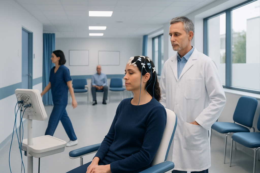
An electroencephalogram, often called an EEG, measures electrical activity in the brain using scalp electrodes. The test is painless, does not send electricity into the brain, and is generally safe for adults and children.
Key Takeaways
- An electroencephalogram, often called an EEG, measures electrical activity in the brain using scalp electrodes.
- The test is painless, does not send electricity into the brain, and is generally safe for adults and children.
- Doctors commonly use EEG to help assess seizures, fainting-like episodes, sleep disorders, and unexplained changes in awareness or behavior.
- A normal EEG does not always rule out a seizure disorder, and an abnormal EEG must be interpreted together with symptoms and medical history.
- Different EEG formats, including routine, sleep-deprived, ambulatory, and video EEG, may be chosen depending on the clinical question.
An electroencephalogram is a noninvasive test that records the brain’s electrical activity through small sensors placed on the scalp. It helps doctors investigate seizures, episodes of altered awareness, sleep-related symptoms, and some other neurologic concerns, but it does not by itself diagnose every brain condition.
What an electroencephalogram is and what it shows
An electroencephalogram is a test that records the brain’s natural electrical activity. Small metal discs called electrodes are placed on the scalp, and these sensors detect patterns of brain waves. The test is usually abbreviated as EEG. It is widely used in neurology because it can provide real-time information about how the brain is functioning during wakefulness, drowsiness, and sleep.
One common myth is that an EEG can “read thoughts” or measure intelligence. It cannot. Instead, it captures electrical patterns that may help a doctor recognize abnormalities linked with certain medical conditions. Another misconception is that the test sends electricity into the body. In routine clinical EEG, the electrodes record activity; they do not shock the brain.
EEG findings are only one part of the diagnostic picture. A doctor interprets them alongside symptoms, examination findings, and often brain imaging or laboratory tests. For example, when a person has episodes that may be seizures, an EEG can support the evaluation, but it is not used in isolation. In some cases, further assessment for conditions such as epilepsy may be recommended.
Why doctors order an EEG
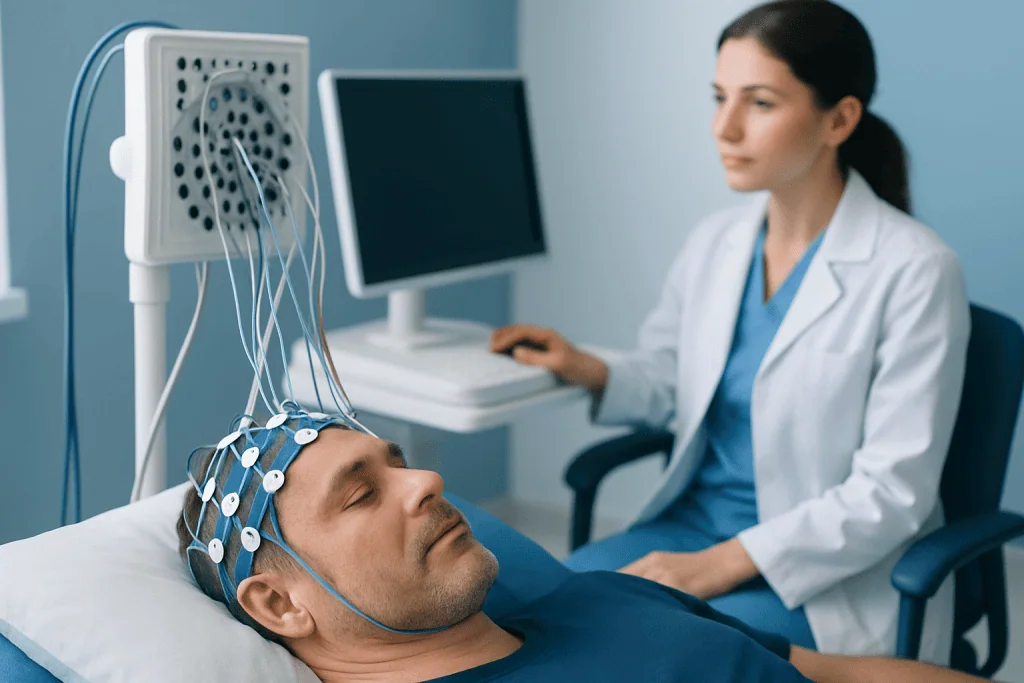
The most familiar reason for ordering an EEG is to investigate suspected seizures. If a person has convulsions, brief staring spells, unexplained blackouts, repetitive jerking, or episodes of confusion, an EEG may help identify electrical patterns that suggest a seizure tendency. It may also assist with classification of seizure types, which can guide management.
EEG is also used in people who have unexplained episodes that are not clearly seizures, such as sudden loss of awareness, unusual movements, temporary memory gaps, or nighttime events. Because some symptoms can have several possible causes, EEG helps doctors understand whether the brain’s electrical activity is involved.
In addition, EEG can be useful in selected cases of sleep disorders, altered mental status, encephalopathy, or after certain brain injuries. It may be part of a broader neurologic workup that also includes imaging. When symptoms suggest a structural problem, doctors may pair EEG findings with studies such as MRI scanning or CT imaging to get a more complete picture.
- Possible seizure or epilepsy evaluation
- Unexplained fainting-like or blackout episodes
- Sleep-related events or unusual nighttime behaviors
- Confusion, altered awareness, or fluctuating mental status
- Monitoring of known neurologic conditions in specific situations
How the test is done
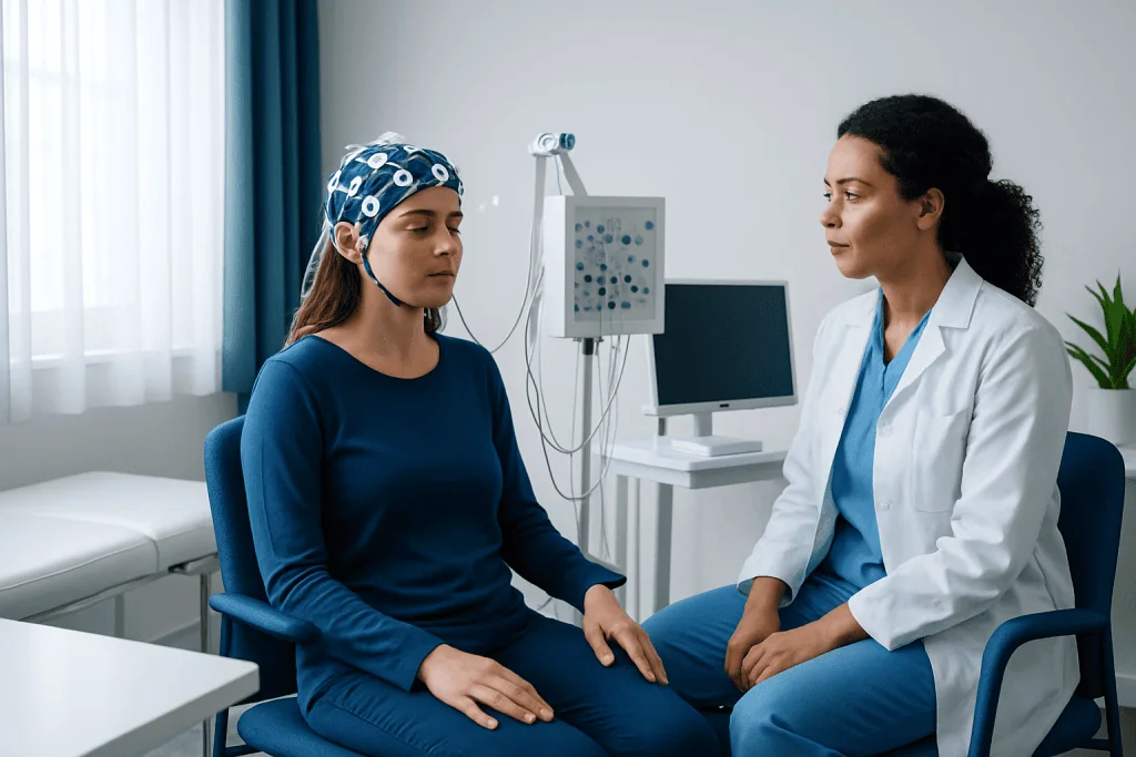
A routine electroencephalogram is usually performed in a hospital or outpatient neurophysiology unit. A technician measures the scalp and places electrodes in standard positions using a special paste or adhesive. The person then sits or lies comfortably while the recording begins. The procedure is painless, and most routine studies are completed within about 20 to 60 minutes, although preparation may add some time.
During the test, the person may be asked to open and close the eyes, breathe deeply for several minutes, or look at flashing lights. These simple activation methods can sometimes bring out brain wave patterns that are helpful for diagnosis. In some circumstances, the doctor may request sleep deprivation beforehand because drowsiness and sleep can increase the chance of detecting certain abnormalities.
Not every EEG is the same. If symptoms happen infrequently, a routine recording may miss them. In that situation, the doctor may recommend longer monitoring, such as ambulatory EEG at home or video EEG in hospital. Video EEG combines a synchronized camera with continuous brain-wave recording, which can be especially helpful when trying to match symptoms with electrical changes. Some patients being assessed more fully may later need neurology evaluation and follow-up as part of an individualized care plan.
Preparing for an electroencephalogram and what to expect afterward
Preparation instructions can vary slightly depending on the type of EEG, so it is important to follow the guidance from the care team. In general, patients are often advised to wash the hair the night before or the morning of the test and avoid oils, sprays, gels, or conditioners that can make it harder for electrodes to stick well. For a sleep-deprived study, the doctor may ask the person to sleep less than usual.
Food and regular medicines should only be changed if the clinician gives specific instructions. In many cases, usual meals and most medications are continued, but some substances can affect the recording, so the technician or doctor may ask about prescription drugs, caffeine use, alcohol, and supplements. Patients should never stop seizure medication or any other prescribed treatment unless a qualified doctor specifically advises it.
After a routine EEG, most people can return to normal activities right away. There may be a little paste left in the hair, but there is typically no recovery period. If sedation is used, which is uncommon for standard EEG and more likely in young children or special situations, extra observation and assistance with travel home may be needed.
Understanding EEG results without myths or oversimplification
EEG results are usually described as normal or showing particular abnormalities, such as slowing or epileptiform discharges. A normal EEG means that no clearly abnormal electrical pattern was captured during the recording. However, this does not always rule out a seizure disorder. Some people with epilepsy have normal routine EEGs between events, especially if seizures are infrequent.
Abnormal results also need careful interpretation. Not every abnormality means a person has epilepsy, and some changes may be nonspecific. The significance depends on the person’s age, symptoms, medical history, medicines, state of wakefulness, and whether the changes match the episodes being investigated. This is why EEG should be read by a trained clinician in context rather than treated as a stand-alone answer.
Sometimes the report leads to further testing rather than a final diagnosis. A doctor may recommend repeat EEG, longer video monitoring, sleep studies, blood tests, or imaging. If a more detailed structural assessment of the brain is needed, the evaluation may include brain MRI to look for causes that an EEG cannot show, such as certain scars, lesions, or developmental differences.
Limitations, safety, and common concerns
EEG is a valuable tool, but it has limits. It records electrical activity near the surface of the brain and may not detect every abnormal event, especially if it occurs deep in the brain or not during the recording. A short EEG can miss intermittent problems, which is one reason longer or repeated studies are sometimes needed.
The test is generally very safe. The electrodes record activity and do not injure the brain. Flashing lights or deep breathing may trigger a seizure in a small number of people who are already susceptible, but the test is performed in a controlled setting by trained staff who know how to respond. Mild scalp irritation from adhesive is possible but usually temporary.
People often wonder whether EEG can diagnose conditions like dementia, stroke, migraine, anxiety, or brain tumors on its own. Usually it cannot. It may contribute useful information in some situations, but these conditions are usually evaluated with a combination of history, examination, and other tests. This distinction matters because EEG is best understood as one evidence-based piece of the neurologic assessment, not a universal brain scan.
When to seek medical care
Medical attention is important if a person has a first-time seizure, repeated unexplained blackouts, episodes of lost awareness, new involuntary movements, or sudden confusion without a clear reason. Urgent evaluation is also needed after head injury, during pregnancy, or if these symptoms occur together with fever, severe headache, weakness, trouble speaking, or difficulty breathing.
For nonemergency concerns, a routine appointment is reasonable when someone has recurrent brief staring spells, unusual nighttime episodes, unexplained memory gaps, or fainting-like events that are not fully understood. A doctor can decide whether an EEG, heart testing, sleep evaluation, or another assessment is most appropriate.
If care is being arranged internationally, Acibadem International’s multidisciplinary specialists and JCI-accredited hospitals evaluate neurologic symptoms with evidence-based testing and treatment planning for international patients. The safest next step is always a qualified medical review, especially if symptoms are new, recurrent, or worsening.
Frequently asked questions
Is an electroencephalogram painful?
No. An electroencephalogram is usually painless because the electrodes only record signals from the scalp and do not send electricity into the brain. Some people notice mild discomfort from adhesive or from sitting still, but the test itself is not painful.
How long does an EEG take?
A routine EEG often takes about 20 to 60 minutes for the recording itself, with extra time for preparation and electrode placement. Longer studies, such as ambulatory or video EEG monitoring, may last many hours or several days depending on the goal of testing.
Can a normal EEG rule out epilepsy?
No. A normal EEG does not completely rule out epilepsy because abnormal electrical activity may not occur during the recording. If symptoms strongly suggest seizures, the doctor may recommend repeat testing, sleep-deprived EEG, or longer video EEG monitoring.
What is the difference between an EEG and an MRI?
An EEG records the brain’s electrical activity over time, while an MRI creates detailed pictures of the brain’s structure. They answer different questions, and doctors often use them together when evaluating seizures or other neurologic symptoms.
Do patients need to stop medications before an EEG?
Usually, medications should only be changed if the doctor specifically instructs it. Stopping seizure medicines or other prescribed drugs without guidance can be unsafe, so patients should follow the preparation plan given by their care team.
Why might a doctor request a sleep-deprived EEG?
Sleep deprivation can make certain brain wave abnormalities more likely to appear during testing. For some patients, drowsiness and sleep increase the chance that the EEG will capture findings that help explain their symptoms.
This article is for general information only and is not a substitute for professional medical advice. Please consult a qualified doctor about your individual situation.
Hair transplant in Turkey — costs, graft counts & a free assessment
JCI-accredited · board-certified surgeons · reply within 24h
Add us as a Preferred Source to see more of our trusted health content across Google Search, AI Overviews and Discover.

More from the Health Library

Blood Pressure Chart: An Evidence-Based Guide for Patients

Blocked Tear Duct — Explained by Medical Evidence, Not Myths

Late Term Abortion: An Evidence-Based Guide for Patients

Ear Canal: A Complete Medical Overview

Chorea disease: A Complete Medical Guide for Patients