Pictures of Moles: What Patients Need to Know
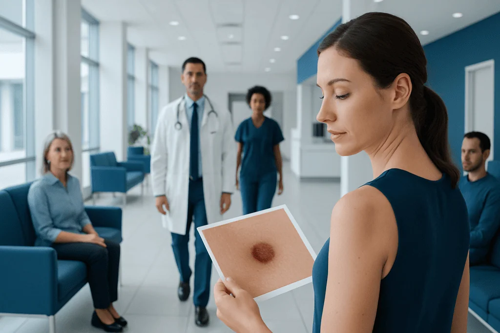
Pictures of moles are educational, but they do not replace a skin examination by a doctor. Many moles are benign and may be flat or raised, light or dark, and round or oval.
Key Takeaways
- Pictures of moles are educational, but they do not replace a skin examination by a doctor.
- Many moles are benign and may be flat or raised, light or dark, and round or oval.
- Changes in size, shape, color, border, or symptoms such as bleeding or itching should be evaluated.
- The ABCDE rule can help patients notice warning signs of melanoma.
- People with fair skin, many moles, family history, or high sun exposure may need closer monitoring.
Pictures of moles can be useful for learning what common moles often look like, but they cannot confirm whether a spot is harmless or cancerous. The safest approach is to use images as a guide, watch for changes, and have any suspicious mole checked by a qualified doctor.
Overview: What Pictures of Moles Can and Cannot Show
Pictures of moles can help patients recognize the wide range of appearances that ordinary moles may have. Some are flat, some are raised, and colors may range from tan to dark brown. Looking at examples may make it easier to notice when a spot looks different from the rest of a person’s skin.
At the same time, pictures of moles have limits. Lighting, camera quality, skin tone, and image angle can change how a mole appears. More importantly, skin cancer and benign moles can sometimes look similar in photographs, so no image gallery can diagnose a lesion with certainty.
For that reason, patients are usually advised to think of mole pictures as a learning tool rather than a diagnostic test. A dermatologist or other qualified doctor may use the spot’s appearance, the patient’s history, and sometimes dermoscopy or biopsy to decide whether a mole is benign or needs further evaluation.
How Common Moles May Look in Pictures
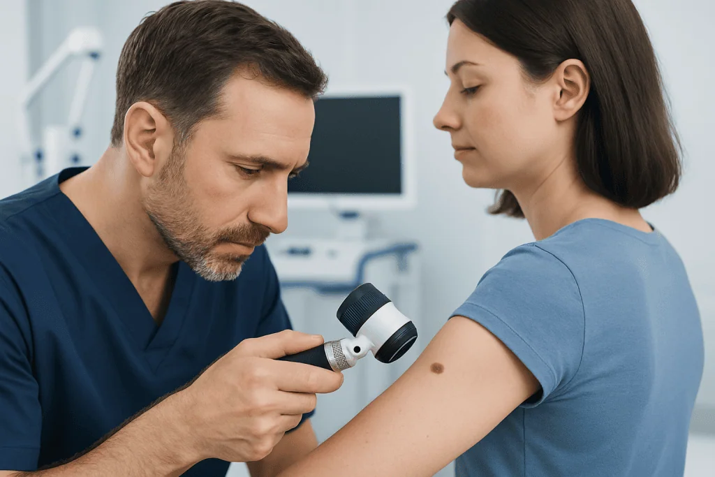
In pictures, common benign moles often appear as small, round or oval spots with a fairly even color and a clear border. They may be flat like a freckle or slightly raised above the skin. Some develop in childhood, while others appear later, especially with sun exposure.
Not all harmless moles look identical. Some may be pink, tan, brown, or nearly skin-colored. Others may contain hair, and some may darken slightly over time without meaning cancer is present. Because normal moles vary, comparison to a single “ideal” image is not always helpful.
Doctors often describe a few broad mole patterns patients may see in photos:
- Junctional nevi: usually flat and evenly pigmented.
- Compound nevi: often slightly raised with a smooth surface.
- Dermal nevi: often more raised and lighter in color.
- Congenital moles: present at birth or soon after.
- Atypical moles: larger or less regular than average, but not always cancerous.
Even when a mole looks ordinary in a picture, context still matters. A new mole in adulthood, a lesion that keeps changing, or one that looks very different from nearby spots may need professional assessment.
Signs That a Mole May Need Attention
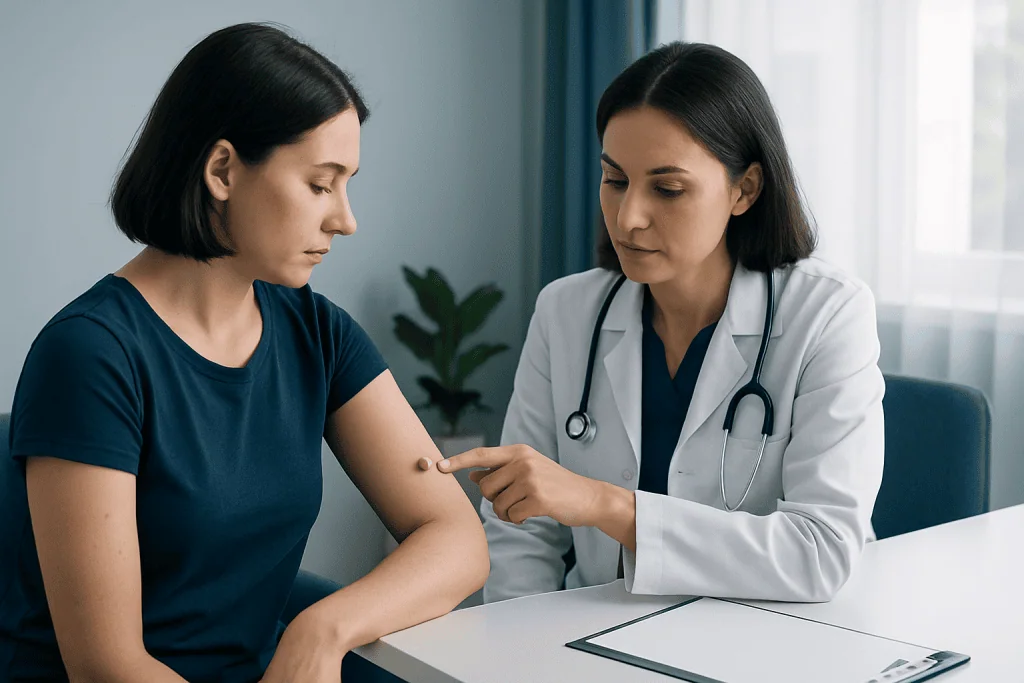
When patients search for pictures of moles, they are often trying to understand whether a spot looks concerning. A commonly used guide is the ABCDE rule. It is not perfect, but it can help people notice changes that deserve medical review.
The ABCDE rule includes:
- A for asymmetry: one half does not match the other.
- B for border: edges are irregular, blurred, or notched.
- C for color: more than one shade is present, such as brown, black, red, white, or blue.
- D for diameter: the lesion is larger than about 6 mm, although smaller melanomas can occur.
- E for evolving: the mole changes in size, shape, color, or symptoms.
There are also other warning signs that may not be obvious in standard online pictures. A mole that bleeds, crusts, becomes painful, itches persistently, or does not heal should be examined. Doctors may also look for the “ugly duckling” sign, meaning a mole that looks noticeably different from a person’s other spots.
These features do not automatically mean cancer, but they do mean a medical opinion is important. Early assessment is especially relevant when there is concern about melanoma or another form of skin cancer.
Who Has a Higher Risk of Problematic Moles?
Anyone can develop a suspicious mole, but some people have a higher risk of skin cancer or atypical moles. Risk tends to increase in those with fair skin, light eyes, freckles, and a history of sunburns. Repeated ultraviolet exposure from sunlight or tanning beds is also an important factor.
People who have many moles, large congenital moles, or atypical moles may need more careful skin monitoring. A personal or family history of melanoma can raise concern further. Immune suppression and older age can also affect skin cancer risk, although melanoma can occur at younger ages too.
Pictures of moles may be especially useful for these higher-risk groups as part of self-awareness. Still, higher risk generally means self-checks should be combined with regular professional skin examinations rather than relying on images alone.
How Doctors Evaluate a Suspicious Mole
If a mole looks unusual in pictures or has changed over time, a doctor will usually begin with a full history and skin examination. The patient may be asked when the spot appeared, whether it has changed, and whether there are symptoms such as itching or bleeding. Family history and sun exposure habits are also relevant.
Dermatologists often use a dermatoscope, a handheld instrument that magnifies and lights the skin. This can reveal structures that are not visible in ordinary photographs. In some clinics, body mapping or serial photography may be used to monitor people with many moles.
If the doctor thinks a mole could be problematic, a biopsy may be recommended. This means removing all or part of the lesion so it can be examined under a microscope. Biopsy is the only way to confirm whether a suspicious mole is benign, precancerous, or cancerous.
When needed, evaluation may include dermatology assessment and skin-focused diagnostic follow-up. If skin cancer is diagnosed, treatment planning may involve specialists in oncology depending on the type and stage.
Treatment Options and What Happens Next
Most benign moles do not need treatment. If a mole is harmless, a doctor may simply advise observation. Some patients choose removal for irritation, repeated friction, or cosmetic reasons, but this is different from removal for medical concern.
When a mole is suspicious, the first step is often excision or biopsy. If testing shows a benign lesion, no further treatment may be necessary other than routine skin checks. If an atypical mole is found, the doctor may recommend follow-up or complete removal depending on the pathology findings.
If the lesion is confirmed to be skin cancer, treatment depends on the exact diagnosis. Management may include surgical removal and, in selected cases, additional specialist care. Related conditions can include skin cancer, which should be managed based on pathology results rather than appearance alone.
Near the end of the care pathway, patients may benefit from a multidisciplinary approach. Acibadem International’s multidisciplinary specialists and JCI-accredited hospitals diagnose and treat skin conditions and skin cancers for international patients when further evaluation or treatment is needed.
Self-Checks, Skin Photography, and Prevention
Patients can use pictures of moles most effectively by comparing them with their own skin over time instead of comparing themselves only with strangers’ photos online. A monthly self-check in good lighting can help identify new lesions or changes. It is helpful to examine the scalp, back, soles, nails, and other easily missed areas, sometimes with help from a mirror or a family member.
Taking clear photographs at regular intervals can make change easier to detect. For consistency, it helps to use the same lighting, distance, and angle. However, home photos should support monitoring, not delay medical advice when a lesion appears suspicious.
Sun protection remains one of the most important preventive steps. Useful measures include:
- Using broad-spectrum sunscreen as directed.
- Wearing protective clothing, hats, and sunglasses.
- Seeking shade during strong midday sun.
- Avoiding tanning beds.
- Protecting children from sunburn.
These steps cannot remove existing moles, but they can lower further ultraviolet damage and support long-term skin health.
When to Seek Medical Care
Medical care is advisable if a mole is new in adulthood, changes noticeably, or looks very different from nearby moles. Prompt assessment is also important if a spot bleeds, forms a crust, becomes painful, itches persistently, or does not heal. Patients should not wait for a lesion to become large before asking for an evaluation.
People with many moles, atypical moles, previous skin cancer, or a family history of melanoma may benefit from routine skin examinations even if no single lesion seems alarming. A doctor can advise how often checks are needed based on individual risk.
If there is uncertainty, it is usually better to arrange a non-urgent medical review than to rely on online comparisons. Early assessment can clarify whether the spot is benign and provide peace of mind, or identify a condition that should be treated sooner.
Frequently asked questions
Can pictures of moles tell if a mole is cancerous?
No, pictures of moles cannot confirm whether a mole is cancerous. They can help patients recognize patterns and warning signs, but diagnosis requires examination by a qualified doctor and sometimes a biopsy.
What does a normal mole usually look like?
A normal mole is often round or oval, with a relatively even color and a clear border. It may be flat or raised, and harmless moles can vary in shade from skin-colored to dark brown.
What changes in a mole should worry patients?
Changes in size, shape, border, or color should be checked, especially if the mole becomes asymmetrical or develops multiple shades. Bleeding, crusting, pain, or persistent itching are also reasons to seek medical advice.
Are raised moles always dangerous?
No, many raised moles are benign and remain stable for years. What matters more is whether the mole is changing, has irregular features, or causes symptoms that are new or unusual.
How often should someone check their moles?
A monthly self-check is a practical routine for many adults, especially those with several moles. People at higher risk, such as those with atypical moles or a family history of melanoma, may also need regular professional skin examinations.
When is a biopsy needed for a mole?
A biopsy may be recommended when a mole looks suspicious on examination or shows concerning changes over time. It allows the tissue to be examined under a microscope, which is the most reliable way to confirm the diagnosis.
References
- American Academy of Dermatology
- National Cancer Institute
- Centers for Disease Control and Prevention
- National Health Service
- World Health Organization
This article is for general information only and is not a substitute for professional medical advice. Please consult a qualified doctor about your individual situation.
Explore treatments in Turkey — costs, top hospitals & a free quote
JCI-accredited · board-certified surgeons · reply within 24h
Add us as a Preferred Source to see more of our trusted health content across Google Search, AI Overviews and Discover.

More from the Health Library


Related Specialists

Dt. Beril Karakaya Gökçimen
Oral & Dental Health
Prof. Dr. Ayşe Korkmaz Toygar
Neonatal Intensive Care Unit
Asst. Prof. Dr. Özer Öztürk
Orthopedic Surgery & Traumatology
Prof. Dr. Cem Akbal
Urology