Tibialis Muscle — Explained by Medical Evidence, Not Myths
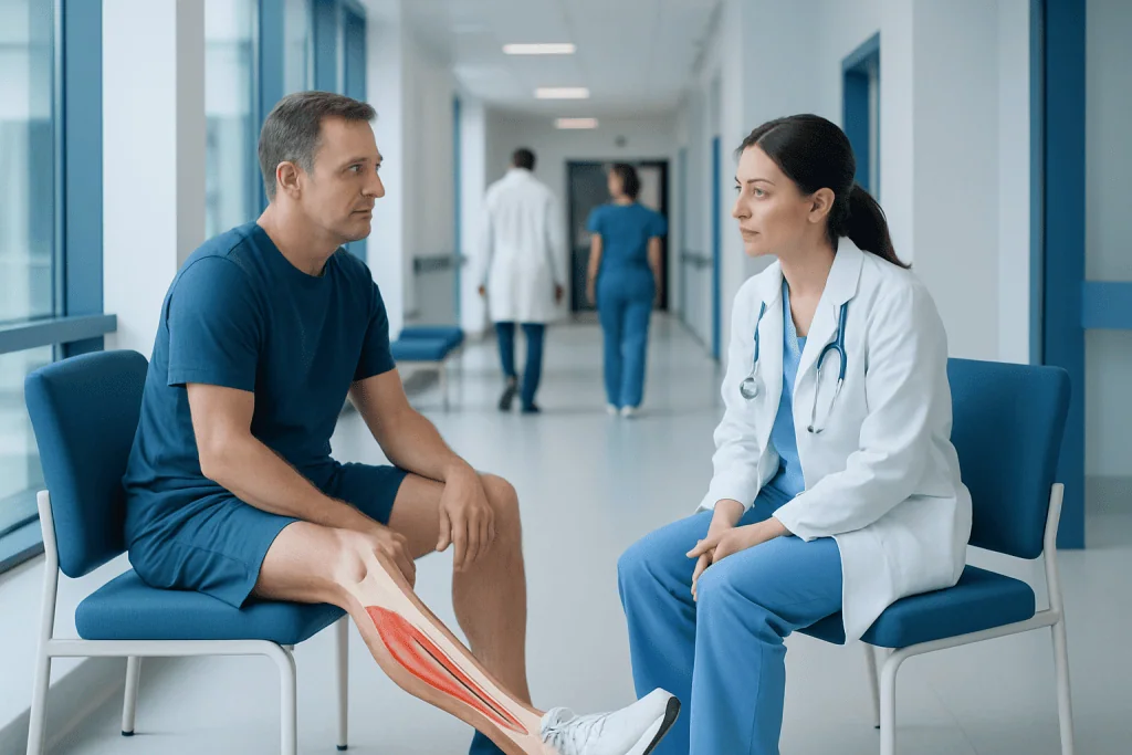
The tibialis anterior lifts the foot and helps control the foot as it contacts the ground. The tibialis posterior supports the inner arch and helps turn the sole inward during walking.
Key Takeaways
- The tibialis anterior lifts the foot and helps control the foot as it contacts the ground.
- The tibialis posterior supports the inner arch and helps turn the sole inward during walking.
- Gradual increases in activity, supportive footwear and strength training can help reduce overuse problems.
- Persistent pain, swelling, weakness, a changing foot shape or difficulty walking should be assessed by a clinician.
- Sudden severe pain after injury, inability to bear weight, numbness or a cold/pale foot requires urgent medical attention.
The tibialis muscle group consists mainly of the tibialis anterior at the front of the shin and the tibialis posterior deeper in the lower leg. These muscles support controlled walking, running, balance and the arch of the foot; pain usually relates to overload, tendon irritation, injury or changes in foot mechanics rather than a single cause.
Tibialis muscle: what it is and why it matters
The tibialis muscle group includes two important lower-leg muscles: the tibialis anterior and tibialis posterior. Although they sit in different areas of the leg and have different roles, both connect the lower leg to the foot through tendons. Together, they help make walking, climbing stairs, running and maintaining balance more efficient.
The tibialis anterior lies along the outer-front part of the shin. Its tendon crosses the front of the ankle and attaches near the inside of the midfoot. The tibialis posterior lies deeper in the calf, behind the shin bone. Its tendon travels around the inside of the ankle and attaches to several bones in the foot, including structures that help support the arch.
These muscles work with the calf muscles, small foot muscles, ligaments and nerves. Symptoms in the shin, ankle or arch may therefore involve more than one structure. A qualified assessment is useful when symptoms do not settle, since similar pain patterns can have different causes.
How the tibialis anterior and posterior work
The tibialis anterior primarily pulls the foot upward toward the shin, a movement called dorsiflexion. It also helps turn the sole of the foot inward, known as inversion. During walking, it helps clear the toes from the ground during the swing phase and then controls the foot as the heel first contacts the floor.
The tibialis posterior also contributes to inversion, but it has a particularly important role in supporting the medial, or inner, arch of the foot. It helps stabilize the foot as body weight moves over it and assists with pushing off the ground. This is why weakness or tendon dysfunction in this muscle may be associated with pain along the inner ankle or arch and, in some people, progressive flattening of the arch.
Neither muscle works in isolation. Hip strength, calf flexibility, foot alignment, running surface, footwear and training load can all affect how much demand is placed on the tibialis muscles and their tendons.
Common tibialis muscle symptoms and patterns

Tibialis anterior problems can cause aching, tenderness or tightness at the front or outer side of the shin. Discomfort may also be felt at the front of the ankle or over the top-inner part of the foot. Symptoms may become more noticeable after walking uphill, running, repeated stair climbing or an abrupt increase in activity.
Tibialis posterior tendon irritation often causes pain or swelling behind and below the inner ankle bone, sometimes extending into the arch. Standing or walking for long periods, especially on uneven ground, may worsen symptoms. Some people notice fatigue in the foot, reduced ability to rise onto the toes on one foot, or a gradual change in the way the foot aligns.
Not all shin or ankle pain is caused by the tibialis muscles. Stress injuries, nerve problems, arthritis, ligament injury, circulation concerns and other tendon conditions can produce overlapping symptoms. A clinician considers the location, timing, activity triggers, injury history and physical examination findings before reaching a diagnosis.
- Muscle soreness is commonly diffuse and improves with rest and recovery.
- Tendon symptoms are often more localized and can be associated with tenderness or swelling.
- Weakness that makes the foot slap, drag or catch may indicate a nerve or tendon issue and deserves prompt assessment.
Causes and risk factors for tibialis pain
Overuse is a frequent reason for tibialis muscle and tendon symptoms. A rapid rise in running distance, hill work, walking volume, jumping activity or time spent standing can exceed the tissues’ current capacity to recover. New shoes, worn-out footwear, a change in surface or returning to activity after a break can also alter loading.
Foot mechanics may contribute in some people. A foot that rolls inward substantially during weight-bearing can increase demand on the tibialis posterior, while limited ankle mobility or calf tightness may affect how the foot moves. These factors are not automatically harmful, but they can be relevant when pain develops.
Age-related tendon changes, prior ankle injury, inflammatory joint disease, diabetes and higher body weight can raise the likelihood of posterior tibial tendon problems in some individuals. Less commonly, a direct blow, forceful ankle movement or laceration can injure a tendon. Sudden severe pain, bruising or loss of function after trauma should not be managed as routine exercise soreness.
How tibialis conditions are assessed
Assessment usually begins with a medical history. The clinician may ask when symptoms began, whether there was an injury, which activities bring on pain, how footwear has changed and whether there are symptoms such as numbness, weakness, swelling or night pain. Medical conditions and current medicines may also be relevant.
A physical examination can include observing standing posture and gait, checking ankle and foot movement, feeling for tenderness or swelling, and testing strength. For suspected tibialis posterior dysfunction, the clinician may assess the arch while standing and ask the person to perform a single-leg heel raise.
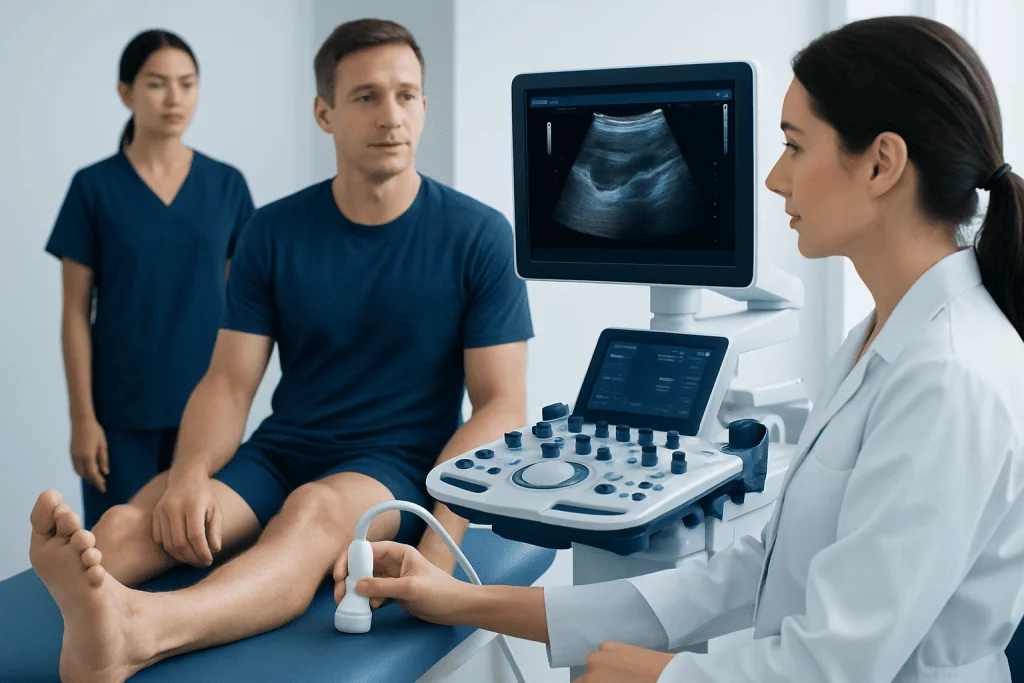
Imaging is not always necessary for mild, improving overuse symptoms. However, X-rays may help evaluate bone alignment or exclude fracture, while ultrasound or magnetic resonance imaging may be used when a tendon tear, significant inflammation or another structural problem is suspected. Persistent shin pain after repetitive impact activity may also require assessment for a bone stress injury.
Treatment and rehabilitation options
Treatment depends on the cause, severity and functional impact of symptoms. For many mild overuse problems, relative rest from the activity that provokes pain, followed by a gradual return, is appropriate. This does not always mean avoiding all movement; lower-impact activities may be suitable if they do not increase symptoms.
Supportive, well-fitting shoes can help, and a clinician may recommend an ankle brace, walking boot or orthotic device for selected tendon conditions. Physiotherapy commonly focuses on restoring comfortable ankle movement, progressively strengthening the tibialis muscles and calf, improving balance, and reviewing movement patterns and training progression. For medically appropriate patients, non-prescription pain relief may be considered with advice from a doctor or pharmacist.
More severe tendon damage, ongoing functional limitation or progressive foot deformity may require specialist orthopedic or foot-and-ankle evaluation. Treatment can range from more structured bracing and rehabilitation to surgery in carefully selected cases. Physical therapy and rehabilitation can help patients rebuild strength and return to daily activity safely after injury or treatment.
At Acibadem International, multidisciplinary specialists in JCI-accredited hospitals can assess lower-leg and foot concerns and coordinate diagnostic, orthopedic and rehabilitation care for international patients.
Prevention and everyday self-care
Gradual progression is one of the most practical ways to reduce the risk of overuse injury. When starting a new exercise program or returning after time away, increasing duration, distance, intensity and hill work step by step gives muscles and tendons time to adapt. Recovery days are also part of a sustainable routine.
Footwear should fit comfortably, suit the planned activity and be replaced when it no longer provides reliable support or cushioning. People with recurring symptoms may benefit from an individualized footwear and foot-support assessment rather than relying on a one-size-fits-all product.
Regular exercises that build calf, ankle and foot strength, together with balance training, may support lower-limb function. Gentle mobility work can be useful when it feels comfortable. Exercises should not create sharp pain or lead to worsening symptoms that continue into the next day; a physiotherapist can tailor a program to the person’s diagnosis and goals.
When to seek medical care
Medical assessment is advisable for pain that persists beyond a short period of activity modification, repeatedly returns, causes visible swelling, or interferes with walking, work, sleep or usual exercise. It is also important to seek advice if the inner ankle or arch is painful and the foot appears to be flattening or changing shape.
Prompt care is needed after a significant injury, especially if there is a snap or pop, marked bruising, inability to bear weight, or sudden weakness lifting the foot. New numbness, tingling, a foot that feels cold or looks pale or blue, rapidly increasing swelling, fever, or severe pain out of proportion to the apparent injury should be assessed urgently.
Early evaluation can clarify whether symptoms are related to muscle strain, tendon irritation, a stress injury or another cause. A timely, accurate diagnosis supports an appropriate plan and can help avoid prolonged limitation.
Frequently asked questions
Is the tibialis muscle one muscle or two?
The term usually refers to two muscles: the tibialis anterior and tibialis posterior. They have different positions and functions, but both help control foot movement and stability.
Why does the tibialis anterior hurt after running?
Pain at the front of the shin after running may result from increased training load, hills, speed work, footwear changes or muscle fatigue. Persistent focal pain, swelling or pain that worsens with impact should be evaluated to exclude other causes, including a bone stress injury.
Can tibialis posterior problems cause flat feet?
The tibialis posterior tendon helps support the inner arch. When the tendon becomes weakened or dysfunctional, some people can develop progressive arch flattening and a change in foot alignment. Early assessment is useful if this change is noticed.
Should a person exercise through tibialis muscle pain?
Sharp pain, worsening pain or pain that changes walking mechanics should not be pushed through. Gentle, pain-limited movement may be appropriate in some cases, but the right approach depends on the cause and should be guided by a clinician or physiotherapist when symptoms persist.
How long does tibialis tendon irritation take to improve?
Recovery varies according to severity, continued loading, overall health and adherence to rehabilitation. Mild overuse symptoms may improve over weeks with appropriate activity changes, while more significant tendon problems can take longer and may need structured treatment.
Can tight calves affect the tibialis muscles?
Yes. Limited calf flexibility or ankle movement can change how forces are distributed through the ankle and foot during walking and running. Addressing mobility and strength as part of an individualized program may reduce unnecessary strain.
This article is for general information only and is not a substitute for professional medical advice. Please consult a qualified doctor about your individual situation.
Explore treatments in Turkey — costs, top hospitals & a free quote
JCI-accredited · board-certified surgeons · reply within 24h
Add us as a Preferred Source to see more of our trusted health content across Google Search, AI Overviews and Discover.

More from the Health Library

Nuggets of Chicken: What Patients Need to Know

Flakes — Explained by Medical Evidence, Not Myths

Lozenges: What Patients Need to Know

Phosphenes: An Evidence-Based Guide for Patients

Pincer Grasp — Explained by Medical Evidence, Not Myths

Anosognosia — Explained by Medical Evidence, Not Myths
Physical Medicine & Rehabilitation Specialists at Acibadem

Fzt. Canberk Altan
Physical Medicine & Rehabilitation
Dr. Cem Tangay
Orthopedic Surgery & Traumatology
Dr. Cem Tekin Baran
Orthopedic Surgery & Traumatology
Fzt. Ceren Kandemir Gençsoylu
Physical Medicine & Rehabilitation