What Does a Cataract Look Like: An Evidence-Based Patient Guide

Cataracts develop when the normally clear lens inside the eye becomes cloudy. Early cataracts commonly affect vision before they create a visible change in the eye’s appearance.
Key Takeaways
- Cataracts develop when the normally clear lens inside the eye becomes cloudy.
- Early cataracts commonly affect vision before they create a visible change in the eye’s appearance.
- Blurred vision, glare, halos, reduced contrast, and faded colors are common symptoms.
- Cataract surgery removes the cloudy lens and replaces it with an artificial intraocular lens.
- Sudden vision loss, eye pain, marked redness, flashes, or a curtain-like shadow need prompt medical assessment.
A cataract is a clouding of the eye’s natural lens. Although an advanced cataract can sometimes make the pupil look gray, white, or yellowish, early cataracts are usually not visible in a mirror and are found during a dilated eye examination.
Overview: What Does a Cataract Look Like?
What does a cataract look like? A cataract is clouding of the natural lens located behind the colored part of the eye (iris) and pupil. In early stages, it usually cannot be seen from the outside; a person may notice changes in vision long before anyone can see a difference in the eye.
When a cataract becomes more advanced, the pupil may appear dull gray, milky white, or sometimes yellow-brown rather than black in certain lighting. This visible change is not present in every person and does not reliably show how much vision is affected. An eye specialist uses a slit-lamp microscope and dilated eye examination to see the lens clearly and assess the type and extent of clouding.
Cataracts are common with aging, but they may also occur after eye injury, long-term corticosteroid use, some health conditions, eye surgery, or radiation exposure. They are not contagious and do not spread from one eye to another, although they often develop in both eyes over time at different rates.
What Does an Actual Cataract Look Like?
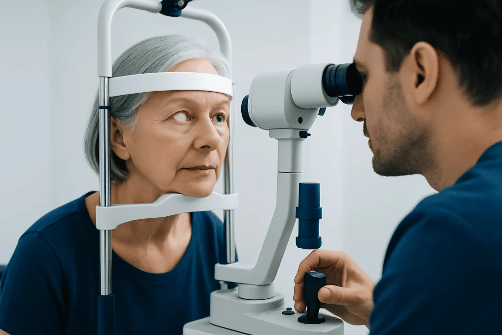
An actual cataract is not a film on the surface of the eye. It is a change within the lens, which sits behind the pupil. In a clinical examination, an ophthalmologist may see areas of haze, spokes, dots, or a dense central opacity in the lens depending on the cataract type.
For example, a nuclear cataract affects the central lens and may make it look yellow or brown. A cortical cataract can create wedge-shaped, spoke-like clouding around the outer part of the lens. A posterior subcapsular cataract forms near the back of the lens and can be especially troublesome for reading, bright lights, and night driving even when it is relatively small.
Photographs showing a completely white pupil often depict a very mature cataract. However, a white pupil can also have other causes, particularly in children, so it should always be assessed urgently by an eye doctor. Visual appearance alone cannot confirm a diagnosis or rule out other eye conditions.
What Do Stage 1 Cataracts Look Like?

“Stage 1 cataract” is not a formal medical classification used consistently by all eye specialists. It generally refers to an early cataract: mild lens changes that may be detected during an eye examination but may cause little or no disruption in daily life.
From the outside, an early cataract usually looks like a normal eye. The pupil remains dark, and there may be no visible white or cloudy spot. Under magnification, a clinician may see subtle lens haze, early yellowing, small spokes, or tiny central changes.
Early symptoms can include needing brighter light to read, greater sensitivity to glare, halos around lights, reduced contrast, colors that seem less vivid, or frequent changes in glasses prescription. Some people have no symptoms at all. Regular eye exams help identify cataracts and other causes of gradual vision change.
Why Cataracts Develop and Who Is at Risk
The lens focuses light onto the retina at the back of the eye. With age, lens proteins can gradually clump together and alter the lens structure, reducing its transparency. This causes light to scatter rather than focus cleanly, producing blur and glare.
Age is the leading risk factor. Other factors include diabetes, smoking, substantial sunlight exposure over many years, previous eye trauma or inflammation, earlier eye surgery, radiation treatment, and prolonged use of corticosteroid medicines. A family history of cataracts can also contribute.
Protective habits cannot prevent every cataract, but they can support eye health. These include not smoking, managing diabetes and other long-term conditions, wearing sunglasses that block ultraviolet radiation outdoors, using eye protection for work or sports, and attending recommended eye examinations.
- Blurred or hazy vision may occur gradually.
- Glare from sunlight or headlights may become more noticeable.
- Night driving and reading in dim light may become harder.
- Double vision in one eye can occasionally occur.
Diagnosis and Decisions About Treatment
An ophthalmologist diagnoses cataracts through a complete eye assessment. This may include checking visual acuity, measuring refraction, examining the eye with a slit lamp, dilating the pupils to inspect the lens and retina, and measuring eye pressure when appropriate. The assessment also looks for other conditions, such as glaucoma, retinal disease, or dry eye, which can affect vision.
Not every cataract requires immediate surgery. If symptoms are mild, updated glasses, brighter task lighting, anti-glare measures, or changes to driving habits may help. Surgery is usually considered when cataract-related vision problems interfere with important activities, independence, work, hobbies, or safe driving.
There is no proven eye drop, supplement, or exercise that reverses an established cataract. The only treatment that removes a cataract is surgery. The timing is individualized and should be based on symptoms, eye findings, overall health, and personal goals rather than the cataract’s outward appearance alone.
Cataract Surgery: How It Works, Candidacy, and Steps
Cataract surgery removes the cloudy natural lens and replaces it with a clear artificial intraocular lens (IOL). Most people with visually significant cataracts are potential candidates, but the eye surgeon considers general health, medications, corneal health, retina and optic nerve function, and whether other eye diseases may limit the expected improvement in vision.
The usual procedure is performed with local anesthetic drops, often with medication to help the person relax. Through a very small incision, the surgeon opens the thin lens capsule, breaks up and removes the cloudy lens using ultrasound energy, and places a folded IOL into the remaining capsule. The procedure is commonly done as day surgery, and the person goes home with postoperative instructions.
Different IOLs may be selected to support distance vision, near vision, astigmatism correction, or a range of vision. No lens choice is perfect for every person. The surgeon explains potential benefits and trade-offs, including whether glasses may still be needed for some activities. A detailed consultation for cataract surgery can help align the surgical plan with visual needs and eye health.
What Doctors Don't Tell You About Cataract Surgery?
Most clinicians discuss the major benefits and risks before surgery, but people may find it helpful to know that visual recovery is not identical for everyone. Vision often begins improving within days, yet it may take several weeks for the eye to stabilize, especially if there is pre-existing dry eye, corneal disease, retinal disease, or inflammation.
A new lens can improve clarity, but it cannot reverse vision loss caused by conditions outside the lens, such as macular degeneration, glaucoma, diabetic retinal disease, or optic nerve damage. Some people also notice temporary glare, halos, light sensitivity, or fluctuating vision while healing. These concerns should be discussed with the surgeon rather than assumed to be a permanent result.
Months or years after successful surgery, some people develop clouding of the capsule behind the implanted lens. This is called posterior capsule opacification and is not the cataract returning. It can often be treated with a brief laser procedure when it meaningfully affects vision.
Recovery, What Is Forbidden After Cataract Surgery, and When to Seek Medical Care
After cataract surgery, the eye may feel mildly scratchy or sensitive to light for a short time. Prescription eye drops are generally used to prevent infection and control inflammation. Follow-up appointments allow the surgeon to monitor healing, eye pressure, and vision. Many routine activities can resume soon, but recovery instructions should always take priority because they vary by individual.
What is forbidden after cataract surgery? For the period advised by the surgical team, patients should avoid rubbing or pressing on the eye, getting water or soap directly into it, swimming or using hot tubs, strenuous exercise, heavy lifting, and dusty or dirty environments. They should also avoid driving until their vision meets legal and practical safety requirements. A protective shield may be recommended while sleeping, and eye drops should be used exactly as prescribed.
Medical care should be sought promptly for worsening rather than improving vision, significant or increasing pain, rapidly increasing redness, a discharge from the eye, new flashes or many floaters, nausea with eye pain, or a curtain-like shadow in vision. These symptoms are uncommon but can indicate complications that require timely assessment. Acibadem International’s multidisciplinary specialists and JCI-accredited hospitals provide assessment and treatment for international patients with cataracts and related eye concerns.
Frequently asked questions
Can someone see a cataract by looking in the mirror?
Usually not when it is early or moderate. Cataracts are inside the eye, behind the pupil, and need an eye examination to be assessed accurately. A very advanced cataract may make the pupil appear gray or white.
Does a cataract look like a cloudy layer on the eye?
No. A cataract is clouding of the lens inside the eye, not a layer over the cornea or the white of the eye. Surface problems such as dry eye or corneal conditions can also cause blurred vision but require different evaluation and treatment.
How do people know they have cataracts?
Common clues are gradually blurred vision, glare, halos around lights, difficulty seeing at night, and colors that appear less bright. These symptoms can also occur with other eye conditions, so an eye examination is important for diagnosis.
When should cataract surgery be done?
Surgery is generally considered when cataract-related vision changes interfere with daily activities, safety, work, or quality of life. It does not have to be delayed until the cataract is visibly advanced, and the decision is made jointly with an ophthalmologist.
Can cataracts be treated without surgery?
Glasses changes, improved lighting, and glare reduction may help manage early symptoms. However, these measures do not remove or reverse the cloudy lens. Surgery is the only treatment that removes a cataract.
What should a person do if one pupil suddenly looks white?
A newly noticed white or unusual pupil should be assessed promptly by an eye specialist, particularly in a child. Although a mature cataract can cause this appearance, other conditions may also be responsible and need timely diagnosis.
References
- American Academy of Ophthalmology
- National Eye Institute
- Mayo Clinic
- World Health Organization
This article is for general information only and is not a substitute for professional medical advice. Please consult a qualified doctor about your individual situation.
Explore treatments in Turkey — costs, top hospitals & a free quote
JCI-accredited · board-certified surgeons · reply within 24h