CT Scan Lung Nodules: Preparation, Procedure and Results
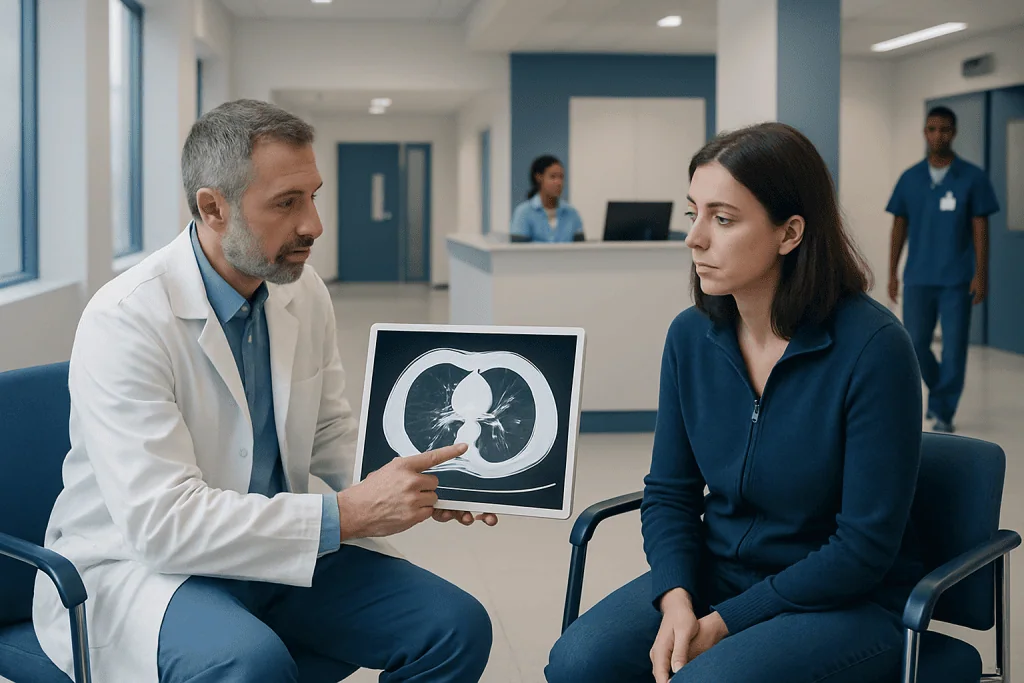
CT scans show lung nodules in much greater detail than standard chest X-rays. A CT scan cannot always confirm whether a nodule is cancerous, but it helps estimate risk and guide next steps.
Key Takeaways
- CT scans show lung nodules in much greater detail than standard chest X-rays.
- A CT scan cannot always confirm whether a nodule is cancerous, but it helps estimate risk and guide next steps.
- Many nodules are harmless scars, healed infections or non-cancerous growths.
- Follow-up timing depends on nodule size, appearance, growth and personal risk factors.
- Most people can return to normal activities immediately after a non-contrast lung CT scan.
A CT scan for lung nodules is a detailed imaging test that helps clinicians measure a nodule, examine its features and compare it with earlier scans. Most lung nodules are not cancer, but CT findings help determine whether monitoring, additional tests or specialist assessment is appropriate.
CT Scan Lung Nodules: What the Scan Shows
A CT scan for lung nodules uses X-rays and computer processing to create detailed cross-sectional images of the lungs. It can identify very small nodules and describe important features, including their size, location, edges, density and whether calcium is present. These details help a clinician decide if the nodule is likely to be benign, needs monitoring or requires further assessment.
A lung nodule is a small rounded or irregular spot in the lung. It may be found during imaging performed for cough, chest symptoms, injury or another health concern, or during lung cancer screening. Finding a nodule does not mean a person has cancer. In fact, nodules are common, and many result from previous infections, inflammation, scarring or benign tissue changes.
The most useful aspect of CT is often comparison over time. A radiologist may compare the new scan with prior chest X-rays or CT scans to see whether a nodule has stayed stable, become smaller or changed. Stability over an appropriate follow-up period can be reassuring, while growth or suspicious features may lead to additional imaging, a PET scan, biopsy or referral to a lung specialist.
How a Lung Nodule CT Scan Works and Who May Need One
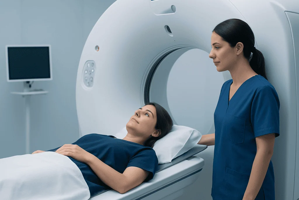
During CT imaging, the person lies on a narrow table that moves through a large ring-shaped scanner. The machine rotates around the body and takes many X-ray images in seconds. A computer combines these images into detailed views that allow the radiologist to examine the lungs layer by layer.
A clinician may recommend CT after a nodule is seen on a chest X-ray, when symptoms or medical history suggest a need for closer assessment, or as part of a scheduled surveillance plan. CT may also be used in people undergoing lung cancer screening when they meet local eligibility criteria based on age and smoking history.
Factors that influence the evaluation include age, current or past tobacco exposure, family history, previous cancer, occupational exposures and the appearance of the nodule itself. People with a known nodule may also be assessed alongside related respiratory conditions, such as lung cancer, although a nodule alone is not a diagnosis of cancer.
A standard low-dose chest CT often does not require contrast material. Contrast may be used when the clinician needs more information about blood vessels, lymph nodes or other structures in the chest. The imaging team will explain which type of scan has been requested and why.
Preparing for a CT Scan for Lung Nodules
Preparation is usually simple. For a CT scan without contrast, most people can eat, drink and take usual medicines unless their healthcare team gives different instructions. Comfortable clothing without metal fasteners is helpful, and jewelry, removable dental appliances, hearing aids or metal accessories may need to be removed before the scan.
If contrast is planned, the imaging department may ask the person not to eat for several hours beforehand. They should tell the care team about kidney disease, diabetes, thyroid conditions, pregnancy or possible pregnancy, prior reactions to contrast material, and all medicines and supplements they use. The team may arrange a blood test or make other adjustments when appropriate.
What not to do before a lung CT scan? A person should not ignore pre-scan instructions, conceal a past contrast reaction or arrive wearing metal objects that could affect image quality. If contrast is planned, they should not eat or drink against the instructions provided by the imaging center. Smoking does not usually prevent the scan, but avoiding smoking before the appointment may make breath-holding easier and supports overall lung health.
It is useful to bring prior imaging reports or ensure that scans from another facility can be shared. Comparison with older images can prevent unnecessary testing by showing that a nodule has been unchanged for a long time.
What Happens During the Procedure and Afterward
Before the scan, a radiographer confirms the person’s identity, reviews relevant safety questions and explains the process. If contrast is needed, it is usually administered through a vein in the arm or hand. A temporary warm sensation, metallic taste or feeling of needing to urinate can occur after contrast injection and usually passes quickly.
The person lies on their back, often with arms raised above the head. They will be asked to remain still and may be asked to hold their breath for a few seconds while images are taken. The actual imaging portion is brief, commonly lasting less than a minute, although the full appointment can take longer because of registration, preparation and safety checks.
For a non-contrast scan, normal activities can generally resume immediately. After contrast, the team may advise drinking fluids unless fluid intake is restricted for another medical reason. Serious contrast reactions are uncommon, but people should seek urgent help for breathing difficulty, swelling, widespread rash or severe dizziness after leaving the imaging department.
CT offers fast, highly detailed evaluation and is especially effective for tracking small changes in nodules. It does involve a small dose of ionizing radiation, so clinicians aim to use the lowest appropriate dose and avoid scans that are not medically needed. The benefit of accurate evaluation and appropriately timed follow-up generally outweighs this risk when CT is recommended.
Can a CT Scan Tell if Lung Nodules Are Cancerous?
A CT scan cannot always tell with certainty whether a lung nodule is cancerous. However, it can show features that make cancer more or less likely. These include nodule size, shape, border characteristics, internal density, calcification pattern and whether the nodule changes on repeat imaging.
For example, a smooth, densely calcified nodule that has been unchanged over time is often more consistent with a benign cause. A larger nodule with irregular or spiculated edges, or one that grows over time, may need more investigation. These imaging findings are considered together with the person’s age, smoking history, medical history and symptoms.
If further clarification is needed, a clinician may recommend repeat CT at a defined interval, PET-CT imaging, bronchoscopy, needle biopsy or surgery in selected cases. A biopsy is the test that can provide tissue for a definitive diagnosis when it is appropriate and safe. Evaluation may involve respiratory medicine, radiology, pathology, oncology and thoracic surgery specialists.
When treatment is needed, options are individualized according to the diagnosis, stage, lung function and overall health. Depending on the findings, a team may discuss lung cancer treatment or another approach suited to the underlying cause.
How Fast Do Lung Nodules Grow if They Are Cancerous?
Cancerous lung nodules do not all grow at the same rate. Some grow relatively quickly, while others grow slowly over months or years. Growth depends on the cancer type and biology, which is why a single CT scan may not provide a complete answer and carefully timed follow-up imaging can be important.
Radiologists assess growth by comparing measurements and, when possible, the estimated volume of a nodule on separate scans. Even a small change in diameter can represent a more meaningful change in volume. At the same time, very small measurement differences may reflect scan technique, breathing position or reader variation rather than true growth.
Some nodules linked with infection or inflammation can change rapidly as well, so growth does not automatically mean cancer. Conversely, certain slow-growing cancers may show only subtle changes. The follow-up plan should therefore be based on established clinical guidance and individual risk rather than assumptions about a particular growth rate.
People should attend planned surveillance scans even if they feel well. This approach allows the care team to identify meaningful changes at the right time while avoiding invasive procedures for nodules that remain low risk.
How Serious Is a Nodule on Your Lungs?
The seriousness of a lung nodule depends on its cause and individual features, not simply on its presence. Many nodules are benign and do not need treatment. Common non-cancerous explanations include healed granulomas from prior infection, small scars, benign tumors and inflammatory conditions.
A nodule may be more concerning when it is larger, has an irregular appearance, grows on follow-up imaging or occurs in a person with relevant risk factors. Even then, the finding requires careful evaluation rather than immediate conclusions. The goal is to identify people who need additional tests while safely monitoring people whose nodules are unlikely to cause harm.
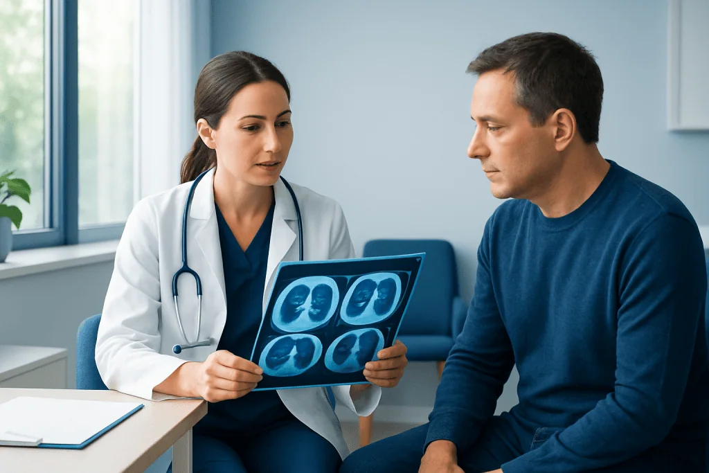
Clinicians commonly use guideline-based follow-up recommendations to determine whether another CT is needed and when. Very small, low-risk nodules may need no routine follow-up, while other nodules may be monitored over time or assessed with additional tests. The report should be discussed with the clinician who requested the scan, as they can interpret it in the context of the person’s full health history.
For international patients, Acibadem International’s multidisciplinary specialists and JCI-accredited hospitals can assess lung nodules and coordinate imaging, respiratory evaluation and treatment planning when needed.
When to Seek Medical Care
A person should arrange follow-up with the clinician who ordered the CT scan to review the result and understand the recommended plan. This is important even when the report appears reassuring, because the appropriate next step depends on previous imaging, risk factors and the reason the scan was performed.
Prompt medical assessment is advisable for persistent or worsening cough, coughing up blood, unexplained shortness of breath, persistent chest pain, repeated chest infections, unexplained weight loss or ongoing fever. These symptoms can have many causes and do not necessarily indicate cancer, but they deserve medical attention.
Urgent care is appropriate for severe breathing difficulty, sudden chest pain, fainting, blue or gray lips, or coughing up a large amount of blood. Anyone who develops symptoms of an allergic reaction after contrast, such as facial swelling, wheezing or difficulty breathing, should seek emergency help.
People should avoid delaying recommended surveillance. Timely review helps clinicians distinguish harmless stable nodules from those that need further investigation, while providing a clear and proportionate plan for care.
Frequently asked questions
How long does a CT scan for lung nodules take?
The image-taking portion is usually very brief, often less than a minute. The full visit may take longer for registration, changing clothes, reviewing safety information and, if needed, administering contrast material.
Do I need contrast for a lung nodule CT scan?
Not always. Many CT scans used to detect or monitor lung nodules are performed without contrast. A clinician may request contrast when additional detail about chest structures, blood vessels or lymph nodes is needed.
Can a lung nodule disappear on a CT scan?
Yes. Nodules caused by temporary inflammation or infection can become smaller or disappear on follow-up imaging. This is one reason why a repeat scan may be recommended instead of immediate invasive testing.
What size lung nodule is concerning?
Size is one important factor, but there is no single size that determines whether a nodule is cancerous. Clinicians also consider the nodule’s appearance, growth, location and the person’s individual risk factors.
Will I receive my CT scan results immediately?
The images are usually reviewed by a radiologist, who prepares a report for the requesting clinician. Timing varies by imaging center and clinical urgency, so patients should ask when and how they will receive results.
Is repeated CT scanning safe for lung nodule follow-up?
CT uses radiation, but follow-up plans are designed to balance this exposure against the value of monitoring a potentially important lung finding. Low-dose CT techniques may be used when appropriate, and scans should be performed only when clinically justified.
References
- American College of Radiology
- Fleischner Society
- American Lung Association
- National Cancer Institute
- Radiological Society of North America
This article is for general information only and is not a substitute for professional medical advice. Please consult a qualified doctor about your individual situation.
Oncology care in Turkey — second opinion and treatment plan
JCI-accredited · board-certified surgeons · reply within 24h
Add us as a Preferred Source to see more of our trusted health content across Google Search, AI Overviews and Discover.