What Can an MRI Find: Preparation, Procedure and Results
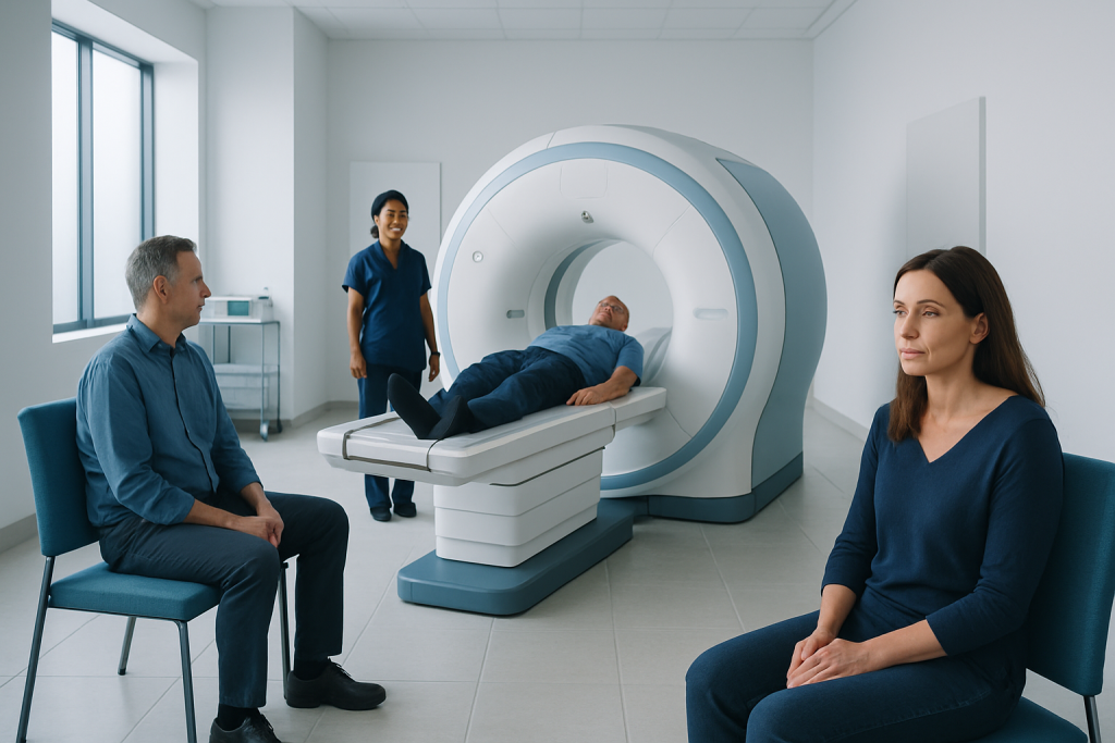
MRI uses a powerful magnet and radio waves, not ionizing radiation, to create detailed images. It can reveal many abnormalities involving the brain, spinal cord, joints, muscles, organs and some blood vessels.
Key Takeaways
- MRI uses a powerful magnet and radio waves, not ionizing radiation, to create detailed images.
- It can reveal many abnormalities involving the brain, spinal cord, joints, muscles, organs and some blood vessels.
- Preparation depends on the body area being scanned and whether contrast material is needed.
- A radiologist interprets MRI images, and the referring clinician explains what the findings mean in the context of symptoms and other tests.
- Urgent MRI results may be communicated quickly, while routine reports commonly take longer depending on the clinical setting.
MRI is a radiation-free imaging test that creates detailed pictures of soft tissues, organs, bones and blood vessels. It can help doctors investigate symptoms, clarify a suspected condition, plan treatment and monitor changes over time.
What can an MRI find?
MRI, or magnetic resonance imaging, can find or help assess many changes inside the body, especially in soft tissues that may not be as clearly seen on standard X-rays. Depending on the area examined, it may show inflammation, swelling, infection, bleeding, tumors, structural differences, tissue injury, degenerative changes and problems affecting blood flow.
Doctors may request an MRI to investigate persistent symptoms, such as unexplained pain, weakness, numbness, headaches, dizziness or changes in movement. It can also be used after an injury, to help plan treatment, or to monitor a known condition. An MRI finding is not automatically a diagnosis: the images must be considered alongside the person’s symptoms, examination and other test results.
MRI is particularly useful for imaging the brain and spinal cord, discs and nerves in the spine, ligaments and cartilage in joints, muscles and tendons, pelvic organs, the liver and other abdominal organs, breasts, heart, and some blood vessels. For example, a clinician may use MRI while evaluating possible brain tumor changes, spinal disc problems, joint injuries or abnormalities in internal organs.
How MRI works and who may need one
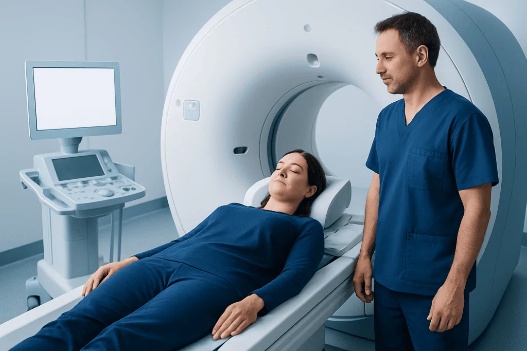
An MRI scanner uses a strong magnetic field and radiofrequency energy to temporarily align particles in the body. When these particles return to their usual state, they release signals that a computer turns into highly detailed images. Unlike CT scans and X-rays, MRI does not use ionizing radiation.
Different MRI sequences highlight different types of tissue. This allows radiologists to compare structures such as fluid, fat, nerves, muscles, cartilage and areas of inflammation. In some examinations, a gadolinium-based contrast agent is injected into a vein to make certain tissues, blood vessels or abnormal areas more visible.
MRI may not be suitable for everyone without additional planning. People should tell the imaging team about any implanted device, metal fragment, previous surgery, medication patch, hearing device, dental work, pregnancy, kidney disease, allergies, or possible pregnancy. Many modern implants are MRI-conditional, meaning scanning may be possible under specified conditions, but this must always be checked before the examination.
What will not show up on an MRI?

MRI is highly detailed, but no single test shows everything equally well. It is generally less useful than CT for quickly identifying certain bone injuries, especially small fractures, and for evaluating some lung conditions because air-filled lungs do not produce MRI signals in the same way as soft tissues. CT may also be preferred in some urgent situations because it is faster and more widely available in emergency settings.
MRI does not directly measure how well an organ works, identify every cause of pain, or reliably distinguish harmless from harmful changes without clinical context. Very small abnormalities may be below the scan’s resolution, may not be visible in a particular sequence, or may require another type of imaging, laboratory test or tissue sampling for clarification.
Some symptoms can arise from functional, chemical or microscopic changes that do not appear on MRI. A normal result can be reassuring, but it does not mean symptoms are unimportant. The doctor may recommend follow-up, another test or a different specialty assessment when symptoms persist.
Do I need to do anything in preparation for an MRI?
What preparation is needed for an MRI depends on the examination. For many scans, no special dietary preparation is required. The imaging center will provide instructions before the appointment, particularly for MRI of the abdomen or pelvis, an examination involving sedation, or a scan using contrast material.
For routine MRI procedure preparation, patients should bring relevant referral information and previous scans or reports if available. They should wear comfortable clothing without metal fasteners when possible and remove jewelry, watches, hairpins, eyeglasses, removable dental devices, hearing aids, credit cards and other metal or magnetic items. Makeup, tattoos and some skin patches can occasionally contain metallic substances, so the team should be told about them.
People wondering what preparation do you need for an MRI should focus on safety screening. They should report pacemakers, neurostimulators, cochlear implants, aneurysm clips, insulin pumps, metal fragments from prior injuries, and any history of kidney problems or contrast reactions. If claustrophobia is a concern, discussing it in advance allows the care team to consider practical support, relaxation techniques, a wider-bore scanner where available, or clinician-prescribed medication when appropriate.
- Follow fasting instructions exactly if they are provided.
- Continue usual medicines unless a clinician advises otherwise.
- Arrive early enough to complete the screening questionnaire.
- Ask beforehand whether a companion may attend, particularly if anxiety or mobility needs are relevant.
What happens during an MRI procedure?
Before the scan, a radiology professional confirms the person’s identity, reviews the safety questionnaire and explains the process. If contrast is needed, an intravenous line may be placed. The patient lies on a padded table, and a receiving device called a coil may be positioned around the body part being scanned to improve image quality.
The table moves slowly into the scanner. During image acquisition, the machine makes loud tapping, knocking or humming sounds. Earplugs or headphones are usually offered. It is important to remain as still as possible, because movement can blur the images. The technologist watches from a nearby room and can communicate through an intercom; patients are also given a call device to use if they need assistance.
The length of an MRI varies by body area and the number of images needed. Some scans take less than half an hour, while more detailed studies can take longer. If contrast is used, a cool sensation at the injection site may occur briefly. Most people can return to normal activities after the examination, unless they received sedating medication or were given specific instructions.
Advanced MRI imaging may be part of a broader diagnostic pathway. At Acibadem International, multidisciplinary specialists and JCI-accredited hospitals assess and treat patients from abroad using appropriate imaging and follow-up care plans.
Benefits, risks and recovery after MRI
The main benefit of MRI is its ability to provide detailed images without ionizing radiation. It can help reduce uncertainty, guide further testing, support treatment decisions and, in some cases, avoid more invasive procedures. The best scan is the one matched to the clinical question, which is why a doctor selects the area and type of MRI carefully.
MRI is generally considered safe when screening procedures are followed. The magnetic field can be dangerous around certain metal objects or devices, which makes accurate pre-scan screening essential. Some people find the noise, confined space or need to lie still uncomfortable. The care team can explain options that may make the experience easier.
Contrast reactions are uncommon, but any previous reaction should be reported. Gadolinium contrast is used selectively, and clinicians take additional care in people with significant kidney disease. After a standard MRI, there is usually no recovery period. If sedation was used, the patient may need someone to take them home and should avoid driving, alcohol and important decisions until the medication has worn off.
When to worry about MRI results?
An MRI report can contain unfamiliar medical terms, including words such as lesion, signal change, degeneration, cyst or enhancement. These terms describe what is visible on the images and do not always mean a serious disease. Some findings are incidental, meaning they are unrelated to the reason for the scan and may not require treatment.
It is reasonable to ask the referring doctor what each finding means, whether it explains the symptoms, whether it needs further tests, and what follow-up is recommended. Results are more concerning when the report identifies a change that needs urgent assessment, but the urgency should be determined by the treating clinician rather than by trying to interpret the report alone.
Patients should seek prompt medical advice if they have worsening neurological symptoms, such as sudden weakness, difficulty speaking, loss of vision, severe new headache, seizures, confusion, or loss of bladder or bowel control. These symptoms require timely assessment whether or not MRI results are available.
How quickly do you get MRI results if something is wrong?
Timing depends on why the MRI was ordered, how urgently it is needed, the complexity of the images and local reporting processes. A radiologist reviews the scan and prepares a report for the referring clinician. In emergency or urgent hospital situations, important findings may be communicated promptly to the clinical team so that care can proceed without waiting for a routine report.
For outpatient scans, results may be available within a few days, although timelines vary. A faster result does not necessarily mean that something is wrong; it may simply reflect the type of referral or the service’s usual workflow. Likewise, a longer wait does not indicate that a serious problem has been found.
Patients who have not received expected follow-up can contact the ordering clinician or imaging department for guidance. The clinician who requested the MRI is best placed to explain how the result relates to symptoms and whether additional evaluation, such as neurology assessment, orthopedic evaluation or another specialist review, is appropriate.
When to seek medical care
Medical care should be sought for symptoms that are severe, sudden, worsening or interfering with daily life. This includes a new severe headache, fainting, stroke-like symptoms, chest pain, sudden shortness of breath, major injury, severe back pain with weakness or bladder/bowel changes, or persistent unexplained pain. Emergency symptoms should be assessed urgently rather than waiting to arrange an MRI independently.
A non-urgent medical appointment is appropriate for ongoing symptoms such as recurrent headaches, persistent joint pain, tingling, numbness, weakness, prolonged dizziness, unexplained changes in balance, or abdominal and pelvic symptoms that do not improve. A clinician can decide whether MRI is likely to help and whether another examination or test would be more suitable.
Patients should not delay care because they are worried about the MRI experience or its results. Clear communication about what to do in preparation for an MRI, safety concerns and expected timing can make the process more manageable and help ensure the examination answers the right clinical question.
Frequently asked questions
Can an MRI detect cancer?
MRI can identify abnormalities that may be suspicious for cancer and can help show their location, size and relationship to nearby tissues. However, MRI alone cannot confirm every cancer diagnosis. Further imaging, follow-up or a biopsy may be needed.
Is MRI painful?
MRI is not usually painful. The main challenges for some people are lying still, the loud sounds and the enclosed scanner space. If discomfort, anxiety or pain makes this difficult, the imaging team should be informed before the scan.
Can I eat and drink before an MRI?
Many MRI examinations do not require fasting, so people can often eat and drink normally. Some abdominal, pelvic, contrast-enhanced or sedation-related scans have specific instructions. The imaging center’s instructions should always take priority.
Can I have an MRI if I have metal in my body?
Possibly, but the type of metal, its location and any implanted device must be assessed before the scan. Many implants can be scanned under defined conditions, while others may require a different imaging test. Never assume an implant is safe without confirming it with the MRI team.
Does a normal MRI mean nothing is wrong?
A normal MRI can rule out or make many structural problems less likely, which can be helpful. However, some causes of symptoms do not appear on MRI, and results must be interpreted alongside the medical history and examination. Persistent or worsening symptoms should be discussed with a clinician.
Why might contrast be used for an MRI?
Contrast can make certain tissues, blood vessels, inflammation and abnormal areas easier to distinguish on images. It is not needed for every scan. The clinician and radiology team decide whether its expected value outweighs any potential risk for the individual.
References
- Radiological Society of North America
- American College of Radiology
- National Institute of Biomedical Imaging and Bioengineering
- U.S. Food and Drug Administration
- National Health Service
This article is for general information only and is not a substitute for professional medical advice. Please consult a qualified doctor about your individual situation.
Explore treatments in Turkey — costs, top hospitals & a free quote
JCI-accredited · board-certified surgeons · reply within 24h
Add us as a Preferred Source to see more of our trusted health content across Google Search, AI Overviews and Discover.