Tethered Cord MRI: Preparation, Procedure and Results
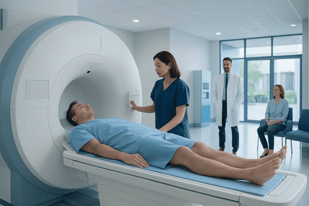
MRI is the main imaging test used to assess a suspected tethered spinal cord. The scan is painless and does not use ionising radiation.
Key Takeaways
- MRI is the main imaging test used to assess a suspected tethered spinal cord.
- The scan is painless and does not use ionising radiation.
- MRI findings are interpreted together with symptoms, physical examination and, when needed, urological or neurological tests.
- Some children and adults need contrast dye, while many routine spinal MRI scans do not.
- Surgery is considered when tethering is causing symptoms or progressive nerve dysfunction, not from an MRI finding alone.
A tethered cord MRI is a non-invasive imaging test that provides detailed pictures of the spinal cord, nerves and surrounding tissues. It helps doctors investigate whether the spinal cord is abnormally attached or under tension and supports decisions about monitoring, further testing or treatment.
Tethered Cord MRI: What It Shows and Why It Is Used
A tethered cord MRI is a magnetic resonance imaging scan focused on the spine, usually the lower back and sacral area. It uses a strong magnetic field and radio waves to create detailed images of the spinal cord, nerve roots, spinal canal, bones and soft tissues. Unlike X-rays and CT scans, MRI does not use ionising radiation.
The test is commonly requested when a doctor suspects tethered cord syndrome, a condition in which the spinal cord is held down or attached to tissues in a way that may limit its normal movement. This tension can affect nerve function over time. MRI can identify anatomical features that may be related to tethering, including a low-lying conus medullaris, a thickened filum terminale, fatty tissue, scar tissue or a spinal lipoma.
An MRI result does not diagnose symptoms on its own. Some people have an anatomical variation on imaging without developing problems, while others have symptoms that need careful assessment even if the findings are subtle. Doctors therefore consider the images alongside a person’s medical history, neurological examination and bladder or bowel function.
Who May Need a Tethered Cord MRI?
A doctor may recommend tethered cord MRI for infants, children or adults with symptoms that could involve the lower spinal cord or nerves. These may include back or leg pain, weakness, numbness, walking changes, foot deformities, scoliosis, reduced sensation, or new difficulty controlling the bladder or bowel.
In babies, a scan may be considered when there are skin signs over the lower back, such as a deep or unusual dimple, a patch of hair, a fatty lump, a birthmark or a small opening in the skin. Many dimples are harmless, but certain features may prompt further evaluation. Ultrasound can sometimes be used in young infants, while MRI gives a more complete assessment when needed.
MRI may also be used after previous spinal surgery, spinal injury or infection, or when a person has a known congenital spinal difference. The scan can help identify related conditions such as spina bifida or a spinal lipoma, and it can assist specialists in planning follow-up care.
How to Prepare for a Tethered Cord MRI

Most people can eat, drink and take their regular medicines before a standard MRI appointment. The imaging team should be told about implanted medical devices, previous surgery, metal fragments, hearing aids, medication patches, pregnancy, kidney disease and any past reaction to MRI contrast. Some implants are MRI-safe or MRI-conditional, but they must be checked before the scan.
People should also tell the team if they experience claustrophobia, anxiety or difficulty lying still. The scanner is shaped like a short tunnel, and the examination can take time. A radiology department may offer reassurance strategies, music where available, a support person according to local policy, or medication prescribed in advance by the treating clinician.
Young children and some adults may require sedation or general anaesthesia so that they can remain still enough for clear images. If sedation is planned, fasting instructions are important and will be provided by the clinical team. A parent or caregiver should arrange transport home if sedating medication or anaesthesia has been used.
Tethered Cord MRI Procedure: Step by Step
Before the scan, the person removes jewellery, watches, hairpins, removable dental appliances and other metal items. They change into clothing suitable for MRI and complete a safety screening questionnaire. An intravenous line may be placed if contrast material is required, although contrast is not needed for every tethered cord MRI.
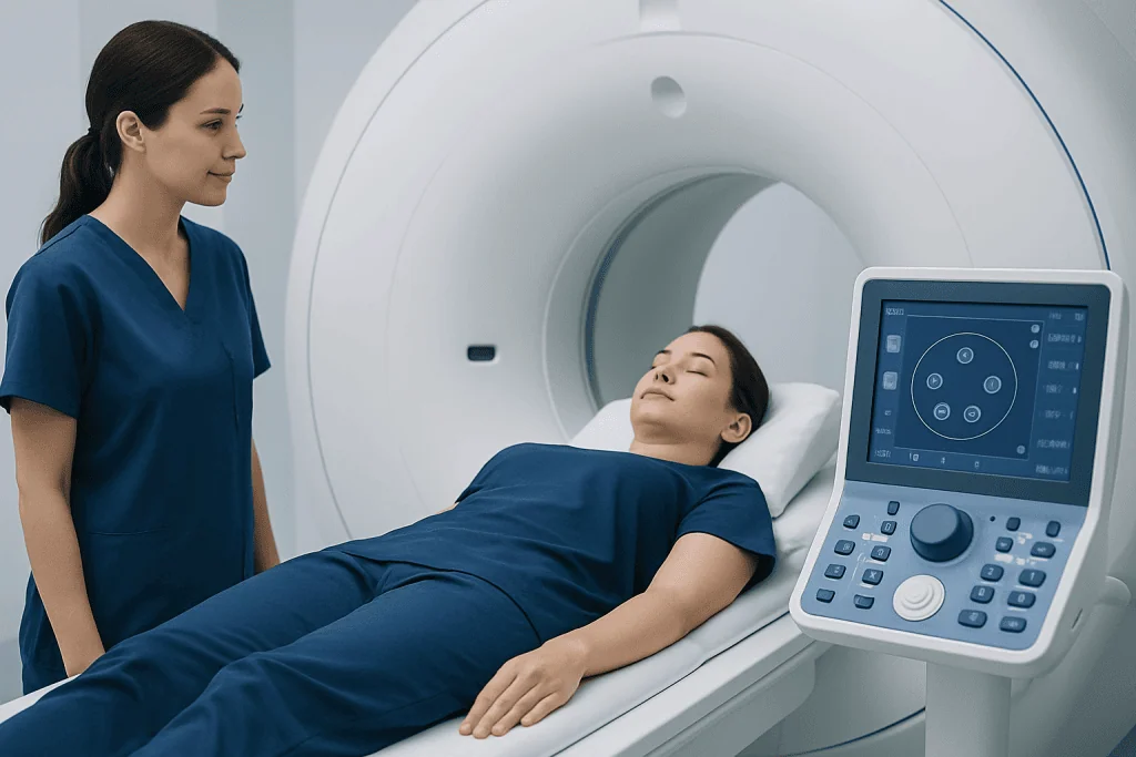
During the examination, the person lies on a padded table, usually on their back. A specialised receiving device called a coil is positioned around the area being scanned. The table moves into the MRI scanner, and the radiographer communicates through an intercom throughout the examination. The machine makes repeated loud knocking or humming sounds, so ear protection is provided.
It is important to lie as still as possible because movement can blur the images. The scan commonly takes about 30 to 60 minutes, although the duration varies with the body areas examined, whether contrast is used and whether additional sequences are needed. The procedure itself is painless, but remaining still may be uncomfortable for some people.
Images are reviewed by a radiologist, often with input from neurology, neurosurgery, orthopaedics or paediatric specialists when appropriate. If a symptomatic tethered cord is confirmed, tethered cord surgery may be discussed as one possible treatment pathway.
MRI Results, Benefits and Possible Risks
The radiology report describes the position and appearance of the spinal cord and surrounding structures. A low conus medullaris, often below the expected level in the lumbar spine, may support the possibility of tethering. Other possible findings include a thickened or fatty filum terminale, lipoma, split cord malformation, dermal sinus tract, postoperative scarring, syringomyelia or signs of associated spinal dysraphism.
One important benefit of MRI is its ability to show soft tissues and nerves in detail without radiation exposure. It can help clarify anatomy, guide specialist referral, establish a baseline for future comparison and support surgical planning when treatment is indicated. MRI can also help rule out other explanations for symptoms.
MRI is generally very safe when screening procedures are followed. The main practical challenges are noise, the enclosed scanner space and the need to remain still. Gadolinium-based contrast, if used, has a low risk of allergic reaction and is selected carefully in people with significant kidney impairment. Sedation and anaesthesia have their own risks, which are discussed individually with the anaesthesia team.
Results are usually reviewed at a follow-up appointment rather than interpreted from the report alone. A specialist may recommend observation, symptom monitoring, urodynamic testing, physiotherapy, pain management or referral for neurosurgical assessment depending on the complete clinical picture.
Recovery Timeline and Next Steps After MRI
After a non-sedated MRI, most people can return to normal activities immediately. If contrast was given, normal hydration is usually appropriate unless a clinician has advised otherwise. There is typically no recovery period from the scan itself.
After sedation or general anaesthesia, the person is monitored until awake and stable. Temporary sleepiness, nausea, irritability or poor coordination can occur. Driving, operating machinery, returning to school or work, and making important decisions should wait until the care team confirms it is safe.
If the MRI raises concern for tethered cord syndrome, the next step is not automatically surgery. Clinicians consider whether symptoms are present, worsening or affecting function. Assessments may include neurological examination, bladder testing, orthopaedic review and discussion of the potential benefits and limitations of treatment.
Acibadem International’s multidisciplinary specialists and JCI-accredited hospitals evaluate and treat tethered spinal cord conditions for international patients, coordinating imaging, neurosurgical assessment and follow-up when needed.
When to Seek Medical Care
Medical assessment is advisable for persistent or worsening low-back pain with leg weakness, altered sensation, balance changes, frequent falls, progressive foot changes, or new bladder or bowel difficulties. These symptoms can have many causes, but timely evaluation helps identify conditions that may need treatment.
Urgent medical care is important for sudden inability to pass urine, new loss of bowel control, rapidly progressing weakness, numbness around the buttocks or genital area, or severe back pain accompanied by fever or feeling unwell. These symptoms may indicate a serious spinal or neurological problem and should not be managed at home.
For children, parents or caregivers should speak with a paediatrician if there are concerns about delayed walking, unusual gait, leg stiffness, repeated urinary infections, changes in continence, scoliosis or a concerning lower-back skin mark. Early specialist guidance can provide reassurance and determine whether imaging is appropriate.
Frequently asked questions
What are the MRI findings of a tethered cord?
MRI findings that can be associated with a tethered cord include a low-lying conus medullaris, a thickened or fatty filum terminale, spinal lipoma, scar tissue or other congenital spinal abnormalities. The scan may also show related conditions such as split cord malformation or syringomyelia. These findings must be interpreted with symptoms and examination results, because imaging alone does not always establish tethered cord syndrome.
Is tethered cord surgery painful?
Tethered cord surgery is performed under general anaesthesia, so the person is asleep and does not feel pain during the operation. Pain or discomfort can occur during recovery, particularly around the incision and back muscles, but it is usually managed with an individualised pain-control plan. The surgical team explains expected recovery, activity restrictions and warning signs before discharge.
At what age is a tethered spinal cord typically diagnosed?
A tethered spinal cord can be diagnosed at any age. Some cases are identified before birth or during infancy because of skin findings or known spinal differences, while others become noticeable in childhood, adolescence or adulthood when symptoms develop. The timing depends on the underlying cause and whether nerve tension affects function.
How serious is a tethered spinal cord?
The seriousness of a tethered spinal cord varies widely. Some people have no symptoms and are monitored, while others develop pain, weakness, walking difficulties or bladder and bowel changes that may worsen without treatment. A neurological and imaging assessment helps determine the likely significance for an individual person.
Does a tethered cord MRI require contrast?
Many tethered cord MRI examinations can be completed without contrast. Contrast may be useful when doctors need to evaluate a mass, inflammation, infection, scar tissue, prior surgery or another specific concern. The radiologist and referring clinician decide whether contrast adds useful information.
Can an adult have a tethered cord?
Yes. Although tethered cord is often linked with congenital spinal differences, symptoms can first become apparent in adulthood. Adults may seek assessment for back or leg pain, sensory changes, weakness, gait problems or bladder and bowel symptoms, and MRI can help evaluate the spinal anatomy.
References
- National Institute of Neurological Disorders and Stroke
- American College of Radiology
- Radiological Society of North America
- Spina Bifida Association
- National Health Service
This article is for general information only and is not a substitute for professional medical advice. Please consult a qualified doctor about your individual situation.
Joint and spine care in Turkey — expert assessment & treatment
JCI-accredited · board-certified surgeons · reply within 24h
Add us as a Preferred Source to see more of our trusted health content across Google Search, AI Overviews and Discover.