Myelomeningocele Repair: Procedure, Recovery and Results

Myelomeningocele is the most severe form of spina bifida and requires prompt specialist assessment. Repair can be performed after birth and, for selected pregnancies, before birth through fetal surgery.
Key Takeaways
- Myelomeningocele is the most severe form of spina bifida and requires prompt specialist assessment.
- Repair can be performed after birth and, for selected pregnancies, before birth through fetal surgery.
- Surgery closes and protects exposed spinal tissues but cannot reverse nerve damage already present.
- Recovery includes wound care, monitoring for hydrocephalus, bladder and bowel support, and rehabilitation.
- Long-term outcomes vary and depend on the level of the spinal lesion and associated conditions.
Myelomeningocele repair is surgery to close an opening in a baby’s spine where the spinal cord and nerves are exposed. It may be performed during pregnancy in carefully selected cases or shortly after birth, with the aim of protecting nerve tissue and supporting the child’s long-term health.
Overview: What Is Myelomeningocele Repair?
Myelomeningocele repair is an operation that closes a defect in the developing spine. In myelomeningocele, a form of spina bifida, part of the spinal cord, nerves and surrounding membranes protrude through an opening in the back. Repair protects these delicate tissues, lowers the risk of infection and creates skin coverage over the area.
The operation does not restore nerve function that was affected before surgery. However, timely repair is an essential part of care and may help prevent further injury to exposed nerves. A child will usually need coordinated follow-up with specialists in neurosurgery, urology, orthopedics, rehabilitation and developmental care.
Most babies undergo repair shortly after delivery. In selected pregnancies, fetal surgery may be considered before birth at a specialist center. The most appropriate approach depends on the baby’s anatomy, the pregnancy, the family’s circumstances and a detailed discussion of potential benefits and risks.
How the Procedure Works and Who May Be a Candidate
Myelomeningocele develops early in pregnancy when the neural tube does not close completely. It is often diagnosed on prenatal ultrasound and may be assessed further with fetal MRI and other tests. Families are referred to a multidisciplinary fetal and pediatric team to understand the findings and plan care.
Postnatal repair is generally recommended soon after birth when myelomeningocele is present. The timing is individualized, but early closure is important because exposed tissues can become damaged or infected. Delivery planning may involve maternal-fetal medicine, neonatology, pediatric neurosurgery and anesthesia teams.
Fetal myelomeningocele repair may be an option for a limited group of pregnant patients. Eligibility commonly depends on the location and type of spinal defect, gestational age, fetal imaging findings, maternal health and the absence of certain fetal or pregnancy-related concerns. It requires thorough counseling because fetal surgery also carries important risks for the pregnant person and the pregnancy.
Evaluation may also identify related issues, including hydrocephalus, Chiari II malformation, clubfoot, hip concerns and differences in bladder or bowel function. These findings help the team prepare for care after delivery and provide a clearer outlook.
Step by Step: What Happens During Repair

For repair after birth, the baby receives general anesthesia and is positioned carefully to allow the surgical team to reach the spinal opening. The surgeon examines the exposed sac and neural tissue, then gently repositions and covers the tissues where possible. The membranes and surrounding layers are closed to create a protective barrier.
The skin is then closed over the repaired area. Some larger or complex defects may require assistance from reconstructive surgeons to achieve safe tissue coverage. The exact surgical technique varies according to the size, location and characteristics of the lesion.
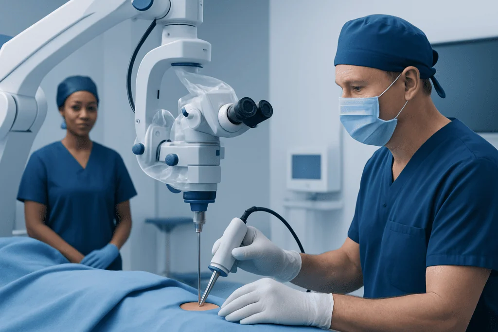
Fetal repair is performed during pregnancy in highly specialized settings. Depending on the planned approach, surgeons access the uterus through an open incision or a minimally invasive fetoscopic technique. The fetal back is repaired, and the uterus and abdominal incision are closed so that the pregnancy can continue under close monitoring.
After either approach, the baby is monitored for breathing, feeding, wound healing, signs of infection and changes that could suggest pressure buildup in the brain. If hydrocephalus develops or progresses, some children may need treatment such as a shunt or an endoscopic procedure to manage cerebrospinal fluid.
Recovery Timeline and Ongoing Care
After postnatal surgery, the baby usually remains in a neonatal intensive care or specialized pediatric unit for observation. Positioning may be adjusted to protect the surgical wound, and the care team checks the incision regularly. Feeding and bonding can continue with support from neonatal staff, depending on the baby’s medical needs.
The length of hospital stay varies. It may be longer when a baby has hydrocephalus, wound concerns, feeding difficulties, premature birth or other health needs. Before discharge, parents and caregivers are taught how to care for the incision, recognize concerning changes and attend planned follow-up visits.
Recovery after fetal surgery includes monitoring for both the pregnant patient and the fetus. The pregnant patient may remain in hospital initially and then have frequent prenatal visits. Because the uterine incision needs time to heal, delivery planning and future pregnancy counseling are important parts of follow-up.
Long-term care focuses on each child’s individual needs. This may include pediatric neurosurgery follow-up, physical and occupational therapy, mobility assessment, orthopedic treatment and regular urologic evaluation. Early support helps children build skills, protect kidney health and participate as fully as possible in daily life.
Potential Benefits, Risks and Expected Results
The main benefit of postnatal myelomeningocele repair is protecting exposed spinal tissues from infection and additional trauma. Fetal repair may reduce the likelihood that a child will need treatment for hydrocephalus in some selected cases and may improve some mobility-related outcomes. These potential benefits must be weighed carefully against the risks of fetal surgery.
Possible surgical risks for the baby include bleeding, infection, cerebrospinal fluid leakage, wound separation, scarring and the need for further procedures. Neurological function can vary significantly, and surgery cannot fully predict or change all effects of the underlying spinal cord difference.
Fetal surgery has additional maternal and pregnancy-related risks, such as premature rupture of membranes, preterm birth, bleeding, infection and complications involving the uterus. A prior fetal surgery incision can also influence delivery recommendations and management of future pregnancies.
Results depend largely on the level of the spinal lesion and the child’s associated medical needs. Some children walk independently, while others use braces, walkers or wheelchairs. Bladder and bowel programs are common, and many children benefit from ongoing therapy and educational support. Regular review allows the care plan to change as the child grows.
Prevention, Self-Care and Family Support
Not all neural tube defects can be prevented, but adequate folic acid before conception and during early pregnancy lowers the risk. People who may become pregnant should discuss folic acid supplementation and individual risk factors with a qualified healthcare professional, especially if there has been a previous neural tube defect-affected pregnancy or certain medication exposures.
After repair, caregivers should follow the surgical team’s instructions for wound care, positioning, bathing and follow-up. They should not apply creams, dressings or other products to the incision unless advised. Safe handling techniques and a clear plan for feeding, sleep and transport can make the early period at home more manageable.
As the child grows, regular care may include physical therapy, adaptive equipment, skin checks and bladder or bowel management. Support from social workers, nurses, therapists and patient support organizations can help families coordinate appointments, schooling and community resources.
Acibadem International’s multidisciplinary specialists and JCI-accredited hospitals support international patients requiring assessment and treatment planning for complex pediatric neurosurgical conditions.
When to Seek Medical Care
During pregnancy, prompt medical assessment is appropriate after an abnormal prenatal scan or if a clinician raises concern about a spinal abnormality. A maternal-fetal medicine specialist and pediatric neurosurgical team can explain the diagnosis, confirm findings and discuss delivery and treatment planning.
After myelomeningocele repair, caregivers should contact the surgical team promptly if the incision becomes increasingly red, swollen, warm, draining fluid or opening. Fever, poor feeding, unusual sleepiness, persistent vomiting, irritability, a rapidly enlarging head size or a bulging soft spot also need urgent medical advice because they may indicate infection or changes in cerebrospinal fluid pressure.
Emergency care is needed for breathing difficulty, seizures, loss of responsiveness or other severe symptoms. Families should keep scheduled appointments even when the child seems well, as some complications and developmental needs are best recognized through regular monitoring.
Frequently asked questions
What is the purpose of myelomeningocele repair?
The purpose is to close and protect the spinal opening where nerves and membranes are exposed. This helps reduce the risk of infection and further injury to the exposed tissue. It does not reverse nerve damage that occurred before the repair.
Is myelomeningocele repair done before or after birth?
Most repairs are performed shortly after birth. Fetal repair during pregnancy may be considered for selected pregnancies at specialist centers. The best option depends on detailed fetal imaging, maternal health and informed discussion of risks and benefits.
How long does recovery take after myelomeningocele surgery?
The first recovery period takes place in hospital and varies according to wound healing, feeding, prematurity and related conditions such as hydrocephalus. Follow-up continues throughout childhood because mobility, bladder and bowel function, and developmental needs can change as a child grows.
Will a child be able to walk after myelomeningocele repair?
Walking ability varies widely and is influenced mainly by the level and severity of the spinal lesion. Some children walk independently, while others need braces, walking aids or a wheelchair. Physical therapy and orthopedic support can help maximize mobility and independence.
Can myelomeningocele cause bladder and bowel problems?
Yes. The nerves that control the bladder and bowel may be affected, which can lead to difficulties with emptying, continence or constipation. Regular urology follow-up and an individualized bladder and bowel program can help protect health and improve daily comfort.
Does myelomeningocele repair cure spina bifida?
Repair treats the open spinal defect, but it does not cure the underlying spina bifida or eliminate all associated effects. Ongoing care is often needed for neurological, orthopedic, urologic and developmental concerns. With coordinated care, many children and adults with myelomeningocele lead active, fulfilling lives.
References
- Centers for Disease Control and Prevention
- National Institute of Neurological Disorders and Stroke
- Spina Bifida Association
- American College of Obstetricians and Gynecologists
- Children's Hospital of Philadelphia
This article is for general information only and is not a substitute for professional medical advice. Please consult a qualified doctor about your individual situation.
Joint and spine care in Turkey — expert assessment & treatment
JCI-accredited · board-certified surgeons · reply within 24h
Add us as a Preferred Source to see more of our trusted health content across Google Search, AI Overviews and Discover.