Accessory Navicular Surgery: Procedure, Recovery and Results

An accessory navicular is an extra bone or cartilage piece on the inner side of the foot, present from birth. Surgery is usually considered only when symptoms continue despite appropriate non-surgical treatment.
Key Takeaways
- An accessory navicular is an extra bone or cartilage piece on the inner side of the foot, present from birth.
- Surgery is usually considered only when symptoms continue despite appropriate non-surgical treatment.
- The most common operation removes the accessory bone; tendon repair or repositioning may be added when needed.
- Initial recovery involves immobilization and limited weight-bearing, while return to higher-impact activity often takes several months.
- Results are commonly favorable for carefully selected patients, but recovery requires patience, follow-up and rehabilitation.
Accessory navicular surgery is an option for persistent inner-foot pain caused by an accessory navicular bone when footwear changes, orthotics, activity adjustment and other non-surgical care have not provided enough relief. The operation usually removes the painful extra bone and may also restore support from the posterior tibial tendon, followed by protected healing and rehabilitation.
Overview: what is accessory navicular surgery?
Accessory navicular surgery is performed to treat ongoing pain from an accessory navicular: an extra piece of bone or cartilage located on the inner side of the midfoot, near the arch. Many people have this normal anatomical variation without symptoms. In others, pressure from footwear, irritation of nearby tissues or strain on the posterior tibial tendon can cause tenderness, swelling and difficulty with activity.
The goal of surgery is to remove the painful accessory bone and address any associated tendon problem while preserving stable, comfortable foot function. It is generally not the first treatment. Clinicians usually begin with shoe modifications, activity changes, supportive orthotics, immobilization when appropriate, physical therapy and pain-relief measures before discussing an operation.
Symptoms related to this bone are sometimes called accessory navicular syndrome. A foot and ankle specialist can distinguish this problem from other causes of medial foot pain, including tendon disorders, stress injuries and flatfoot-related strain.
How rare is an accessory navicular?
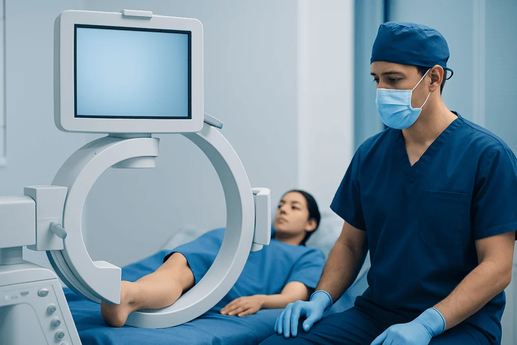
An accessory navicular is not exceptionally rare. It is a congenital variation, meaning it develops as the foot forms, and it may be present in one or both feet. It is often noticed in adolescence or early adulthood because growth, sports participation, footwear pressure or an injury can make an otherwise painless area symptomatic.
Only a proportion of people with an accessory navicular develop symptoms. The most common symptomatic form is located close to the main navicular bone and the attachment of the posterior tibial tendon. This location can be irritated by shoe rubbing or repeated pull from the tendon during walking and running.
Having an accessory navicular does not automatically mean surgery is needed. Imaging may identify it incidentally, and treatment decisions should be based on the person’s symptoms, examination findings, daily needs and response to conservative care rather than the X-ray appearance alone.
Candidacy and preparation for surgery
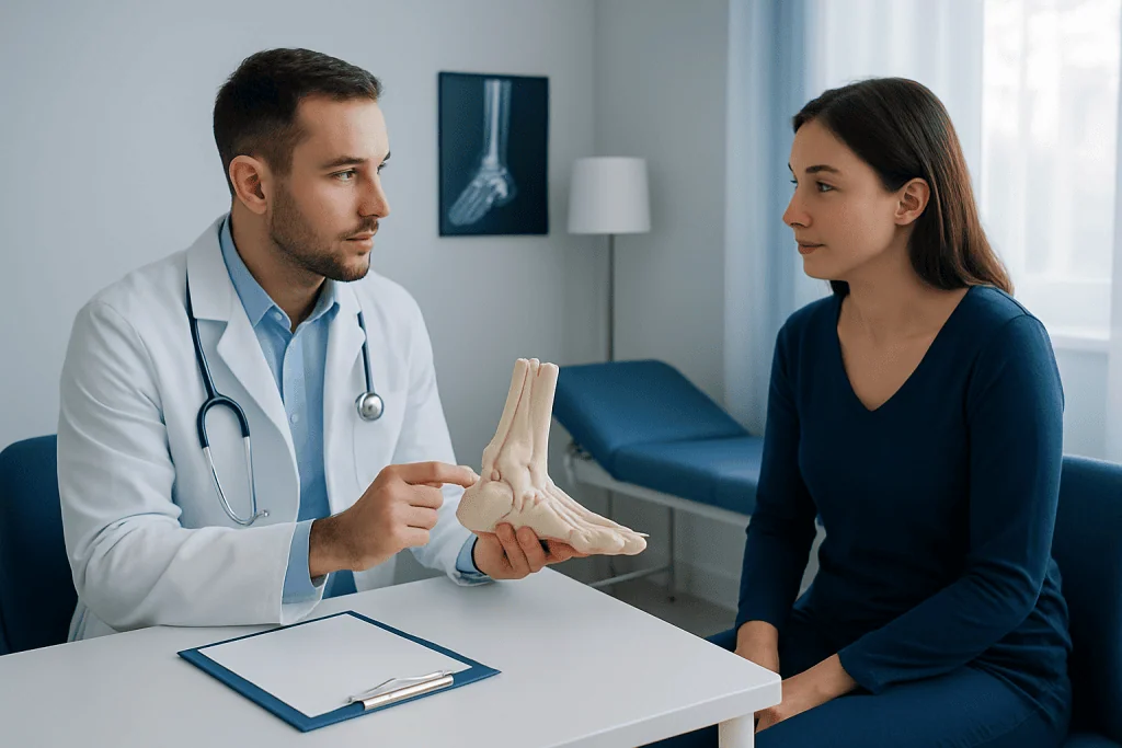
A surgeon may consider accessory navicular surgery when pain remains limiting after a well-planned course of non-surgical treatment. Typical concerns include ongoing tenderness over the bony prominence, recurrent swelling, inability to tolerate shoes, pain during sports or walking, or posterior tibial tendon irritation linked to the accessory bone.
Assessment usually includes a detailed history and physical examination. X-rays confirm the anatomy and may show the type of accessory navicular. Ultrasound or MRI can be helpful when the clinician needs to assess the posterior tibial tendon, inflammation or other possible sources of pain. Foot alignment, arch shape, flexibility and walking pattern are also important because they may influence the procedure and recovery plan.
Before surgery, patients should discuss work demands, mobility at home, smoking or nicotine use, medical conditions, regular medicines and plans for travel. The team may advise stopping certain medicines only under medical guidance. Preparing crutches, a knee scooter or other mobility support in advance can make the early recovery period easier.
How the procedure works: step by step
The operation is typically carried out under anesthesia in a hospital or surgical setting. The surgical team makes an incision over the inner midfoot, carefully protects nearby nerves and soft tissues, and identifies the accessory navicular and the posterior tibial tendon attachment.
In a simple excision, the surgeon removes the symptomatic accessory bone and smooths the area as needed. When the tendon attachment requires additional support, the procedure may include repairing, advancing or reattaching the posterior tibial tendon to the main navicular bone. This combined approach is often referred to as a Kidner-type procedure, although the exact technique is individualized.
After the repair, the incision is closed and the foot is placed in a splint, cast or protective boot. The operation and anesthesia plan vary with the anatomy and whether tendon work is needed. The surgeon will explain the expected restrictions and the specific post-operative plan before the procedure.
Orthopedic foot and ankle care may involve imaging specialists, anesthesiologists, physiotherapists and rehabilitation professionals. When surgery is appropriate, orthopedic surgery planning focuses on both pain relief and safe restoration of walking function.
Benefits, limitations and possible risks
The expected benefit of accessory navicular surgery is reduced pain from the prominent bone and improved ability to wear footwear, walk and participate in desired activities. When symptoms have clearly been traced to the accessory navicular and non-surgical care has not helped, surgery can provide meaningful improvement for many patients.
However, no operation can guarantee complete relief of every type of foot pain. Recovery may be slower when there is significant tendon irritation, altered foot alignment, stiffness, deconditioning or another untreated source of discomfort. Persistent symptoms can occasionally require further assessment or treatment.
Possible complications include infection, bleeding, wound-healing problems, scar sensitivity, numbness or nerve irritation, blood clots, stiffness, ongoing swelling, tendon weakness, recurrent pain and delayed return to activity. Risks are usually discussed in relation to the individual’s health, smoking status, circulation, medications and surgical technique. Following weight-bearing and wound-care instructions helps reduce preventable complications.
How long does it take to recover from accessory navicular surgery?
Accessory navicular surgery recovery time varies, especially according to whether the posterior tibial tendon was repaired or repositioned. The first phase commonly involves a splint or cast and avoiding or limiting weight-bearing for several weeks. Patients then usually transition to a removable boot and begin gradual loading as directed by the surgeon.
Over the following weeks, rehabilitation focuses on restoring ankle and foot movement, reducing swelling, rebuilding calf and foot strength, and improving balance. Many people can transition toward supportive shoes after the protected healing phase, but comfortable walking may continue to improve gradually. Return to running, jumping, field sports or other high-impact exercise commonly takes several months and should be guided by healing, strength and symptoms.
An accessory navicular surgery rehab protocol is individualized rather than fixed. It may include range-of-motion exercises, progressive strengthening of the posterior tibial muscle and calf, gait training, balance work and temporary orthotic support. Doing too much too soon can prolong swelling or irritate healing tissues, while consistent clinician-approved rehabilitation supports recovery.
Accessory navicular recovery time can also be affected by age, overall health, foot alignment, the demands of work or sport, and adherence to restrictions. Follow-up appointments allow the care team to monitor the incision, assess healing and adjust the plan safely.
What are the worst days after foot surgery?
For many patients, the most uncomfortable period is often the first few days after surgery, once anesthesia has worn off and swelling is most active. Keeping the foot elevated as instructed, protecting the dressing or splint, taking prescribed or recommended pain relief safely, and resting can make this period more manageable.
The first one to two weeks can also feel challenging because mobility is limited and everyday tasks may require planning. Sleep, bathing, stairs, transport and work arrangements may need temporary adjustments. Some people find that swelling increases later in the day for several weeks, particularly after the foot is lowered or activity increases.
Severe or worsening pain that is not controlled by the agreed plan, new numbness, toes that become unusually pale or blue, fever, increasing redness, drainage, calf pain or shortness of breath requires urgent medical advice. Patients should use the surgical team’s post-operative contact instructions rather than waiting for a routine appointment.
Is accessory navicular surgery worth it?
Whether accessory navicular surgery is worth it depends on the individual. It may be a reasonable option when pain has been clearly linked to the accessory bone, non-surgical approaches have been tried adequately, and symptoms meaningfully interfere with walking, footwear, work, school or valued activities.
The decision should balance the likely benefits against the recovery commitment and surgical risks. A patient whose pain is mild, intermittent or improving with orthotics and activity modification may prefer continued non-surgical care. In contrast, someone with persistent, well-localized symptoms and limitations despite these measures may find the potential improvement worthwhile.
A foot and ankle surgeon can help set realistic expectations by reviewing imaging, tendon function, foot alignment and activity goals. Asking about the planned technique, expected period of non-weight-bearing, rehabilitation needs and signs of delayed healing can support informed decision-making.
When to seek medical care
Medical assessment is appropriate for persistent pain or swelling on the inner side of the foot, especially if a bony prominence becomes painful in shoes or symptoms limit walking, exercise or daily activities. Evaluation is also useful after a twist, fall or increase in training when pain does not settle with rest and sensible self-care.
Prompt care is important for inability to bear weight, marked swelling, an open wound, fever with a red or hot foot, new weakness, or changes in skin color or sensation. These symptoms can have causes other than an accessory navicular and should not be self-diagnosed.
Acibadem International’s multidisciplinary specialists and JCI-accredited hospitals assess and treat foot and ankle conditions for international patients. A qualified clinician can determine whether symptoms are related to an accessory navicular and whether conservative care, rehabilitation or surgery is the most suitable next step.
Frequently asked questions
What is accessory navicular surgery?
Accessory navicular surgery treats persistent pain caused by an extra bone on the inner side of the foot. The surgeon usually removes the symptomatic bone and may repair or reattach the nearby posterior tibial tendon when it is involved. It is generally considered after non-surgical treatment has not relieved limiting symptoms.
Will I need a cast or boot after accessory navicular surgery?
Most patients need a splint, cast or protective boot after surgery to protect the incision, bone area and tendon repair if one was performed. The duration and weight-bearing restrictions vary by procedure and surgeon. Patients should follow their individualized instructions closely.
When can I walk after accessory navicular surgery?
Walking usually resumes gradually rather than immediately. Some patients must avoid weight-bearing at first, then progress in a boot before moving to supportive footwear. The timing depends on healing and whether tendon work was needed.
Can an accessory navicular come back after surgery?
When the accessory bone is removed, it does not grow back. However, pain can persist or develop from tendon irritation, scar sensitivity, foot alignment issues or another foot condition. Follow-up assessment is important if symptoms do not improve as expected.
How long before I can return to sport after surgery?
Return to sport is based on wound healing, strength, balance, comfortable walking and the demands of the activity. Higher-impact sports often require several months of recovery and rehabilitation. A surgeon or physiotherapist should clear return to running, jumping or contact sport.
Is physical therapy needed after accessory navicular surgery?
Physical therapy is often recommended, particularly when the posterior tibial tendon has been repaired or repositioned. Rehabilitation may restore range of motion, strength, balance and walking mechanics. The timing and exercises should be tailored to the surgical procedure and healing stage.
References
- American Orthopaedic Foot & Ankle Society
- American Academy of Orthopaedic Surgeons
- OrthoInfo
- National Health Service
This article is for general information only and is not a substitute for professional medical advice. Please consult a qualified doctor about your individual situation.
Joint and spine care in Turkey — expert assessment & treatment
JCI-accredited · board-certified surgeons · reply within 24h
Add us as a Preferred Source to see more of our trusted health content across Google Search, AI Overviews and Discover.