Craniectomy Surgery: Procedure, Recovery and Results

A craniectomy removes a bone flap temporarily; it differs from a craniotomy, where the bone is replaced during the same operation. The operation can be lifesaving when swelling raises pressure inside the skull, but recovery depends mainly on the underlying brain injury or illness.
Key Takeaways
- A craniectomy removes a bone flap temporarily; it differs from a craniotomy, where the bone is replaced during the same operation.
- The operation can be lifesaving when swelling raises pressure inside the skull, but recovery depends mainly on the underlying brain injury or illness.
- Patients commonly need intensive monitoring, rehabilitation and, later, a cranioplasty to protect the brain and restore the skull contour.
- Possible complications include infection, bleeding, seizures, fluid-balance problems and neurological disability.
- New or worsening neurological symptoms after surgery need urgent medical assessment.
Craniectomy surgery is a major neurosurgical procedure in which a section of skull is temporarily removed to give a swollen brain more room and reduce harmful pressure. It is usually performed urgently after severe brain injury or stroke, although it may also be used in selected other neurological conditions.
Overview: what is craniectomy surgery?
Craniectomy surgery is an operation that temporarily removes a section of the skull, called a bone flap. Its main purpose is to make space for a swollen brain. Because the skull is rigid, swelling inside it can increase pressure on delicate brain tissue and reduce blood flow. Removing part of the skull may lower this pressure while the brain recovers.
Most craniectomies are decompressive procedures performed in an emergency. They may be needed after a severe traumatic brain injury, a large stroke, bleeding in or around the brain, or severe swelling caused by another condition. The operation treats the pressure problem; it does not reverse the original injury, so outcome varies widely.
A craniectomy is different from a craniotomy. In a craniotomy, the surgeon removes and replaces the bone flap during the same procedure. In a craniectomy, the bone is left off temporarily and is commonly replaced later with a separate operation called cranioplasty.
How craniectomy works and who may need it
Brain swelling can occur when injured tissue accumulates fluid or when bleeding and inflammation take up space inside the skull. Rising intracranial pressure can compress brain structures and may become life-threatening. A decompressive craniectomy creates extra volume so swollen tissue can expand outward rather than being squeezed within the skull.
Doctors may consider the procedure when medical treatments, such as careful ventilation, sedation, drainage of cerebrospinal fluid or medicines to reduce pressure, are not enough. It can be used for selected patients with severe head injury, a major ischemic stroke with extensive swelling, certain brain hemorrhages, infections that cause swelling, or complications following tumor surgery.
Candidacy is decided quickly but carefully by a neurosurgical and critical-care team. They consider the cause and severity of swelling, brain scans, neurological examination, age and overall health, the timing of symptoms, and the person’s previously expressed wishes when known. Families are supported in understanding both the potential benefit of preventing fatal pressure and the possibility of significant long-term disability.
- Severe traumatic brain injury with uncontrolled pressure
- Large stroke causing dangerous swelling
- Bleeding, infection or other conditions that substantially raise intracranial pressure
- Selected cases requiring access to treat the underlying brain problem
What happens during the procedure?

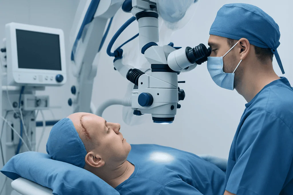
Craniectomy surgery takes place under general anesthesia, so the patient is unconscious and does not feel pain. The surgical team positions the head securely, cleans the scalp and makes an incision. The location and size of the opening depend on where swelling or injury is located; a large opening on one side of the skull is often called a hemicraniectomy.
The neurosurgeon removes a section of skull and may open the tough protective covering around the brain, called the dura, to provide further space. A dural patch may be placed to allow expansion without constriction. If needed, the surgeon may also remove a blood clot, control bleeding, repair damaged tissue or place a drain or pressure-monitoring device.
The removed bone flap may be stored under controlled conditions or preserved according to hospital practice for possible later replacement. In some circumstances it cannot be reused, and a custom synthetic implant may be planned for later cranioplasty. The scalp is closed over the opening, and the patient is transferred to intensive care for close observation.
Neurosurgery teams coordinate with neurocritical care, anesthesiology, neuroradiology, rehabilitation specialists and other clinicians throughout this process. This multidisciplinary approach helps address both the immediate pressure emergency and the effects of the original neurological condition.
Benefits, risks and expected results
The potential benefit of craniectomy surgery is rapid relief of dangerously high intracranial pressure. In appropriate emergency circumstances, it can prevent brain herniation, support blood flow to the brain and improve the chance of survival. It may also give surgeons access to remove a clot or address another urgent cause of compression.
It remains major surgery with important risks. These include bleeding, infection of the wound or brain coverings, blood clots, seizures, stroke, fluid accumulation, leakage of cerebrospinal fluid, wound problems and complications related to anesthesia. The unprotected area of brain is also vulnerable until cranioplasty is completed, so a protective helmet is often advised during mobility if the care team recommends it.
Results cannot be predicted from the operation alone. Some people regain substantial independence, while others have lasting difficulties with movement, speech, thinking, behavior, vision or swallowing. The original cause of injury, the extent and location of brain damage, treatment timing, complications and access to rehabilitation all influence recovery.
Before surgery, clinicians explain the known risks, likely goals and uncertainties as clearly as possible. When the operation is urgent and the patient cannot participate in decisions, the team discusses choices with the legally authorized representative or closest available family member according to local requirements.
Recovery timeline and rehabilitation
Recovery begins in the intensive care unit. During the first hours and days, clinicians monitor consciousness, pupil responses, blood pressure, oxygen levels, temperature, wound drainage and, when used, intracranial pressure. Repeat CT or MRI scans may be used to assess swelling, bleeding and healing. Mechanical ventilation, nutrition support, infection prevention and seizure management may be necessary.
Once medically stable, the patient may move to a neurosurgical ward and then to inpatient or outpatient rehabilitation. Physical therapy can address strength, balance and walking; occupational therapy supports daily activities; speech and language therapy may help with communication, cognition or swallowing. Emotional support, neuropsychological assessment and family education are also important parts of recovery.
The scalp and soft tissues typically heal over several weeks, but recovery of brain function may continue for months or longer. Cranioplasty is often considered after swelling has settled and there is no active infection, although the timing is individualized. The procedure may use the person’s stored bone flap or a manufactured implant to restore skull protection and contour.
At Acibadem International, multidisciplinary specialists and JCI-accredited hospitals support international patients needing neurosurgical assessment, critical care and rehabilitation planning after complex brain conditions.
Can you live a normal life after craniectomy?
Some people can return to independent daily life after a craniectomy, including work, study and social activities. However, this depends much more on the underlying brain injury or disease than on the skull operation itself. A person whose swelling is controlled with limited brain damage may recover very well, while someone with extensive stroke or traumatic injury may need continuing support.
Recovery is often gradual. Fatigue, headaches, reduced concentration, mood changes, weakness or balance problems can improve with time and targeted rehabilitation, but some effects may be permanent. Follow-up with neurosurgery, neurology, rehabilitation and primary care helps identify practical goals and treat ongoing symptoms.
Until cranioplasty has been completed, the person should follow the team’s advice about protecting the unshielded area of the head, activity restrictions and helmet use. Driving, return to work, contact sports and travel should be discussed individually because safety depends on neurological function, seizure risk and healing progress.
How long does it take to heal from a craniectomy?
Initial wound healing often takes several weeks, but there is no single timetable for full recovery. The duration of hospitalization can range from days to weeks or longer, depending on the reason for surgery, level of consciousness, complications and rehabilitation needs. Brain recovery commonly continues over many months.
The period before cranioplasty also varies. Surgeons generally wait until brain swelling has improved and infection risk is acceptably low, but the safest timing differs from person to person. After cranioplasty, additional healing and follow-up are needed before the care team clears a patient for more demanding activities.
Families can support recovery by attending follow-up visits, helping the person follow medication and rehabilitation plans, reporting new symptoms promptly and maintaining a calm, structured environment. Rest, balanced nutrition, avoiding alcohol or non-prescribed substances, and gradual activity as approved by clinicians can also support general recovery.
Is a craniectomy a major surgery, and when to seek medical care
Yes. A craniectomy is major brain surgery, usually performed in a hospital with neurosurgical operating facilities and intensive care. It requires general anesthesia, careful monitoring and coordinated follow-up. Although it is frequently an emergency treatment, it is undertaken because the risk from uncontrolled brain pressure may be greater than the risks of surgery.
After discharge, urgent medical care is needed for a seizure, fainting, sudden severe headache, repeated vomiting, new weakness or numbness, difficulty speaking, confusion, worsening drowsiness, vision changes, fluid leaking from the wound, or redness, swelling, fever or discharge around the incision. A significant head impact should also be assessed urgently, particularly before the skull has been reconstructed.
Routine follow-up is equally important. Patients should contact their care team about persistent headaches, mood or memory changes, increasing imbalance, medication side effects, concerns about the wound or questions about rehabilitation. Prompt assessment can help clinicians identify treatable complications and adjust the recovery plan safely.
Frequently asked questions
What are the chances of survival after craniectomy?
Survival after craniectomy varies greatly because the operation is performed for different serious conditions, including severe head injury, major stroke and brain bleeding. The severity and location of the underlying brain damage, age, other health conditions, treatment timing and complications all affect prognosis. The treating neurosurgical and intensive-care teams are best placed to discuss an individual outlook using examination findings and brain imaging.
Does the skull grow back after a craniectomy?
In adults, the removed skull section does not usually grow back on its own. A later procedure called cranioplasty is commonly used to replace the bone flap or fit a custom implant once swelling has resolved. This helps restore protection to the brain and the shape of the skull.
Will a person need to wear a helmet after craniectomy?
Many patients are advised to wear a protective helmet when out of bed until cranioplasty, because the brain is less protected where the bone flap was removed. The exact recommendations depend on the size and location of the craniectomy, mobility, fall risk and the surgeon’s guidance. The care team should provide clear instructions about when and how to use it.
Can craniectomy cause personality or memory changes?
Changes in memory, concentration, emotions or personality can occur after a serious brain injury, stroke or brain surgery. These effects are often related primarily to the underlying brain condition and the affected area of the brain, rather than the removal of skull bone itself. Neuropsychological assessment and rehabilitation can help identify needs and guide treatment.
When is cranioplasty done after craniectomy?
Cranioplasty is planned after the brain swelling has settled and the surgical team considers the risk of infection or other complications to be acceptable. The timing varies between patients and may be influenced by wound healing, need for further treatment and overall medical stability. The neurosurgeon will explain the recommended timing and implant options.
Can a person exercise after craniectomy?
Light activity may begin gradually when the treating team considers it safe, often as part of rehabilitation. Strenuous exercise, heavy lifting and activities with a risk of falling or head impact should be avoided until specifically cleared by the neurosurgeon. Activity plans should be individualized, especially before cranioplasty or if seizures, weakness or balance problems are present.
References
- National Institute of Neurological Disorders and Stroke
- American Association of Neurological Surgeons
- Brain Trauma Foundation
- National Health Service
- Mayo Clinic
This article is for general information only and is not a substitute for professional medical advice. Please consult a qualified doctor about your individual situation.
Explore treatments in Turkey — costs, top hospitals & a free quote
JCI-accredited · board-certified surgeons · reply within 24h
Add us as a Preferred Source to see more of our trusted health content across Google Search, AI Overviews and Discover.