Ntd Ultrasound: Preparation, Procedure and Results

An NTD ultrasound uses sound waves to assess the fetal brain, spine and surrounding anatomy without radiation. The detailed anatomy scan, commonly performed around 18 to 22 weeks of pregnancy, is the main ultrasound assessment for neural tube defects.
Key Takeaways
- An NTD ultrasound uses sound waves to assess the fetal brain, spine and surrounding anatomy without radiation.
- The detailed anatomy scan, commonly performed around 18 to 22 weeks of pregnancy, is the main ultrasound assessment for neural tube defects.
- A screen-positive blood test or ultrasound finding does not confirm a diagnosis and usually requires targeted imaging and specialist review.
- Folic acid before conception and in early pregnancy helps reduce the risk of neural tube defects.
- Earlier specialist care is important when screening results are abnormal, there is a prior affected pregnancy or symptoms need urgent assessment.
An NTD ultrasound is a prenatal imaging examination that evaluates the developing baby's brain, spine and related structures for signs of neural tube defects. It is noninvasive, does not use radiation and is most informative during the detailed second-trimester anatomy scan, although some findings may be seen earlier.
Overview: What Is an NTD Ultrasound?
An NTD ultrasound is a prenatal ultrasound examination used to look for neural tube defects (NTDs), a group of conditions that affect the early development of the brain, spinal cord or their protective tissues. It is a safe, noninvasive scan that uses high-frequency sound waves rather than radiation. The examination can assess the fetal spine directly and can also identify changes in the shape or appearance of the brain that may be associated with certain spinal defects.
The neural tube forms very early in pregnancy, usually before many people know they are pregnant. When it does not close fully, conditions such as spina bifida or anencephaly can occur. Ultrasound does not cause neural tube defects; it helps clinicians look for features that may suggest one and plan any needed follow-up.
The most comprehensive assessment is usually part of the mid-pregnancy anatomy scan. If a screening blood test is elevated, a family history is present or an earlier scan raises a concern, a clinician may recommend a targeted ultrasound performed by a maternal-fetal medicine specialist. Spina bifida is one of the neural tube defects that can be evaluated in this way.
How the Scan Works and Who May Need It
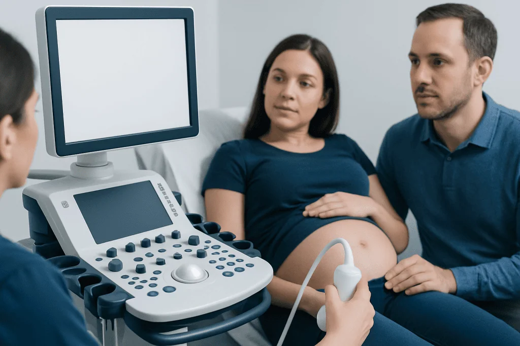
Ultrasound equipment sends sound waves through the abdomen and receives returning echoes to create real-time images. During an NTD-focused examination, the sonographer or doctor obtains views along the full length of the fetal spine and examines the skull and brain from several angles. The position of the baby, the stage of pregnancy and image quality can affect which views are possible at one appointment.
All pregnant patients are generally offered a detailed fetal anatomy ultrasound. A more focused or specialist scan may be particularly helpful after a screen-positive maternal serum alpha-fetoprotein (AFP) result, an uncertain finding on a routine scan, a prior pregnancy affected by an NTD, certain medicines or medical conditions, or a known family history. Having a risk factor does not mean a baby will have an NTD.
Ultrasound is often combined with other prenatal assessments. Depending on the situation, these may include blood screening, genetic counselling, diagnostic testing such as amniocentesis, or fetal MRI later in pregnancy. The appropriate plan is individual and should be discussed with the obstetric team.
Preparation and Step-by-Step Procedure
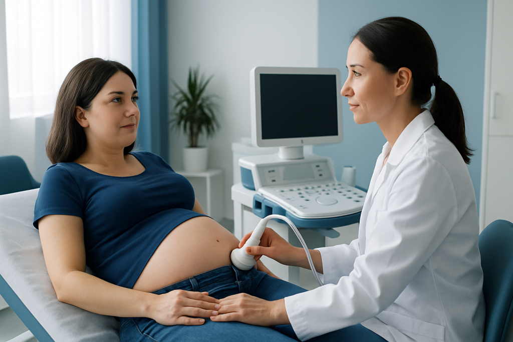
Most patients need little preparation for an NTD ultrasound. The imaging centre may provide specific instructions, including whether to arrive with a comfortably full bladder for an early-pregnancy scan. For the usual second-trimester abdominal examination, special dietary preparation is not typically required. Comfortable two-piece clothing can make the appointment easier.
At the visit, the clinician reviews pregnancy dates, relevant health history and prior screening results. The patient lies on an examination couch while gel is applied to the abdomen. A transducer is moved gently over the skin to obtain images. Mild pressure may be needed to improve a view, particularly if the baby is positioned facing the mother’s back.
The examination includes measurements of fetal growth and a structured review of anatomy. For NTD assessment, the specialist studies the spine in multiple planes and evaluates the head and brain. A transvaginal ultrasound may occasionally be offered in early pregnancy or when it provides clearer images; it is performed with consent and is generally well tolerated.
The scan commonly lasts longer than a brief dating ultrasound because many structures must be assessed. If all required images cannot be obtained because of fetal position, a repeat scan may be arranged. This is common and does not by itself indicate an abnormality.
How Soon Can You See Neural Tube Defects on Ultrasound?
Some major neural tube defects may be visible during first-trimester ultrasound, particularly when a specialised early scan is performed. However, the routine and most detailed ultrasound evaluation for neural tube defects is generally the second-trimester anatomy scan, often carried out between 18 and 22 weeks of pregnancy. At this stage, the fetal spine, skull and brain are usually easier to assess in detail.
Timing varies because fetal size, pregnancy dating, body anatomy and fetal position influence image quality. An early normal scan may not replace the detailed anatomy scan. Likewise, a finding that is unclear on an earlier scan may become easier to interpret after a short interval or with targeted imaging.
Open spina bifida can often produce characteristic brain findings as well as visible changes in the spine. Closed spinal defects may be more difficult to detect prenatally. A specialist can explain what the scan can and cannot show in an individual pregnancy and whether further assessment is appropriate.
Understanding Results and a Screen-Positive NTD Result
What does screen positive for NTD mean?
A screen-positive result means that a screening test has found a result associated with a higher-than-average chance of a neural tube defect. It is not the same as a diagnosis. For example, an elevated AFP blood-screening result can occur for several reasons, including inaccurate pregnancy dating, more than one fetus, placental factors or an open fetal defect.
When screening is positive, the next step is commonly a detailed targeted ultrasound. Depending on the findings and the patient’s preferences, clinicians may also discuss genetic counselling or diagnostic testing. These tests can provide more information, but the decision to have them is personal and should be made after a clear discussion of benefits, limits and possible risks.
A normal targeted ultrasound is reassuring, but no prenatal test can detect every possible condition. The reporting clinician will explain whether the examination was complete, what was seen and whether repeat imaging or routine pregnancy follow-up is recommended.
What are the earliest signs of neural tube defects?
There are usually no symptoms that a pregnant person can feel that reliably indicate a fetal neural tube defect. These conditions develop early, and many are first suggested by prenatal screening or ultrasound rather than maternal physical changes. Nausea, fatigue, abdominal growth and other common pregnancy experiences do not diagnose or rule out an NTD.
On ultrasound, early signs depend on the specific condition. Major brain and skull abnormalities may sometimes be identified early, while spinal abnormalities may require later detailed views. Promptly attending scheduled prenatal visits and scans is the most practical way to support early detection.
Benefits, Limitations, Risks and Recovery
The principal benefit of NTD ultrasound is timely information. When an abnormality is identified or suspected, families can receive counselling, arrange care with appropriate specialists and consider pregnancy-management options according to local laws, clinical guidance and personal values. It can also provide reassurance when the scan is normal.
Diagnostic ultrasound is considered safe when used appropriately in pregnancy and does not expose the fetus to ionising radiation. The physical risks are minimal; some people experience temporary discomfort from probe pressure or from lying in one position. If a transvaginal scan is used, mild short-lived discomfort may occur, but it should not be painful.
There is no physical recovery period after an ultrasound. Most people can return to usual activities immediately. Emotional recovery may take longer if a result is uncertain or abnormal; it can be helpful to ask for a follow-up consultation, bring a support person and write down questions for the care team.
Ultrasound has limitations. Image quality can be reduced by fetal position, gestational age and other technical factors, and not every NTD can be identified before birth. A suspicious result often needs confirmation or clarification rather than an immediate conclusion.
Pregnancy After an NTD: Follow-up, Prevention and Support
Is a pregnancy after NTD considered high risk?
A pregnancy after a previous neural tube defect is generally offered enhanced preconception and prenatal care because the chance of recurrence is higher than the baseline population chance. This does not mean that another affected pregnancy is expected. Early discussion with an obstetrician, maternal-fetal medicine specialist or genetic counsellor can help create an individual screening and prevention plan.
Folic acid taken before conception and during early pregnancy lowers the chance of neural tube defects. People with a prior affected pregnancy may be advised to take a higher dose than standard prenatal supplementation, but the correct amount should be prescribed by a clinician before pregnancy whenever possible. A balanced diet, management of diabetes, review of medicines and avoidance of overheating in early pregnancy may also be relevant for some individuals.
If an NTD is diagnosed, care may involve maternal-fetal medicine, neonatology, paediatric neurosurgery, urology, rehabilitation and genetic counselling. In selected cases of open spina bifida, specialists may discuss fetal surgery as one possible treatment pathway; suitability depends on detailed medical and pregnancy criteria. Acibadem International’s multidisciplinary specialists and JCI-accredited hospitals can assess and coordinate care for international patients who require prenatal evaluation or treatment planning.
When to Seek Medical Care
Pregnant people should contact their obstetric team promptly after a screen-positive AFP result, an abnormal or incomplete ultrasound report, or if they have a previous pregnancy affected by a neural tube defect and have not yet discussed early pregnancy care. A referral to maternal-fetal medicine can provide focused imaging and counselling without assuming that a diagnosis is present.
Urgent medical assessment is needed for heavy vaginal bleeding, severe or persistent abdominal pain, fluid leakage, fainting, fever, severe headache with visual changes, or a noticeable reduction in fetal movement later in pregnancy. These symptoms are not specific signs of neural tube defects, but they require timely evaluation in pregnancy.
Questions about results are appropriate at any stage. Patients may ask what structures were seen, whether the examination was complete, how certain the findings are and what follow-up is advised. Clear communication and supportive counselling are important parts of prenatal care.
Frequently asked questions
Is an NTD ultrasound the same as the anatomy scan?
An NTD assessment is often included in the standard mid-pregnancy anatomy scan. If there is a screening concern or increased risk, the clinician may request a more detailed targeted ultrasound performed by a specialist.
Does an ultrasound diagnose every neural tube defect?
No. Ultrasound is highly useful for detecting many major neural tube defects, especially open spina bifida and major brain abnormalities, but it cannot identify every condition in every pregnancy. Image quality and the type of defect can affect detection.
Is NTD ultrasound painful?
The abdominal scan should not be painful, although pressure from the probe can be briefly uncomfortable. A transvaginal scan, if needed, may cause mild discomfort but is usually short and well tolerated.
What happens if the ultrasound suggests spina bifida?
The care team usually arranges a detailed consultation with maternal-fetal medicine and may recommend additional imaging, genetic counselling or diagnostic testing. The aim is to clarify the finding and discuss care options in a calm, individualised way.
Can folic acid prevent all neural tube defects?
Folic acid substantially reduces the risk of many neural tube defects, but it cannot prevent every case. Taking it before conception is especially important because the neural tube develops very early in pregnancy.
Can a normal first-trimester scan rule out neural tube defects?
A normal early scan is reassuring but does not replace the detailed second-trimester anatomy ultrasound. Some features are more reliably assessed later, when the fetal brain and spine are larger and easier to visualise.
References
- American College of Obstetricians and Gynecologists
- Society for Maternal-Fetal Medicine
- Centers for Disease Control and Prevention
- National Institute of Child Health and Human Development
- International Society of Ultrasound in Obstetrics and Gynecology
This article is for general information only and is not a substitute for professional medical advice. Please consult a qualified doctor about your individual situation.
Ultrasound in Turkey — costs, top hospitals & a free quote
JCI-accredited · board-certified surgeons · reply within 24h
Add us as a Preferred Source to see more of our trusted health content across Google Search, AI Overviews and Discover.

Check-up & Preventive Medicine
Comprehensive health screenings designed for early detection and prevention.
3 specialists in this unitMore from the Health Library

Related Specialists

Dr. Ertuğrul Tekçe
Radiation Oncology
Dr. Burak Sercan Erçin
Aesthetic Plastic & Reconstructive Surgery
Dr. Türker Kundak
Internal Medicine
Dr. Yusuf Ay
Emergency Service