Cardiac Sarcoid MRI: Preparation, Procedure and Results
Cardiac MRI is an important test for evaluating suspected or known cardiac sarcoidosis. The scan uses magnets rather than ionizing radiation and may include a gadolinium-based contrast agent.
Key Takeaways
- Cardiac MRI is an important test for evaluating suspected or known cardiac sarcoidosis.
- The scan uses magnets rather than ionizing radiation and may include a gadolinium-based contrast agent.
- Preparation instructions vary, especially for people with implanted devices, kidney disease, diabetes or claustrophobia.
- Results are interpreted alongside symptoms, ECG findings, blood tests and other imaging rather than used alone.
- Urgent assessment is important for fainting, sustained palpitations, chest pain or new shortness of breath.
A cardiac sarcoid MRI is a noninvasive scan that helps clinicians assess whether sarcoidosis may be affecting the heart muscle. It can show patterns of inflammation, swelling, reduced heart function and scar tissue, supporting diagnosis and decisions about monitoring or treatment.
Overview: What Is a Cardiac Sarcoid MRI?
A cardiac sarcoid MRI, also called cardiac magnetic resonance imaging (CMR), is a detailed scan of the heart used when clinicians suspect that sarcoidosis may involve the heart. Sarcoidosis is an inflammatory condition in which small clusters of immune cells, called granulomas, can form in different organs. When the heart is affected, the condition is known as cardiac sarcoidosis.
The scan can assess heart size, pumping function, areas of swelling and patterns of scar tissue. A contrast agent may be used to make certain changes in the heart muscle more visible. Cardiac MRI does not use X-rays or other ionizing radiation, although it does involve lying still in a strong magnetic field.
Cardiac sarcoidosis can be difficult to confirm because its effects may be patchy and symptoms can overlap with other heart conditions. MRI findings are therefore considered together with a person’s medical history, symptoms, electrocardiogram (ECG), echocardiogram, blood tests and, when appropriate, other tests such as PET imaging or rhythm monitoring.
How Cardiac MRI Works and Who May Need It
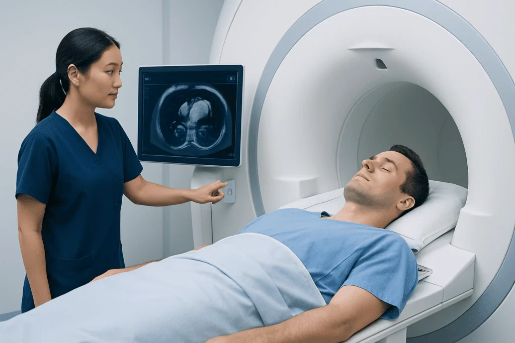
During cardiac MRI, the scanner uses magnetic fields and radio waves to create moving and still images of the heart. ECG leads placed on the chest help synchronize image collection with the heartbeat. This allows the radiology and cardiology teams to evaluate the heart chambers, valves and heart muscle in detail.
When contrast is given through a vein, delayed images can show areas where contrast remains within damaged or scarred heart muscle. This is commonly called late gadolinium enhancement. In suspected cardiac sarcoidosis, the location and pattern of enhancement may provide useful information, but it is not specific to sarcoidosis and must be interpreted by experienced clinicians.
A doctor may recommend cardiac MRI for a person with sarcoidosis who has palpitations, unexplained fainting, changes on an ECG, reduced heart function or other concerning findings. It may also be used to assess a known diagnosis over time. People with certain pacemakers, defibrillators, metal fragments or other implanted devices require a device-safety review before the scan.
Preparation: What Should You Not Do Before a Cardiac MRI?

Patients should follow the instructions provided by the imaging department, as preparation depends on the reason for the scan and whether contrast or medication to control the heart rate may be used. In many cases, patients are asked to avoid caffeine for at least 12 to 24 hours if the examination includes stress imaging. Caffeine may be present in coffee, tea, energy drinks, cola, chocolate and some medicines.
Patients should not stop prescribed medicines unless their clinician specifically advises this. It is important to tell the care team about all medicines, allergies, pregnancy or possible pregnancy, kidney problems, diabetes, asthma, implanted devices, prior surgeries and any history of anxiety in enclosed spaces.
Metal objects must not enter the MRI scanner room. Patients should remove jewelry, watches, hearing aids, removable dental appliances, hair accessories and clothing with metal components when asked. They should also inform staff about any metal injury, especially a past eye injury involving metal, as further screening may be needed.
Can you eat or drink before a cardiac MRI? For a standard cardiac MRI, light food and water may often be allowed, but local instructions should always take priority. Some patients may be asked not to eat for several hours beforehand, particularly if contrast, sedation or stress imaging is planned. Clear fluids may be permitted, but patients should confirm this with the imaging team before the appointment.
Step by Step: What Happens During the Procedure?
Before the scan, a radiographer reviews the safety questionnaire and explains the process. The patient changes into suitable clothing, removes metal items and may have an intravenous line placed in the arm for contrast. Small ECG electrodes are placed on the chest to coordinate images with the heartbeat.
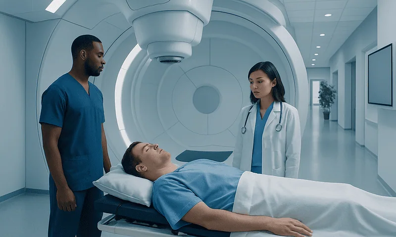
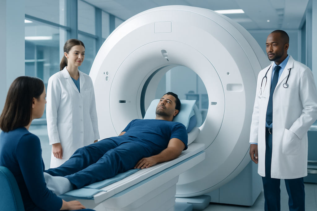
The patient lies on a padded table that moves into the MRI scanner. A coil is positioned over the chest to receive the imaging signals. The scanner makes repeated loud knocking or humming sounds, so earplugs or headphones are provided. The patient can communicate with staff throughout the examination using an intercom and is usually given a call button.
Several image sequences are taken while the patient holds their breath briefly, usually for a few seconds at a time. If contrast is needed, it is injected through the intravenous line partway through the scan. The examination commonly takes about 30 to 60 minutes, although more complex studies may take longer.
The test is painless, but remaining still can be uncomfortable for some people. Patients who are concerned about claustrophobia should discuss this in advance; the team may offer practical support or, in selected situations, arrange medication or an alternative imaging approach.
Results, Benefits and Possible Risks
How long does it take to get results from a cardiac MRI? Preliminary information may sometimes be available on the day of the scan, but a complete cardiac MRI interpretation usually requires review by a specialist radiologist or cardiologist. Results are often sent to the referring clinician within several days, although timing varies by facility and the complexity of the images. The referring clinician explains what the findings mean in the context of the individual’s health.
The main benefit of cardiac MRI is its ability to provide detailed information about heart muscle structure and function without radiation exposure. It may identify changes that warrant closer monitoring, further testing or treatment. It can also help doctors distinguish active inflammation from established scarring when interpreted alongside other clinical information.
MRI is generally considered safe for appropriately screened patients. Gadolinium contrast is usually well tolerated, though mild side effects such as a cool sensation at the injection site, nausea or headache can occur. Severe allergic reactions are uncommon. People with severe kidney impairment need individual assessment because certain contrast agents may not be appropriate.
Results may be normal, may suggest inflammation or scarring, or may show another explanation for symptoms. An abnormal result does not automatically confirm cardiac sarcoidosis, and a normal result does not replace clinical follow-up when symptoms or other tests remain concerning.
How Serious Is Cardiac Sarcoidosis?
How serious is cardiac sarcoidosis? Cardiac sarcoidosis can be serious because inflammation or scar tissue may interfere with the heart’s electrical system or its ability to pump effectively. Some people have no symptoms, while others may develop rhythm disturbances, heart block, reduced heart function or, less commonly, life-threatening arrhythmias. The outlook varies widely and depends on the location and extent of heart involvement, response to treatment and ongoing monitoring.
Early recognition is helpful because specialists can assess the risk of complications and create an individualized care plan. Management may include medicines to control inflammation, treatment for heart failure or rhythm problems, monitoring devices, and in selected cases a pacemaker or implantable cardioverter-defibrillator. Decisions are based on the whole clinical picture rather than MRI findings alone.
Care is often coordinated between cardiology, electrophysiology, pulmonology or rheumatology, radiology and other relevant specialties. At Acibadem International, multidisciplinary specialists and JCI-accredited hospitals support diagnosis and treatment planning for international patients with complex cardiac and inflammatory conditions.
Recovery, Follow-Up and When to Seek Medical Care
Most people can return to usual activities immediately after a cardiac MRI. If contrast was used, drinking fluids afterward may be encouraged unless a clinician has advised fluid restriction. The injection site may be mildly sore for a short time. A person who received sedating medicine should not drive, operate machinery or make important decisions until the effects have worn off and they have followed the discharge instructions.
Follow-up depends on the result and the reason for the scan. The clinician may recommend repeat imaging, a PET scan, ambulatory rhythm monitoring, an echocardiogram, blood testing or referral to a heart rhythm specialist. Keeping scheduled follow-up appointments is important, even when symptoms are mild or absent.
When to seek medical care: A person with known or suspected cardiac sarcoidosis should seek urgent medical assessment for fainting, near-fainting, persistent or severe palpitations, chest pain, new or worsening shortness of breath, marked swelling in the legs, or sudden severe weakness. Emergency services should be contacted for severe chest pain, collapse, severe breathing difficulty or symptoms of a possible heart emergency.
For nonurgent concerns, patients should contact their doctor if they notice new palpitations, reduced exercise tolerance, dizziness or medication side effects. These symptoms can have many causes, but timely assessment helps clinicians decide whether heart rhythm testing or other evaluation is needed.
Frequently asked questions
Does a cardiac sarcoid MRI diagnose cardiac sarcoidosis by itself?
No. Cardiac MRI provides important evidence about heart inflammation, scarring and function, but it is not usually used as a stand-alone diagnostic test. Clinicians interpret it with symptoms, medical history, ECG findings, other imaging and sometimes biopsy results.
Is cardiac MRI painful?
The scan itself is not painful. Patients may feel discomfort from lying still, hear loud scanner noises and notice brief warmth or a cool sensation if contrast is injected. The staff can communicate with the patient throughout the examination.
Can a person with a pacemaker have a cardiac MRI?
Some people with MRI-conditional pacemakers or defibrillators can safely undergo MRI under a specialized protocol. The device type, leads, implantation details and local MRI safety procedures must be reviewed in advance. Patients should never assume that an implanted device is MRI-safe without confirmation.
Why is contrast used for a cardiac MRI?
Gadolinium-based contrast can help reveal patterns of scar tissue or areas of altered heart muscle. These details can add to the assessment of suspected cardiac sarcoidosis and other heart diseases. Contrast is not necessary for every scan and is considered carefully in people with kidney disease.
How should a person prepare if they are claustrophobic?
They should tell the ordering clinician and imaging department before the appointment. The team can explain the scan, offer communication and comfort measures, and discuss whether a prescribed calming medicine or another approach is appropriate. If sedation is used, transport home is usually needed.
What happens if the cardiac MRI shows scarring?
Scarring can have several causes, so the result needs careful interpretation by the treating team. Depending on the pattern and the person’s symptoms, clinicians may recommend additional testing, rhythm monitoring, medication changes or specialist review. A scar finding does not by itself predict the same outcome for every person.
References
- American Heart Association
- American College of Cardiology
- Society for Cardiovascular Magnetic Resonance
- National Heart, Lung, and Blood Institute
- Sarcoidosis Foundation
This article is for general information only and is not a substitute for professional medical advice. Please consult a qualified doctor about your individual situation.
Heart care in Turkey — expert evaluation and treatment
JCI-accredited · board-certified surgeons · reply within 24h
Add us as a Preferred Source to see more of our trusted health content across Google Search, AI Overviews and Discover.
Check your numbers in seconds
BMI, calories, due date, blood pressure and 30+ more clinical calculators — free, instant, doctor-reviewed ranges.