Mediastinoscopy Procedure: An Evidence-Based Patient Guide

Mediastinoscopy is usually performed under general anesthesia through a small incision above the breastbone. The procedure commonly collects lymph node samples to clarify a diagnosis or guide cancer staging and treatment planning.
Key Takeaways
- Mediastinoscopy is usually performed under general anesthesia through a small incision above the breastbone.
- The procedure commonly collects lymph node samples to clarify a diagnosis or guide cancer staging and treatment planning.
- Most people go home the same day or after one night in hospital, with recovery often taking about one to two weeks.
- Bleeding is an uncommon but important complication because major blood vessels are located in the mediastinum.
- Modern imaging and needle-based techniques may be suitable for some patients, but mediastinoscopy remains useful when a surgical tissue sample is needed.
A mediastinoscopy procedure is a surgical test that allows a surgeon to view and take tissue samples from lymph nodes and other structures in the mediastinum, the area between the lungs. It is most often used to investigate enlarged chest lymph nodes or to help stage suspected or confirmed lung cancer.
Overview: What Is a Mediastinoscopy Procedure?
A mediastinoscopy procedure is a minimally invasive surgical examination of the mediastinum, the central space in the chest between the lungs. This space contains lymph nodes, the windpipe, major blood vessels, nerves and other important structures. A surgeon uses a thin, lighted instrument called a mediastinoscope to see selected areas and remove small tissue samples for laboratory analysis.
The procedure is commonly recommended when chest imaging shows enlarged or unusual lymph nodes, or when doctors need more information about a suspected infection, inflammatory condition or cancer. In people with lung cancer, lymph node findings can help establish the stage of disease and support decisions about surgery, radiation therapy, systemic treatment or monitoring. It may be part of an assessment for lung cancer when tissue confirmation is needed.
Although scans such as CT and PET can identify areas that need investigation, they cannot always determine the cause. A biopsy obtained during mediastinoscopy can provide more definite information because a pathologist examines the cells directly. The healthcare team will explain why this approach is being considered and whether a less invasive test may be appropriate.
Why It May Be Recommended and Who May Be Suitable
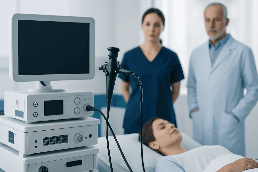
Mediastinoscopy is most often used to biopsy lymph nodes near the trachea and around the main airways. It may help distinguish cancer from conditions such as sarcoidosis, tuberculosis and other infections. It can also be used when previous samples have not provided a clear answer or when the location of a lymph node makes another biopsy technique less suitable.
Before recommending surgery, clinicians review imaging, symptoms, medical history and prior test results. Bronchoscopy with endobronchial ultrasound-guided needle sampling may be considered first for some lymph nodes. However, mediastinoscopy may offer a larger tissue sample or access to areas that cannot be adequately assessed by a needle-based approach.
Suitability depends on individual factors, including previous surgery or radiation in the neck or chest, anatomy, bleeding risk, heart and lung health, and the ability to have general anesthesia. Patients should tell their team about all medicines, supplements, allergies, implanted devices, pregnancy and any history of bleeding or anesthesia-related problems.
For people undergoing an evaluation of chest symptoms or abnormal imaging, clinicians may also assess related respiratory conditions and arrange appropriate lung testing before surgery. The aim is to choose the safest investigation that is likely to answer the clinical question.
How Does the Mediastinoscopy Procedure Work?

The operation is performed in an operating room under general anesthesia, so the patient is asleep and does not feel the procedure. A breathing tube is placed after anesthesia begins, and the surgical team continuously monitors oxygen level, heart rhythm, blood pressure and breathing throughout the operation.
The surgeon usually makes a short incision at the base of the neck, just above the breastbone. The mediastinoscope is gently passed through a natural tissue plane in front of the trachea to reach selected lymph nodes. A camera may display the view on a screen; however, a mediastinoscopy procedure video is an educational illustration and cannot show the individualized decisions and safety checks that take place during an actual operation.
Using small instruments passed through the scope, the surgeon removes samples from one or more lymph nodes. The specimens are sent to a pathology laboratory. Some results may be available quickly during surgery, but detailed testing commonly takes several days and, in some cases, longer if specialized studies are required.
After the samples are collected, the scope is removed and the incision is closed. The procedure itself often takes less than an hour, although preparation, anesthesia and postoperative monitoring mean the total time at the hospital is longer.
Mediastinoscopy Procedure Steps and Positioning
The mediastinoscopy steps generally begin with a preoperative review, confirmation of consent and an anesthesia assessment. The patient changes into a hospital gown, an intravenous line is placed, and the team confirms relevant imaging, medication instructions and the planned biopsy sites. Antibiotics may be given when clinically indicated.
For the mediastinoscopy procedure position, the patient is placed on their back, known as the supine position. The neck is typically extended gently by placing support beneath the shoulders, allowing the surgeon to access the area just above the breastbone. The degree of extension is adjusted carefully for comfort, safety and any existing neck condition.
Once anesthesia is established, the skin is cleaned and covered with sterile drapes. The surgeon makes the small incision, inserts the mediastinoscope, identifies the intended lymph node stations and takes tissue samples. The team checks for bleeding before closing the incision and applying a dressing.
Afterward, the patient is transferred to a recovery area while the anesthesia wears off. Nurses monitor breathing, blood pressure, pain, swallowing and the incision site. Some patients have a chest X-ray after surgery, depending on the procedure details and local clinical practice.
Benefits, Risks and the Most Common Complication
The main benefit of mediastinoscopy is obtaining tissue that can provide a reliable diagnosis and, where cancer is present, more accurate staging. This information may prevent unnecessary treatment, help clinicians choose the most appropriate treatment plan and clarify whether surgery is likely to be beneficial.
Like all operations, mediastinoscopy has risks. The most common complication is usually minor bleeding at the incision or biopsy site. Sore throat, temporary hoarseness, neck discomfort and mild swelling can also occur, often related to the breathing tube, surgical area or normal healing.
Less common but more serious complications include significant bleeding from a blood vessel, infection, injury to the recurrent laryngeal nerve that affects the voice, air leakage around a lung, damage to the trachea or esophagus, and complications related to anesthesia. These events are uncommon, but the surgical team is prepared to recognize and manage them.
Patients can lower avoidable risks by following instructions about fasting, smoking, medicines and arrangements for transport home. Blood-thinning medicines should never be stopped independently; the prescribing clinician and surgical team should provide individualized advice. If results lead to a cancer diagnosis, the care team may discuss options such as lung cancer treatment based on the pathology findings and overall health.
Recovery Timeline, Pain and Self-Care
Recovery begins in the postoperative area, where drowsiness, a dry mouth or a sore throat are common after general anesthesia. The incision may feel tender for several days. Pain is usually mild to moderate and can often be managed with the medications recommended by the treating team.
Many patients return home on the day of surgery after a period of observation, while others stay overnight because of their medical history, the extent of sampling or recovery needs. A responsible adult should take the patient home and remain available after outpatient anesthesia. Driving, alcohol, important decisions and strenuous activity should be avoided for the period advised by the medical team.
Most people can resume light daily activity within a few days and return to usual routines within about one to two weeks, depending on their work, overall health and any additional procedures. Heavy lifting, vigorous exercise and swimming are commonly postponed until the incision has healed and the surgeon confirms it is safe.
Wound-care instructions vary, but patients are generally advised to keep the incision clean and dry as directed, avoid picking at skin adhesive or stitches, and attend follow-up appointments. The pathology report is discussed at follow-up, where the clinician explains what the findings mean and whether further tests or treatment are needed.
When to Seek Medical Care
Patients should contact their surgical team promptly if they develop increasing redness, warmth, drainage, swelling or worsening pain at the incision; fever; persistent nausea or vomiting; or a new or worsening cough. They should also report ongoing hoarseness, trouble swallowing or symptoms that do not improve as expected.
Urgent medical care is needed for trouble breathing, chest pain, coughing up blood, fainting, rapidly increasing neck swelling, severe bleeding or any sudden severe symptom. These problems are not expected after routine recovery, but timely assessment is important.
Before surgery, patients should seek medical advice if they develop a respiratory infection, fever or a significant new illness, as the procedure may need to be reassessed. Questions about fasting, diabetes medicines, inhalers, blood thinners or other prescriptions should be addressed before the scheduled date.
Acibadem International’s multidisciplinary specialists and JCI-accredited hospitals diagnose and treat chest conditions for international patients, coordinating surgical assessment, pathology and follow-up when mediastinoscopy is appropriate.
Frequently asked questions
How long is recovery from mediastinoscopy?
Many people go home on the same day or after one overnight stay. Light activities can often resume within a few days, while complete recovery commonly takes around one to two weeks. Recovery may take longer if a person has other health conditions or undergoes additional procedures.
What is the most common complication of mediastinoscopy?
Minor bleeding is generally considered the most common complication of mediastinoscopy. Temporary sore throat, incision discomfort and mild hoarseness can also occur. Serious bleeding and injury to nearby structures are uncommon but important risks discussed during consent.
Is a mediastinoscopy painful?
The procedure itself is not painful because it is performed under general anesthesia. Afterward, patients may have tenderness around the neck incision, a sore throat or mild discomfort when swallowing. The care team can recommend appropriate pain relief and should be contacted if pain becomes severe or worsens.
How should a patient be positioned during a mediastinoscopy?
The patient is generally positioned on their back on the operating table. The shoulders may be supported so the neck can be gently extended, giving the surgeon access to the lower neck and upper chest. Positioning is adjusted to protect the neck, joints and breathing during anesthesia.
What does mediastinoscopy diagnose?
Mediastinoscopy can help diagnose the cause of enlarged lymph nodes or abnormal tissue in the mediastinum. It may identify cancer, inflammatory conditions such as sarcoidosis, or certain infections. The tissue sample is examined by a pathologist, and the final result guides the next steps.
When will mediastinoscopy biopsy results be ready?
Some preliminary information may be available soon after surgery, but final pathology results often take several days. More complex testing, including specialized stains or molecular tests, can take longer. The surgeon or referring clinician will explain when and how results will be communicated.
References
- American Cancer Society
- National Cancer Institute
- Society of Thoracic Surgeons
- MedlinePlus
- National Health Service
This article is for general information only and is not a substitute for professional medical advice. Please consult a qualified doctor about your individual situation.
Oncology care in Turkey — second opinion and treatment plan
JCI-accredited · board-certified surgeons · reply within 24h
Add us as a Preferred Source to see more of our trusted health content across Google Search, AI Overviews and Discover.
Check your numbers in seconds
BMI, calories, due date, blood pressure and 30+ more clinical calculators — free, instant, doctor-reviewed ranges.