Medulloblastoma MRI: Preparation, Procedure and Results
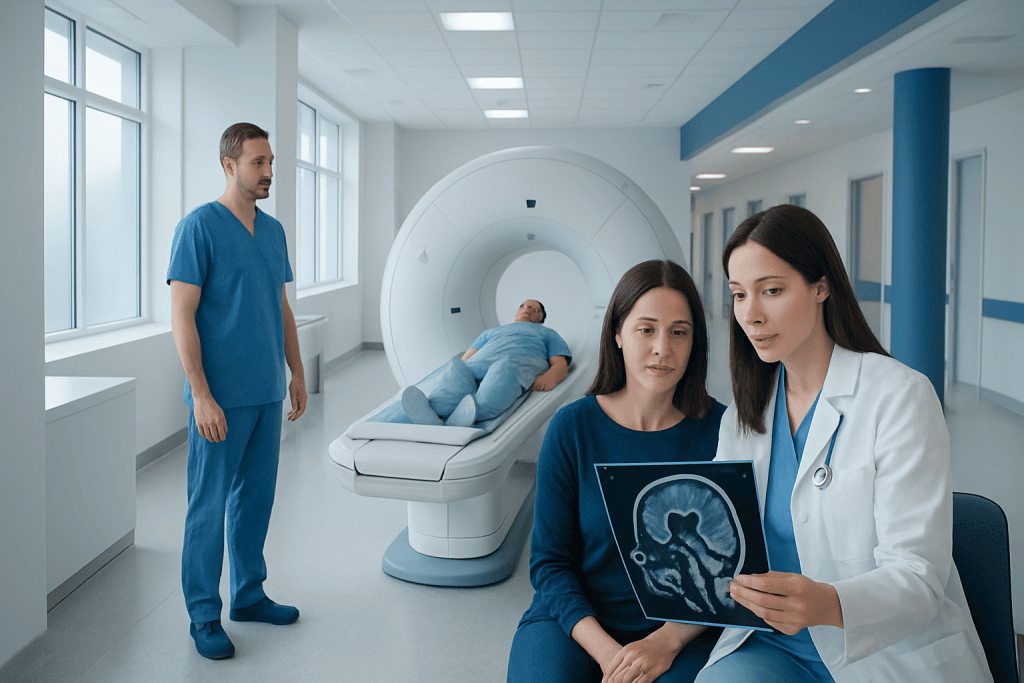
MRI is the main imaging test used to evaluate a suspected medulloblastoma in the cerebellum and assess the spinal fluid pathways. Brain and spine MRI with contrast can help show tumor size, location, effects on fluid flow and possible spread.
Key Takeaways
- MRI is the main imaging test used to evaluate a suspected medulloblastoma in the cerebellum and assess the spinal fluid pathways.
- Brain and spine MRI with contrast can help show tumor size, location, effects on fluid flow and possible spread.
- MRI appearances can strongly suggest a tumor, but they cannot reliably prove whether a brain tumor is malignant or identify its molecular subtype.
- Children may need preparation, comfort support or anesthesia to remain still during a medulloblastoma pediatric MRI.
- Care commonly involves pediatric neuro-oncology, neurosurgery, neuroradiology, radiation oncology and rehabilitation specialists.
A medulloblastoma MRI is a detailed scan of the brain, and often the spine, used to investigate a suspected tumor, guide surgical planning and check whether disease has spread. MRI is painless and does not use ionizing radiation, but tissue testing is needed to confirm the diagnosis and tumor type.
Overview: what a medulloblastoma MRI shows
A medulloblastoma MRI is a magnetic resonance imaging scan used to create detailed pictures of the brain and, in many cases, the entire spine. It is usually performed when symptoms or an earlier scan raise concern for a tumor in the cerebellum, the part of the brain that helps control balance and coordination. The scan helps clinicians understand the lesion’s location, size, relationship to nearby structures and effects on the flow of cerebrospinal fluid.
Medulloblastoma is a fast-growing tumor that most often occurs in children, although it can also affect adults. MRI is central to the assessment, but it does not replace surgery or biopsy when tissue is needed for a definitive diagnosis. Pathology and molecular testing of tumor tissue provide important information that guides treatment and outlook.
Images may be obtained before and after an intravenous contrast medicine is given. A brain MRI is generally followed by an MRI of the spine after diagnosis or strong suspicion, because medulloblastoma can spread through cerebrospinal fluid. The results are interpreted alongside symptoms, neurological examination findings and laboratory or surgical information.
How does a medulloblastoma MRI work, and who may need one?
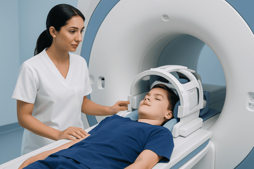
MRI uses a powerful magnet, radio waves and a computer to produce cross-sectional images. Unlike CT scans and X-rays, it does not use ionizing radiation. Different MRI sequences highlight different features, including fluid, swelling, blood products and the way a lesion takes up contrast. A medulloblastoma MRI sagittal view, which shows the brain from the side, can be particularly useful for assessing the posterior fossa and the pathways where cerebrospinal fluid circulates.
A doctor may request MRI for a child or adult with persistent headaches, vomiting that is not explained by a stomach illness, worsening balance, clumsiness, double vision or other neurological changes. In young children, unexplained irritability, developmental regression, an enlarging head size or difficulty walking may also prompt urgent assessment. These symptoms have many possible causes, and an MRI is used to clarify what is happening.
MRI is also used after treatment. Follow-up scans can help detect changes at the original tumor site and monitor the spine for signs of spread or recurrence. The timing of surveillance imaging is individualized by the treating neuro-oncology team.
Preparing for the scan and the step-by-step procedure

Before the appointment, the MRI team checks for implanted devices, metal fragments, certain hearing implants, medication pumps and other items that may be affected by the scanner’s magnet. Families should tell the team about kidney disease, previous contrast reactions, pregnancy and any history of anxiety in enclosed spaces. Most modern implants can be assessed for MRI safety, but this must be confirmed in advance.
During a medulloblastoma pediatric MRI, staying completely still is important for clear images. Older children and adults may manage with explanation, music or headphones and reassurance. Infants, younger children and some people with anxiety or movement difficulties may need sedation or general anesthesia. If anesthesia is planned, the team will provide specific instructions about eating, drinking and medications before the scan.
The person lies on a padded table with the head positioned in a specialized coil. The table moves into the scanner, and the machine makes loud tapping or knocking sounds; ear protection is provided. The radiographer can see and speak with the patient throughout. If contrast is required, it is injected through a small intravenous line during part of the examination. Brain and spine imaging may take longer than a brain-only examination, especially when anesthesia is used.
Medulloblastoma MRI images are not photographs. They are sets of grayscale images produced in several planes and sequences, sometimes before and after contrast. A neuroradiologist examines these images carefully and sends a report to the referring clinical team.
What are the typical MRI findings of medulloblastoma?
Typical MRI findings of medulloblastoma include a solid mass in the posterior fossa, commonly arising in or near the cerebellar midline in younger children. In older children and adults, the tumor may be located more laterally within a cerebellar hemisphere. The mass can press on the fourth ventricle or block normal cerebrospinal fluid flow, leading to hydrocephalus, a buildup of fluid in the brain’s cavities.
On many MRI sequences, medulloblastoma appears relatively dark on T1-weighted images and relatively bright on T2-weighted or FLAIR images, although appearances vary. Restricted diffusion is often present because the tumor is densely cellular. After contrast, it often enhances, meaning it becomes more visible, but the pattern can range from fairly uniform to uneven. These features help radiologists consider medulloblastoma among the possible diagnoses.
A complete assessment often includes contrast-enhanced MRI of the entire spine. Radiologists look for small deposits along the spinal cord or nerve roots, sometimes called leptomeningeal dissemination. Imaging features overlap with other tumors, so even highly characteristic findings must be confirmed through expert clinical and pathological evaluation.
How fast does medulloblastoma grow?
Medulloblastoma is generally considered a high-grade, fast-growing brain tumor. This means symptoms may develop or worsen over weeks to months, particularly if the tumor obstructs cerebrospinal fluid flow and causes pressure to rise inside the skull. However, the exact rate of growth cannot be predicted from symptoms alone and varies between individuals and tumor subtypes.
Comparing MRI scans taken at different times can show whether a lesion has changed, but clinicians do not usually delay appropriate assessment simply to measure growth. When MRI findings suggest a posterior fossa tumor, prompt evaluation by a specialist team is important. Decisions about surgery, fluid management and further testing depend on the person’s clinical condition as well as the imaging findings.
Modern classification also considers molecular features, not only how the tumor looks under a microscope. These features can influence risk assessment and treatment planning, which is why tumor tissue is so important after imaging has identified a suspected lesion.
Can you tell if a brain tumor is malignant from an MRI?
MRI can provide important clues about whether a brain lesion may be aggressive. Features such as rapid growth on serial scans, infiltration of nearby tissue, marked swelling, restricted diffusion, necrosis or a particular enhancement pattern may raise concern. However, MRI alone cannot reliably confirm that a tumor is malignant, determine its exact tumor type or identify its molecular group.
Some noncancerous conditions can look concerning on imaging, while some tumors with malignant behavior may have less dramatic MRI appearances. For this reason, a neuroradiology report generally describes the most likely possibilities rather than treating imaging as a final diagnosis. The report guides the next steps, which may include neurosurgical consultation and planning for safe tissue diagnosis or tumor removal.
For suspected medulloblastoma, pathology confirms the diagnosis. Specialized testing can further characterize the tumor and helps the multidisciplinary team recommend the most appropriate combination of surgery, chemotherapy, radiation therapy and supportive care.
Is medulloblastoma always stage 4?
No. Medulloblastoma is not described with the same stage 1 to stage 4 system that is commonly used for many cancers outside the brain. Instead, specialists assess factors such as the patient’s age, the amount of tumor remaining after surgery, the tumor’s molecular features and whether there is evidence of spread through cerebrospinal fluid.
Spread is often described using metastatic categories, sometimes called M staging. An MRI of the brain and spine, together with cerebrospinal fluid testing when it is safe and appropriate, can help assess this. Finding spread does not mean the condition is automatically called “stage 4”; it means the care team will use a risk-based approach to plan treatment.
It is understandable to seek a simple stage label after an MRI report. The treating team can explain the imaging findings in context and discuss what additional information is needed before making treatment recommendations or discussing prognosis.
Benefits, risks, recovery and when to seek medical care
The main benefit of medulloblastoma MRI is detailed, noninvasive imaging that supports urgent decision-making and long-term monitoring without radiation exposure. Most people return to normal activities immediately after a scan that does not involve sedation. If sedation or anesthesia is used, recovery staff monitor breathing, alertness and nausea until the patient is ready to go home; the person may be sleepy for the rest of the day and should follow the anesthesia team’s instructions.
MRI is considered very safe when standard screening procedures are followed. The main practical challenges are noise, the need to lie still and the enclosed scanner space. Contrast reactions are uncommon, and severe reactions are rare; the team is prepared to manage them. Contrast is used thoughtfully in people with significant kidney problems. Sedation and anesthesia have their own risks, which are discussed with families beforehand and managed by trained clinicians.
Medical care should be sought promptly for persistent or worsening headache, repeated unexplained vomiting, new balance problems, weakness, seizures, confusion, double vision or a marked change in a child’s behavior or walking. Emergency assessment is appropriate for sudden severe neurological symptoms, reduced consciousness or repeated vomiting with severe headache. These symptoms do not necessarily indicate a tumor, but they deserve timely evaluation.
After a suspected or confirmed diagnosis, care is coordinated across specialties. Acibadem International’s multidisciplinary specialists and JCI-accredited hospitals support diagnosis and treatment planning for international patients, including neurosurgical assessment and follow-up imaging.
Frequently asked questions
How long does a medulloblastoma MRI take?
A brain MRI often takes around 30 to 60 minutes, while imaging of both the brain and entire spine can take longer. The total visit may be extended by preparation, intravenous contrast, sedation or anesthesia. The MRI team can give a more precise estimate based on the planned scan.
Does an MRI for suspected medulloblastoma require contrast?
Contrast is commonly used because it can show tumor boundaries and patterns of enhancement more clearly. It may also help evaluate the brain and spinal coverings for possible spread. The radiologist and treating doctor decide whether contrast is appropriate for each patient.
Can MRI diagnose medulloblastoma without a biopsy?
MRI can make medulloblastoma a strong diagnostic consideration, especially when the location and imaging features are typical. It cannot provide complete certainty or define the tumor’s molecular characteristics. Tissue obtained during surgery or biopsy is usually needed to confirm the diagnosis and guide treatment.
Why is a spine MRI performed after medulloblastoma is suspected?
Medulloblastoma can spread through cerebrospinal fluid along the brain and spinal cord coverings. A contrast-enhanced spine MRI checks for signs of this spread and helps with risk assessment and treatment planning. It is often performed as part of initial staging after a suspected or confirmed diagnosis.
Will a child need anesthesia for a medulloblastoma MRI?
Not every child needs anesthesia. Children who can remain still for the required time may complete the scan awake with support from parents and MRI staff. Younger children or those unable to stay still may need sedation or general anesthesia so that the images are accurate and the experience is safe.
When are MRI results available?
A radiologist usually reviews the images after the scan and sends a report to the referring doctor. In urgent situations, preliminary findings may be communicated quickly to the clinical team. The doctor who knows the patient’s symptoms and medical history is best placed to explain what the results mean.
References
- World Health Organization Classification of Tumours of the Central Nervous System
- National Cancer Institute
- American Cancer Society
- Children’s Oncology Group
- Radiological Society of North America
This article is for general information only and is not a substitute for professional medical advice. Please consult a qualified doctor about your individual situation.
Joint and spine care in Turkey — expert assessment & treatment
JCI-accredited · board-certified surgeons · reply within 24h
Add us as a Preferred Source to see more of our trusted health content across Google Search, AI Overviews and Discover.

More from the Health Library



Related Specialists

Dt. İletişim | 444 55 44
Oral & Dental Health
Dt. İletişim | 444 55 44
Oral & Dental Health
Assoc. Prof. Dr. Ali Erhan Kayalar
Neurosurgery
Prof. Dr. Yeşim Eralp
Medical Oncology