Radiation Dose Calculator: How It Works, Results and What to Expect

Radiation dose calculators estimate exposure using the examination, body area, equipment settings and patient-related factors. A reported dose does not by itself predict harm; clinicians interpret it alongside the medical reason for the test or treatment.
Key Takeaways
- Radiation dose calculators estimate exposure using the examination, body area, equipment settings and patient-related factors.
- A reported dose does not by itself predict harm; clinicians interpret it alongside the medical reason for the test or treatment.
- Diagnostic imaging doses and radiation therapy doses are measured and used differently because their clinical purposes are different.
- Radiation therapy planning uses sophisticated software and repeated quality checks to deliver a prescribed dose to a defined target.
- Patients can ask why an examination is needed, whether alternatives are appropriate and how prior imaging is considered.
A radiation dose calculator estimates the amount of ionizing radiation a person may receive from an imaging examination or radiation treatment. It is a planning and communication tool, not a substitute for an individualized medical assessment by a radiology or oncology specialist.
Overview: What Is a Radiation Dose Calculator?
A radiation dose calculator is a tool that estimates radiation exposure from a medical imaging test, such as an X-ray or CT scan, or supports planning for radiation therapy. It uses technical information about the procedure and, in some settings, patient characteristics to provide an estimate that clinicians can interpret in context.
Its result is not a personal prediction of cancer risk, radiation injury or treatment outcome. Medical teams use dose information to ensure that an examination is justified, that equipment is optimized, and that the expected benefit of obtaining important diagnostic information outweighs the small potential risk associated with radiation exposure.
It is important to distinguish diagnostic imaging from cancer treatment. Imaging uses the lowest practical exposure to create useful pictures, while radiotherapy deliberately delivers a carefully prescribed dose to a tumor or treatment area while protecting nearby healthy tissues as much as possible.
How a Radiation Dose Calculator Works
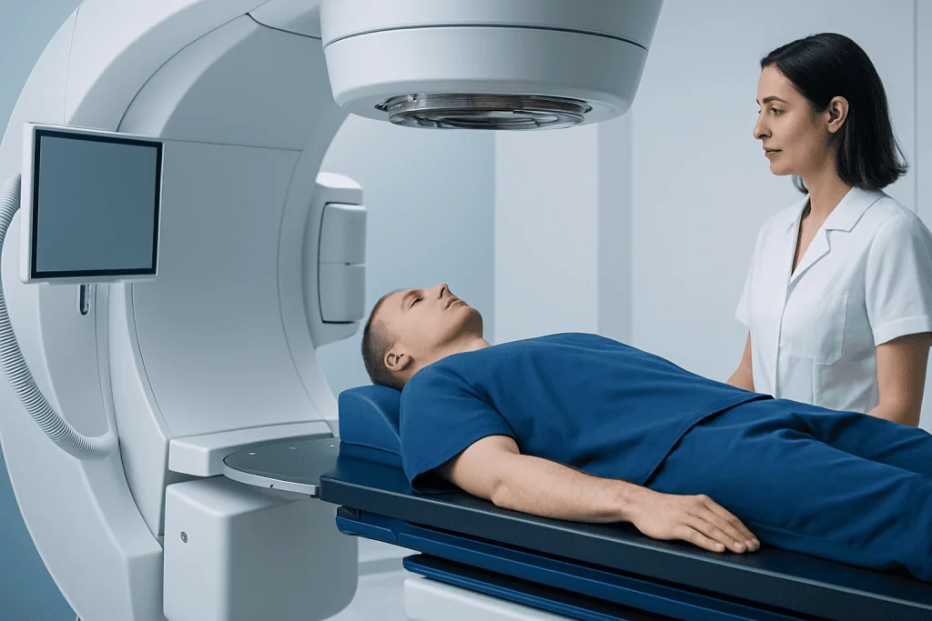
For diagnostic imaging, calculators may use details such as the type of examination, body part scanned, scan length, imaging settings, and machine-reported dose indicators. CT scanners, for example, record technical measures that can be converted into an estimated effective dose. Effective dose is useful for comparing broad categories of exposure, but it is not an exact measurement of radiation absorbed by one individual.
Factors including age, body size, pregnancy status, the reason for imaging and the number of scan phases may affect how a clinician considers exposure. Children are generally more sensitive to radiation than adults, so pediatric imaging protocols are specifically adjusted whenever possible. A calculator cannot account perfectly for every biological difference between people.
For radiation therapy, planning systems work differently. Specialists combine imaging scans with contouring of the tumor and nearby organs, then calculate how radiation beams should be shaped and delivered. Medical physicists and radiation oncologists review the plan to confirm that the intended target receives the prescribed dose and that dose limits for surrounding tissues are respected.
Understanding Results and Dose Units

Radiation results can be reported in different units. The gray (Gy) describes absorbed radiation energy in tissue and is commonly used in radiation therapy. The sievert (Sv), often shown in millisieverts (mSv), is used to express effective dose for radiation protection purposes, particularly when discussing diagnostic imaging exposure.
A dose estimate should always be interpreted with the procedure type in mind. A CT scan generally involves more radiation than a standard X-ray because it produces many detailed cross-sectional images. Nuclear medicine studies involve a radiopharmaceutical and have their own dose calculations, based on the substance used and how it behaves in the body.
In cancer care, a treatment dose may appear much larger than a diagnostic imaging dose because radiation therapy has a different purpose: destroying cancer cells or controlling tumor growth. It is delivered in a planned way, often over multiple sessions, with measures designed to reduce exposure to normal tissues. A patient should discuss any result directly with the referring clinician, radiologist, medical physicist or radiation oncologist rather than relying on an online estimate alone.
Who May Need Dose Assessment and Planning?
Most people do not need to calculate radiation exposure themselves before a routine medical test. The referring clinician and imaging department assess whether the test is appropriate and select a suitable protocol. Dose review can be particularly relevant for people who need repeated imaging, children, pregnant patients, and people undergoing cancer treatment.
Radiation therapy planning is considered for people with certain cancers and, less commonly, selected noncancerous conditions. Suitability depends on the diagnosis, tumor location and stage, prior treatment, general health and personal goals. Radiation may be used alone or alongside surgery, chemotherapy, immunotherapy or other approaches, depending on the individual situation.
Patients should tell their healthcare team about previous imaging or radiation treatment, possible pregnancy, breastfeeding, implanted medical devices and relevant medical conditions. When prior images are available, sharing them can sometimes avoid unnecessary repeat testing. Related conditions, including lung cancer, may require coordinated planning among oncology, radiology, surgery and other specialties.
What to Expect: From Referral to Results
For diagnostic imaging, the process usually begins with a clinical question, such as investigating persistent symptoms, checking an injury or monitoring a known condition. The clinician selects the test most likely to answer that question. Before the examination, staff may ask about pregnancy, allergies, kidney problems, medications and previous procedures; some CT and nuclear medicine examinations require additional preparation.
During the scan, the radiographer positions the patient and uses protocols intended to obtain diagnostic-quality images with an appropriate dose. The radiation exposure itself is painless and cannot usually be felt. Afterward, a radiologist reviews the images and sends a report to the clinician who requested the test; the dose figure is typically recorded as part of the technical examination record.
Radiation therapy has more steps. A planning scan, often called simulation, helps the team establish an accurate treatment position. The care team creates and verifies a personalized plan, and treatment is then delivered in scheduled sessions. The actual beam delivery is brief, although positioning and safety checks may take longer. Follow-up visits monitor response and manage any side effects.
Benefits, Limitations and Possible Risks
The main benefit of dose calculation and optimization is that it supports informed, careful use of radiation. Imaging can identify injuries, infections, bleeding, tumors and other conditions that may not be clear from symptoms or physical examination alone. Accurate diagnosis can guide timely treatment and may prevent more invasive procedures.
The radiation dose from medically justified imaging is generally low, but ionizing radiation is not entirely risk-free. Risk is influenced by total exposure over time, age and the area examined. For this reason, healthcare teams follow the principle of using radiation that is as low as reasonably achievable while still obtaining the needed clinical information.
Radiation therapy can damage cancer cells, but it may also affect healthy tissues near the treatment area. Side effects vary according to the body part treated, total dose and treatment technique; they may include tiredness, skin changes or localized symptoms. The oncology team explains expected effects, prevention strategies and when to contact the clinic. Dose calculators are valuable tools, but they cannot replace professional judgment or guarantee a particular result.
Preparing, Recovering and Supporting Safe Care
Preparation depends on the procedure. Patients should follow instructions about food, fluids, contrast material or medications, and should bring details of prior scans when requested. It is helpful to ask what information the examination is expected to provide and whether a non-radiation alternative, such as ultrasound or MRI, could answer the same clinical question. However, an alternative is not always suitable or equally informative.
There is usually no recovery time after a standard X-ray, CT scan or radiotherapy planning scan. Most people can resume regular activities immediately unless they received sedation, contrast material or specific instructions from the clinical team. After external beam radiation therapy, people are not radioactive and do not expose family members or other people to radiation.
Patients receiving radiation therapy should report new or troublesome symptoms and attend scheduled follow-up appointments. Skin care, nutrition, activity and rest recommendations vary by treatment site and should be tailored by the oncology team. Acibadem International’s multidisciplinary specialists and JCI-accredited hospitals provide diagnostic assessment and radiation treatment planning for international patients when clinically appropriate.
When to Seek Medical Care
A person should contact a doctor promptly for symptoms that are severe, persistent or worsening, especially if there is chest pain, sudden weakness, difficulty breathing, confusion, major injury, unexplained bleeding or a new neurological symptom. The need for imaging is based on the clinical situation, not on a dose calculator result alone.
Patients who are pregnant, think they may be pregnant, or are breastfeeding should inform the imaging or treatment team before any procedure involving ionizing radiation. This does not automatically mean that imaging cannot be performed; it allows clinicians to choose the safest appropriate approach and make necessary adjustments.
After contrast-enhanced imaging, urgent medical advice is needed for signs of a serious allergic reaction, such as trouble breathing, swelling of the face or throat, or widespread hives. During radiation treatment, the care team should be contacted for severe pain, fever, dehydration, rapidly worsening skin reactions or symptoms that interfere with eating, drinking or daily activities.
Frequently asked questions
Can a radiation dose calculator tell whether a scan is safe?
A calculator can estimate exposure, but it cannot determine safety for one person in isolation. A clinician considers the reason for the scan, the expected diagnostic benefit, the patient's age and medical history, and whether another test could provide the needed information.
What is the difference between mSv and Gy?
Millisieverts, or mSv, are commonly used to discuss effective dose and radiation protection in diagnostic imaging. Grays, or Gy, describe absorbed dose and are commonly used in radiation therapy planning. They measure different concepts and should not be directly compared without clinical context.
Are CT scans associated with more radiation than X-rays?
CT scans usually involve a higher radiation dose than a single standard X-ray because CT creates detailed images from many angles. The exact dose varies by body part, scan protocol, equipment and patient factors. CT may still be the most appropriate test when detailed information is needed.
Does radiation therapy make a person radioactive?
External beam radiation therapy does not make a person radioactive. They can generally be around family, friends and children after treatment. Some internal radiation treatments require specific temporary precautions, which the treatment team will explain clearly.
Should patients avoid all medical imaging that uses radiation?
No. Medical imaging is often important for diagnosing conditions, guiding treatment and monitoring recovery. The goal is not to avoid necessary imaging, but to ensure every examination is clinically justified and performed with an optimized protocol.
Can prior scans reduce the need for another radiation test?
Sometimes. Providing previous images and reports may help a clinician determine whether new imaging is needed or whether an existing study can answer the current question. A repeat examination may still be necessary if symptoms have changed or a different type of image is required.
References
- International Atomic Energy Agency
- International Commission on Radiological Protection
- United States Food and Drug Administration
- National Cancer Institute
- Radiological Society of North America
This article is for general information only and is not a substitute for professional medical advice. Please consult a qualified doctor about your individual situation.
Oncology care in Turkey — second opinion and treatment plan
JCI-accredited · board-certified surgeons · reply within 24h
Add us as a Preferred Source to see more of our trusted health content across Google Search, AI Overviews and Discover.