Gonioscopy Procedure: An Evidence-Based Patient Guide
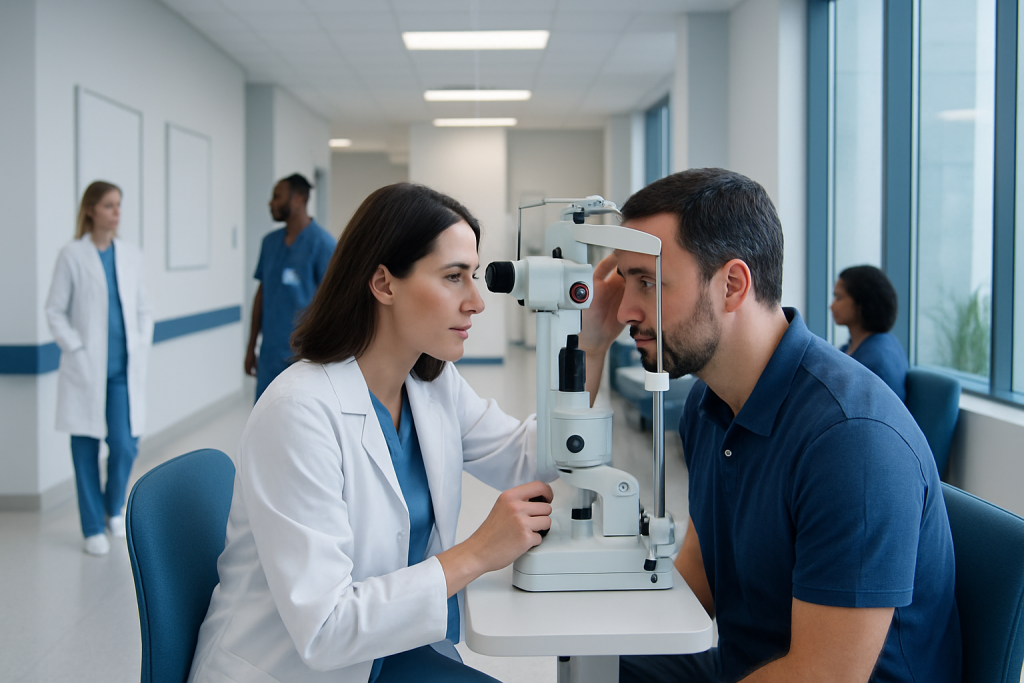
Gonioscopy examines the eye’s drainage angle, which cannot usually be seen clearly during a routine eye exam. It is commonly used to assess glaucoma, narrow angles, eye injury and unusual changes in eye pressure.
Key Takeaways
- Gonioscopy examines the eye’s drainage angle, which cannot usually be seen clearly during a routine eye exam.
- It is commonly used to assess glaucoma, narrow angles, eye injury and unusual changes in eye pressure.
- Numbing drops are used, and most people experience pressure or awareness rather than pain.
- The examination usually takes only a few minutes per eye and generally has no recovery downtime.
- Results help an ophthalmologist decide whether monitoring, preventive laser treatment, medicines or other care may be appropriate.
A gonioscopy procedure is a brief examination in which an eye specialist views the drainage angle between the cornea and iris. It helps determine whether the angle is open, narrow, closed or affected by abnormal tissue, supporting accurate glaucoma assessment and treatment planning.
Overview: what is a gonioscopy procedure?
A gonioscopy procedure is a specialized eye examination used to inspect the drainage angle at the front of the eye. This angle is where the clear cornea meets the colored iris. It contains structures that allow aqueous humor, the fluid naturally made inside the eye, to drain and help maintain healthy eye pressure.
Because the cornea bends light, the drainage angle cannot be fully assessed by looking directly at the eye. During gonioscopy, an ophthalmologist places a small mirrored contact lens called a gonioscopy lens on the numbed eye. The lens allows the clinician to see the angle clearly and record whether it is open, narrow, closed or altered by other findings.
Gonioscopic evaluations are an important part of evaluating glaucoma and conditions that may raise the risk of sudden angle closure. The test itself does not treat glaucoma, but it provides information that can shape the safest next steps for each person.
What does a gonioscopy test for?
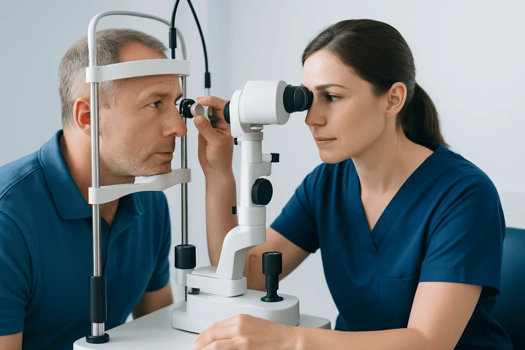
A gonioscopy test primarily assesses how open the drainage angle is and whether fluid can leave the eye normally. It helps distinguish open-angle glaucoma from angle-closure mechanisms, since these conditions can require different approaches to monitoring and treatment.
The examination may also identify peripheral anterior synechiae, which are areas where the iris adheres to the drainage angle. In addition, the specialist may look for pigment deposits, signs of inflammation, new abnormal blood vessels, trauma-related changes, developmental differences or material associated with certain types of glaucoma.
Doctors may recommend gonioscopy when eye pressure is elevated, the optic nerve appears suspicious, visual field testing suggests glaucoma, or the angle looks narrow on another examination. It may also be performed after eye injury or before considering selected eye procedures. A narrow angle does not always mean that an emergency will occur, but it deserves individualized assessment by an eye specialist.
Who may need gonioscopy and how it works
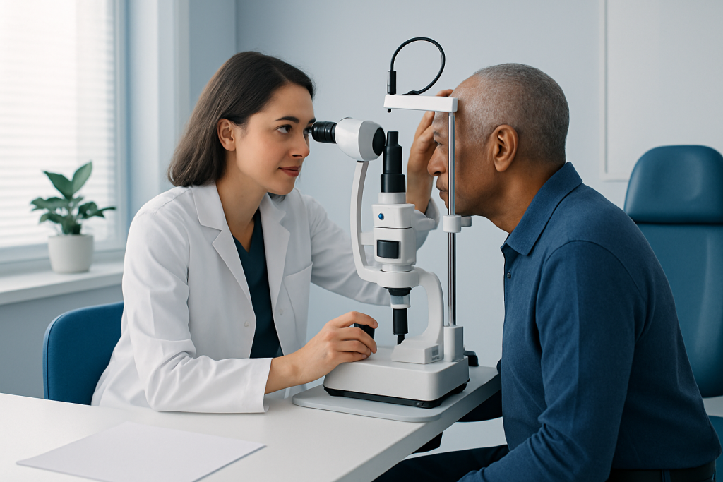
Gonioscopy is often recommended for people being evaluated for glaucoma or followed for known glaucoma. It can be especially useful when an eye specialist suspects narrow or occludable angles, when eye pressure changes unexpectedly, or when there are differences between the two eyes.
People with a family history of angle-closure glaucoma, farsightedness, certain anatomical eye features, prior eye inflammation or eye trauma may benefit from assessment when their clinician considers it appropriate. Risk also tends to increase with age. However, candidacy is based on the complete eye examination rather than one risk factor alone.
The procedure uses optical mirrors in the gonioscopy lens to redirect the view around the curve of the cornea. Some clinicians may gently apply pressure through the lens, called indentation gonioscopy, to help determine whether a narrow angle can open or is closed by adhesions. This is a diagnostic maneuver and is performed carefully after the eye has been anesthetized.
In addition to direct gonioscopy, imaging tests may sometimes help evaluate the front of the eye. These tests can complement, but do not always replace, a clinician’s direct view of the angle. The ophthalmologist interprets all findings alongside eye pressure, optic nerve examination and visual field testing.
Step-by-step: what happens during the examination?
Before the test, the eye care professional explains the examination and asks about relevant symptoms, eye history and medicines. Contact lenses are generally removed. The patient sits at a slit lamp, which is the microscope commonly used in eye clinics, and rests the chin and forehead on supports.
Numbing eye drops are placed in the eye. The clinician may also use a clear gel or lubricating solution on the gonioscopy lens. The patient is asked to keep both eyes open, look in selected directions and avoid squeezing the eyelids while the lens gently touches the surface of the eye.
The clinician examines the angle through the slit lamp and may document findings using an angle grading system, drawings, photographs or electronic health records. Gonioscopy documentation requirements vary by clinical setting, insurer and local regulations, but accurate notes generally describe the angle appearance and relevant abnormalities in each eye.
The examination commonly lasts a few minutes per eye. A person may notice light, mild pressure or the sensation of the lens, but should tell the clinician if there is significant discomfort. Eye drops used for dilation may be needed for other parts of an eye examination, although dilation is not required for every gonioscopy procedure.
Is gonioscopy painful?
Gonioscopy is usually not painful because anesthetic drops numb the surface of the eye. Many people notice the bright examination light and a mild feeling of pressure or contact from the lens. The sensation is typically brief and ends when the lens is removed.
Temporary blurred vision, watering or mild redness can occur, especially if gel was used or the eye was sensitive beforehand. These effects usually settle soon after the visit. People should not rub their eyes immediately after the examination because the numbing drops can briefly reduce normal sensation.
Serious complications from a routine gonioscopy examination are uncommon. Rarely, contact with the eye can cause temporary surface irritation or a small corneal abrasion. Individuals should contact their eye care team promptly if they develop persistent pain, increasing redness, marked light sensitivity, discharge or vision changes after the appointment.
Results, benefits and next steps
The main benefit of gonioscopy is that it gives a direct view of the drainage angle and supports a more precise diagnosis. An open angle may point toward open-angle glaucoma when other findings are present. A narrow or closed angle may lead the ophthalmologist to discuss monitoring, medicines, laser treatment or other options based on the person’s anatomy and level of risk.
If an angle is considered at risk of closure, a specialist may discuss laser peripheral iridotomy, a procedure that creates a small opening in the iris to improve fluid movement between areas of the eye. The decision is individualized and depends on the examination, symptoms and overall glaucoma assessment.
Gonioscopy practice includes repeating the test when clinically necessary. Angle structures can change over time, and a previous normal assessment does not replace future review if symptoms, eye pressure or other examination findings change. Keeping scheduled glaucoma follow-up appointments is an important part of protecting vision.
At Acibadem International, multidisciplinary eye specialists in JCI-accredited hospitals assess glaucoma and related eye conditions for international patients, using examination findings to develop an individualized care plan.
How much does gonioscopy cost?
The cost of gonioscopy varies according to the country, clinic, insurance coverage and whether it is billed as part of a comprehensive eye examination or glaucoma consultation. It may also depend on whether additional tests, such as eye pressure measurement, optic nerve imaging or visual field testing, are needed at the same visit.
People can ask the clinic in advance whether gonioscopy is included in the planned appointment, whether prior authorization is required and what out-of-pocket expenses may apply. For billing purposes, a gonioscopy procedure code may be used in some healthcare systems, but coding practices and reimbursement rules differ by location and insurer.
Cost should not be the only factor in deciding whether to have the test. When an ophthalmologist recommends gonioscopy, it is generally because the result can clarify the type of glaucoma risk or explain an important eye finding. A clinic’s billing team or insurance provider can provide the most accurate personal estimate.
Can an optometrist perform gonioscopy? When to seek medical care
Whether an optometrist can perform gonioscopy depends on professional training, scope-of-practice rules and local regulations. Many optometrists are trained to perform gonioscopy and use it in glaucoma assessment, while ophthalmologists also routinely perform the test. Referral to an ophthalmologist may be appropriate when results are uncertain, the angle is narrow or closed, glaucoma is suspected or treatment is being considered.
People should arrange an eye examination if they have been told they have high eye pressure, suspicious optic nerve findings, a narrow angle or a family history of glaucoma. Regular comprehensive eye care is particularly important because glaucoma can develop without early noticeable symptoms.
Urgent medical assessment is needed for sudden severe eye pain, headache with nausea or vomiting, a red eye, halos around lights, sudden blurred vision or rapid vision loss. These symptoms can occur with acute angle closure and other serious eye problems. They should not be managed by waiting for a routine appointment.
After gonioscopy, most people can return to usual activities straight away unless the clinician gives different instructions. If vision is blurred from dilation or other drops, driving should be avoided until vision is clear and safe for the task.
Frequently asked questions
What does a gonioscopy test for?
A gonioscopy test examines the drainage angle between the cornea and iris. It helps identify whether the angle is open, narrow or closed and can reveal changes linked with different forms of glaucoma, inflammation, trauma or abnormal blood vessels.
How long does a gonioscopy procedure take?
The direct examination usually takes only a few minutes for each eye. The complete appointment may be longer if it includes eye pressure measurement, dilation, optic nerve assessment, imaging or visual field testing.
Is gonioscopy painful?
Gonioscopy is generally well tolerated because numbing drops are used first. A person may feel brief pressure from the lens or notice bright light, but significant pain is not expected.
How much does gonioscopy cost?
Costs vary by country, clinic, insurance plan and whether the test is included in a broader eye or glaucoma assessment. The clinic and insurance provider can explain expected charges, coverage and any authorization requirements before the appointment.
Can an optometrist perform gonioscopy?
In many locations, appropriately trained optometrists can perform gonioscopy within their licensed scope of practice. Local regulations differ, and ophthalmology referral may be needed when glaucoma is suspected, the angle is narrow or treatment decisions are required.
Do I need to prepare for gonioscopy?
Most people need little preparation. They should bring relevant glasses, medication information and details of previous eye conditions or surgery, and they should remove contact lenses unless the clinic gives other instructions. If dilation is planned, arranging transport may be helpful because vision can be temporarily blurred.
References
- American Academy of Ophthalmology
- National Eye Institute
- Glaucoma Research Foundation
- National Health Service
This article is for general information only and is not a substitute for professional medical advice. Please consult a qualified doctor about your individual situation.
Explore treatments in Turkey — costs, top hospitals & a free quote
JCI-accredited · board-certified surgeons · reply within 24h
Add us as a Preferred Source to see more of our trusted health content across Google Search, AI Overviews and Discover.

More from the Health Library
Related Specialists

Dr. Nurşat Erdemli
Pediatrics
Dr. Yasemin Ballıca Özen
Gynecology & Obstetrics
Dr. Nilüfer Yıldırım (m)
General Surgery
Assoc. Prof. Dr. Filiz Gülustan
Ear Nose & Throat