Endo Bronchial Ultrasound: Preparation, Procedure and Results
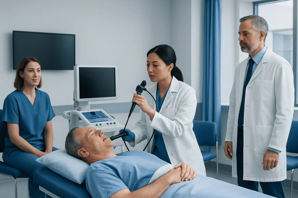
Endo bronchial ultrasound uses a flexible bronchoscope with an ultrasound probe to view structures beyond the airway wall. It is commonly used to sample lymph nodes in the chest and evaluate certain lung or airway findings.
Key Takeaways
- Endo bronchial ultrasound uses a flexible bronchoscope with an ultrasound probe to view structures beyond the airway wall.
- It is commonly used to sample lymph nodes in the chest and evaluate certain lung or airway findings.
- Most people receive sedation, need to fast beforehand, and go home the same day with an accompanying adult.
- Preliminary findings may be discussed soon after the procedure, while biopsy and laboratory results often take several days.
- Temporary sore throat, cough, and mild blood-streaked mucus can occur, but severe symptoms need urgent medical review.
Endo bronchial ultrasound, also called endobronchial ultrasound or EBUS, combines bronchoscopy with ultrasound imaging to examine areas beside the airways and collect tissue samples when needed. It can help doctors investigate enlarged chest lymph nodes, lung abnormalities, infections, inflammatory conditions, and suspected cancer without requiring open surgery.
Overview: What Is Endo Bronchial Ultrasound?
Endo bronchial ultrasound is a procedure that allows a respiratory specialist to see the airways from inside and use ultrasound to assess tissues outside them. The term is often written as endobronchial ultrasound, or EBUS. A thin, flexible tube called a bronchoscope is passed through the mouth, or sometimes the nose, into the breathing passages.
At the tip of the bronchoscope, an ultrasound probe creates real-time images of lymph nodes, blood vessels, and other structures next to the windpipe and larger airways. If an area requires further assessment, the doctor can guide a fine needle through the airway wall to collect cells or small tissue samples. This is called EBUS-guided transbronchial needle aspiration.
EBUS is often used as part of the assessment of a lung mass, enlarged chest lymph nodes, unexplained findings on a chest scan, or possible infection or inflammatory disease. It may also help doctors determine the extent of certain lung cancers by checking whether nearby lymph nodes contain abnormal cells. It is a diagnostic procedure, meaning its purpose is to clarify a diagnosis and guide next steps.
How Endobronchial Ultrasound Works and Who May Need It
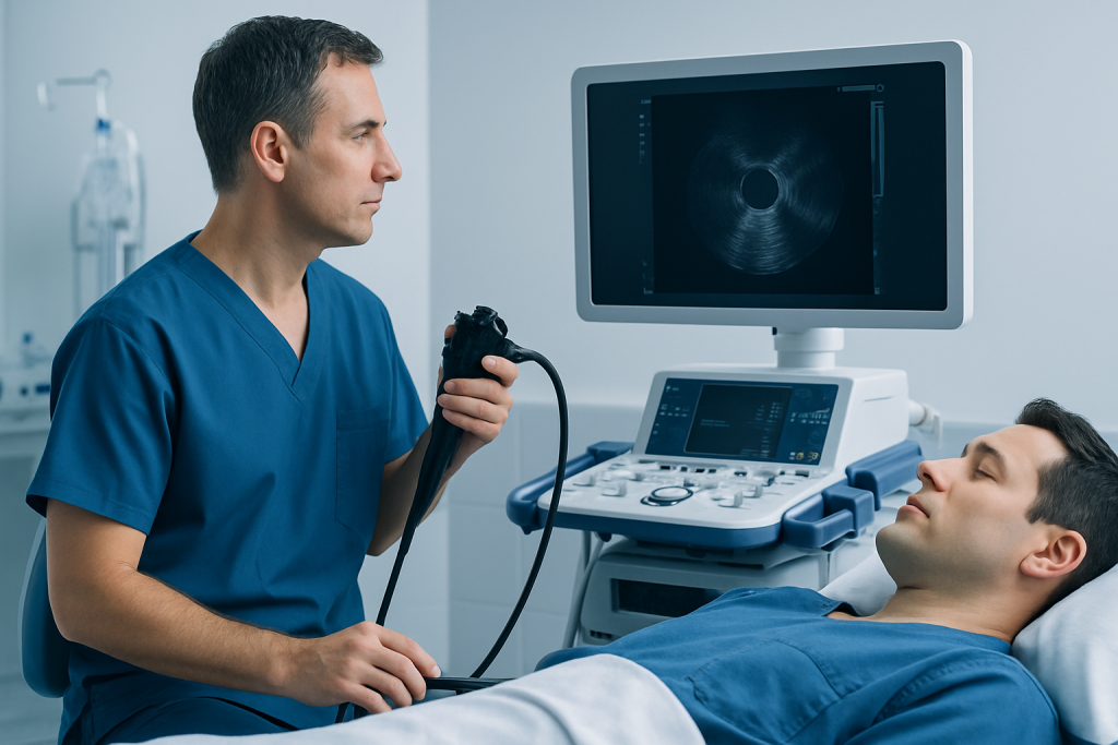
Ultrasound uses sound waves to form images and does not expose the patient to radiation. During EBUS, the ultrasound sensor sits directly against the inside of the airway wall. This close position can provide detailed views of lymph nodes and tissue that may not be visible during standard bronchoscopy alone.
A clinician may recommend endo bronchial ultrasound after a chest X-ray, CT scan, or PET scan shows enlarged lymph nodes, a lung lesion, or another change needing clarification. It can also be used to investigate persistent unexplained symptoms or abnormal imaging in people with suspected tuberculosis, sarcoidosis, lymphoma, or other conditions affecting the chest.
Not every person with an abnormal chest scan needs EBUS. The decision depends on the location and appearance of the finding, medical history, overall health, medications, and whether a tissue sample is likely to change care. For people being evaluated for lung cancer, EBUS can sometimes provide important information for diagnosis and staging while avoiding more invasive surgical sampling.
Before recommending the procedure, the medical team reviews imaging, breathing status, allergies, bleeding risk, and medicines such as anticoagulants, antiplatelet medicines, insulin, and other diabetes treatments. Patients should not stop prescribed medicine on their own; the clinician performing the procedure should give individualized instructions.
Preparing for an EBUS Bronchoscopy

Preparation instructions vary between hospitals and depend on the planned sedation or anesthesia. In many cases, patients are asked not to eat for several hours before EBUS and may be allowed clear liquids until a specified time. Fasting reduces the chance of stomach contents entering the lungs during sedation. The hospital will provide exact instructions, which should take priority over general advice.
Patients should tell the team about all prescription medicines, non-prescription products, supplements, previous reactions to anesthesia, sleep apnea, heart or lung conditions, pregnancy, and any tendency to bleed or bruise. Blood-thinning medicines may need temporary adjustment to reduce bleeding risk, but this should only be done with advice from the prescribing clinician and bronchoscopy team.
It is usually necessary to arrange a responsible adult to take the patient home and stay available after sedation. Driving, operating machinery, signing important documents, drinking alcohol, and making major decisions should be avoided for the rest of the day. Comfortable clothing and a list of current medicines can make the visit easier.
Some people have a chest ultrasound performed through the skin, which is different from endobronchial ultrasound. A standard chest ultrasound generally does not require fasting. However, EBUS is usually performed with sedation, so fasting may be needed because of the sedation rather than the ultrasound itself.
Step by Step: What Happens During the Procedure
On arrival, the care team confirms identity, consent, medical history, and fasting status. A nurse may place an intravenous line, check vital signs, and provide oxygen through a small tube under the nose. The throat is often numbed with local anesthetic spray or liquid to reduce gagging and coughing.
Most EBUS procedures are performed with moderate or deep sedation, although some patients may have general anesthesia depending on their health needs and local practice. The patient is closely monitored throughout. Sedation is intended to keep the person comfortable and relaxed; they may remember little or nothing of the procedure.
The specialist gently advances the bronchoscope into the airways and examines them. The ultrasound probe is then used to locate lymph nodes or other target areas. If sampling is needed, a small needle is passed through the bronchoscope to obtain specimens. Several samples may be taken from one or more lymph nodes to improve the chance of an accurate diagnosis.
Once the examination is complete, the bronchoscope is removed and the patient is observed in a recovery area. The clinical team monitors breathing, blood pressure, oxygen levels, alertness, and swallowing before discharge. Diagnostic bronchoscopy and EBUS are often coordinated with pathology, radiology, respiratory medicine, oncology, and thoracic surgery when results need multidisciplinary interpretation.
How Long Does an Endobronchial Ultrasound Take?
The bronchoscopy and endo bronchial ultrasound itself commonly takes about 30 to 60 minutes. The exact time depends on whether tissue samples are needed, how many lymph nodes are assessed, the complexity of the airway anatomy, and whether additional bronchoscopic tests are performed at the same appointment.
Patients should plan to spend longer at the hospital than the procedure time alone. Registration, preparation, sedation, recovery, and discharge checks can mean a total visit of several hours. If there are medical concerns after the procedure, observation may take longer.
After EBUS, mild throat discomfort, hoarseness, sleepiness, coughing, or a small amount of blood-streaked phlegm can occur. These effects are usually short-lived. The team will provide written discharge instructions tailored to the procedures performed and any medicines given.
Recovery, Benefits and Possible Risks
Many people return home on the day of an EBUS bronchoscopy. Eating and drinking can usually resume once throat numbness has worn off and swallowing feels normal, often after the recovery team confirms it is safe. A quieter day at home is sensible, and most people can resume usual routine activities the following day unless their clinician advises otherwise.
The main benefit of EBUS is that it can obtain targeted samples from chest lymph nodes and nearby structures through the airways. This may help avoid a more invasive surgical procedure and can provide information that guides treatment planning. A sample may identify cancer cells, infection, inflammation, or benign tissue, although occasionally a sample is insufficient or does not fully explain the imaging finding.
EBUS is generally considered safe, but all invasive procedures have risks. Possible complications include sore throat, cough, temporary low oxygen levels, reactions to sedative medication, bleeding, fever, infection, or airway spasm. Rarely, a collapsed lung or more significant bleeding can occur, particularly when additional lung biopsies are performed.
Patients should seek urgent medical advice after discharge for increasing shortness of breath, chest pain, coughing up more than a small amount of blood, persistent fever, severe dizziness, confusion, or symptoms that are worsening rather than improving. These symptoms do not always mean a serious complication, but they require prompt assessment.
What Should You Not Do After a Bronchoscopy?
After a bronchoscopy with sedation, patients should not drive, ride a bicycle in traffic, operate machinery, drink alcohol, take recreational drugs, or make legally important decisions until the next day or for the period advised by their care team. Sedation can affect judgment, coordination, and reaction time even when the person feels awake.
Food and drinks should be avoided until the numbness in the throat has resolved and swallowing is safe. Starting with small sips of water may be recommended. Hot drinks and hard foods may be uncomfortable initially if the throat is sore.
Strenuous exercise and heavy lifting are usually best postponed for the rest of the day. Patients should follow specific instructions about restarting blood thinners and other regular medicines. If a biopsy was taken, the clinician may give additional precautions based on the sampling method and the individual’s health.
How Fast Do You Get Results From a Bronchoscopy?
Some bronchoscopy findings can be discussed shortly after recovery, such as whether the airways looked narrowed, inflamed, or otherwise abnormal. If samples were collected, however, final results usually take longer because laboratory testing is needed. Many routine pathology results are available within several days, while specialized tests may take one to two weeks or longer.
The timeline depends on the type of sample and the tests requested. Cells may be examined under a microscope, tested for infection, or assessed with molecular tests that can help guide cancer treatment. The clinician will explain which results are expected and how the patient will receive them.
A result that is non-diagnostic does not necessarily mean that everything is normal. Sometimes a sample does not contain enough representative cells, or imaging findings require follow-up despite reassuring pathology. The specialist may recommend repeat sampling, a different biopsy method, follow-up imaging, or discussion by a multidisciplinary team. Acibadem International’s multidisciplinary specialists and JCI-accredited hospitals support diagnosis and treatment planning for international patients.
When to Seek Medical Care
A person should arrange medical review for a persistent cough, coughing up blood, unexplained breathlessness, ongoing chest discomfort, recurrent chest infections, unexplained weight loss, or an abnormal chest scan. These symptoms have many possible causes and should be assessed calmly and promptly, especially when they persist or change.
Emergency care is appropriate for severe difficulty breathing, sudden or severe chest pain, blue or gray lips, fainting, or large-volume bleeding from the airways. Anyone who has recently undergone bronchoscopy and develops worsening breathlessness, chest pain, high fever, or significant bleeding should contact the procedure team or seek urgent medical help.
Endo bronchial ultrasound is only one part of respiratory assessment. Depending on symptoms and imaging, a doctor may recommend lung function testing, blood tests, sputum testing, CT imaging, or other investigations. The safest next step is guided by the individual’s clinical picture rather than by symptoms alone.
Frequently asked questions
Is endo bronchial ultrasound painful?
Most people receive sedation and local anesthetic to make the procedure more comfortable. They may feel brief coughing or throat irritation, but significant pain is not expected. Mild sore throat or hoarseness can occur afterward and usually improves within a short time.
How long does an endobronchial ultrasound take?
The EBUS procedure itself often takes around 30 to 60 minutes. The full hospital visit is longer because it includes preparation, sedation, monitoring, and recovery. Sampling several lymph nodes or combining EBUS with other tests can extend the procedure time.
What should you not do after a bronchoscopy?
After sedation, people should not drive, use machinery, drink alcohol, or make important decisions until the following day or as instructed. They should wait to eat and drink until throat numbness has worn off and swallowing is normal. Heavy exercise is commonly avoided for the rest of the day.
Does a chest ultrasound need fasting?
A standard chest ultrasound performed through the skin generally does not require fasting. Endobronchial ultrasound is different because it is usually performed during bronchoscopy with sedation. Fasting instructions for EBUS are mainly intended to make sedation safer.
How fast do you get results from a bronchoscopy?
The doctor may discuss visual findings from the bronchoscopy on the same day. Biopsy, cell, and infection test results usually take several days, while specialized testing can take longer. The care team should explain the expected timeline and arrange follow-up.
Can EBUS diagnose lung cancer?
EBUS can collect tissue or cells from lymph nodes and certain areas near the airways, which may help confirm or rule out lung cancer. It can also help assess whether nearby lymph nodes are involved. Final diagnosis depends on pathology results and the wider clinical assessment.
References
- American Thoracic Society
- European Respiratory Society
- British Thoracic Society
- National Cancer Institute
- Mayo Clinic
This article is for general information only and is not a substitute for professional medical advice. Please consult a qualified doctor about your individual situation.
Ultrasound in Turkey — costs, top hospitals & a free quote
JCI-accredited · board-certified surgeons · reply within 24h
Add us as a Preferred Source to see more of our trusted health content across Google Search, AI Overviews and Discover.

Check-up & Preventive Medicine
Comprehensive health screenings designed for early detection and prevention.
3 specialists in this unit