Targeted Ultrasound Breast: Preparation, Procedure and Results

Targeted breast ultrasound evaluates a particular symptom, lump, or imaging finding rather than screening the whole breast. The examination uses sound waves, involves no radiation, and is usually comfortable and brief.
Key Takeaways
- Targeted breast ultrasound evaluates a particular symptom, lump, or imaging finding rather than screening the whole breast.
- The examination uses sound waves, involves no radiation, and is usually comfortable and brief.
- Most people do not need special preparation, but they should avoid applying products to the breast and underarm area on the day of imaging.
- Results may be discussed on the same day or reported after a radiologist reviews the images and compares prior studies.
- Ultrasound can distinguish many fluid-filled cysts from solid areas, but some findings still require mammography, follow-up imaging, or biopsy.
A targeted ultrasound breast examination is a focused, radiation-free imaging test used to assess a specific area of concern in the breast or underarm. It can help clarify findings from a physical examination or mammogram and may guide a biopsy when tissue sampling is needed.
Overview: what is targeted ultrasound breast imaging?
Targeted ultrasound breast imaging is a diagnostic examination that focuses on one specific area of the breast or armpit. A clinician may request it when a person notices a lump, localized pain, skin or nipple change, or enlarged lymph node, or when a mammogram shows an area that needs closer assessment. Unlike routine breast screening, the examination is directed to the exact location of the concern.
The scan uses high-frequency sound waves to create real-time images of breast tissue. It does not use ionizing radiation and is generally safe during pregnancy and breastfeeding when clinically appropriate. Ultrasound is particularly useful for showing whether an area is fluid-filled, such as a simple cyst, or solid, and for evaluating breast tissue that may appear dense on mammography.
Targeted ultrasound is usually one part of a broader breast assessment rather than a stand-alone test for every concern. The radiologist considers the person’s symptoms, physical examination, age, mammogram findings, and prior breast images to recommend the most appropriate next step.
How targeted breast ultrasound works and who may need it
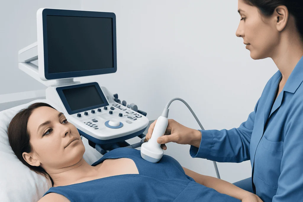
During the examination, a handheld device called a transducer is placed against the skin. The device sends sound waves into the breast and receives returning echoes, which a computer converts into images. The sonographer may scan the breast in several directions and compare the area of concern with nearby tissue or the opposite breast.
A targeted scan may be appropriate for a newly felt lump, focal tenderness that persists, a change in breast contour, nipple discharge, a skin change, or an area identified on a mammogram or breast MRI. It can also assess lymph nodes in the underarm and may be used to monitor a known benign finding when follow-up imaging has been advised.
Ultrasound does not replace mammography for most people who are eligible for breast cancer screening. Some early breast cancers, including certain calcifications, are better detected with mammography. When imaging shows an area that needs tissue confirmation, ultrasound may help guide a needle biopsy accurately. Ultrasound-guided breast biopsy can sample a visible area while allowing the radiologist to watch needle placement in real time.
Preparing for the procedure: what should you not do before a breast ultrasound?
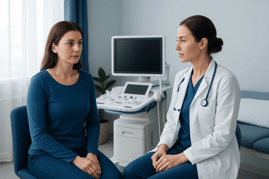
Most people do not need to fast, stop usual medicines, or make major changes to their routine before a breast ultrasound. Unless the healthcare team gives different instructions, eating and drinking normally is acceptable. It is helpful to wear a comfortable two-piece outfit, as only clothing above the waist usually needs to be removed.
On the day of the appointment, it is sensible to avoid lotions, powders, deodorant, perfume, or oils on the breasts and underarms. These products can sometimes leave residue on the skin or interfere with related breast imaging. If a mammogram is scheduled on the same day, this advice is especially important.
People should bring previous mammogram, ultrasound, MRI, and biopsy reports or ensure that the imaging center can access them. Comparing current and earlier images often helps the radiologist determine whether a finding is new, stable, or changing. Patients should also tell the team about pregnancy, breastfeeding, breast implants, prior surgery, and any exact location of symptoms.
What is the best time to do a breast ultrasound?
A targeted breast ultrasound can usually be performed at any point in the menstrual cycle when there is a new lump, persistent focal pain, an abnormal mammogram result, or another concerning change. A potentially important symptom should not be delayed simply to wait for a particular time of the month.
For people who have regular periods and are booking non-urgent breast imaging because of cyclical breast tenderness, the week after menstruation may be more comfortable. Hormonal breast swelling and tenderness are often less pronounced then. However, the best timing depends on the reason for the test and the advice of the referring clinician or breast imaging service.
Breast ultrasound may also be used during pregnancy or breastfeeding when a new breast symptom needs assessment. In these situations, hormonal changes can affect breast tissue, but targeted imaging remains valuable for evaluating a specific concern. The imaging team can tailor the examination to the individual’s circumstances.
What happens during a targeted breast ultrasound?
At the appointment, the patient is usually asked about the symptom, when it began, and where it is felt. They will lie on an examination table, often with one arm raised or placed behind the head. A clear water-based gel is applied to the skin so the transducer can move smoothly and produce clear images.
The sonographer or radiologist moves the transducer over the area of concern and may apply gentle pressure. The procedure is typically painless, although a tender breast area may be mildly uncomfortable when touched. Images and measurements are recorded, and the clinician may scan nearby tissue or underarm lymph nodes if needed.
The scan itself often takes about 15 to 30 minutes, though timing varies with the reason for the examination and whether additional views are required. There are no injections, incisions, or recovery restrictions for a standard ultrasound. The gel is wiped away afterward, and the person can return to normal activities immediately.
How long after a breast ultrasound will I get results?
Timing varies between imaging centers and depends on whether the radiologist can review the images during the appointment. In some settings, preliminary findings may be discussed before the patient leaves. In others, the formal report is sent to the referring clinician within a few days after the radiologist reviews the examination and compares it with earlier images.
Results commonly describe whether the examined area appears normal, benign, probably benign but suitable for follow-up, or suspicious enough to require further assessment. Radiologists often use the Breast Imaging Reporting and Data System (BI-RADS) to communicate the level of concern and recommended next step in a standardized way.
A result that recommends more imaging or a biopsy does not by itself mean cancer is present. Additional testing is used to clarify uncertain findings and reach the most accurate diagnosis. The referring doctor or breast specialist can explain what the report means in the context of the person’s symptoms and medical history.
Benefits, limitations, and possible next steps
The main benefits of targeted ultrasound are that it is radiation-free, noninvasive, and able to assess a precise area in real time. It is especially helpful for evaluating palpable findings and for distinguishing many simple cysts from solid masses. It can also guide a biopsy or drainage procedure with precision.
There are no known major risks from diagnostic breast ultrasound itself. The test may not identify every breast abnormality, and its accuracy can be influenced by the type of finding and the surrounding breast tissue. For this reason, a normal ultrasound does not always end the evaluation if a clinician can still feel a concerning lump or symptoms persist.
Depending on the result, the next step may be reassurance, routine screening, short-interval follow-up imaging, diagnostic mammography, MRI, or biopsy. A biopsy is recommended when imaging cannot confidently show that a finding is benign or when the imaging appearance and physical examination do not match. Breast specialists may coordinate imaging, pathology, surgery, and oncology input when necessary.
How long does it take to recover from an ultrasound-guided breast biopsy?
Recovery after a standard targeted breast ultrasound is immediate because no tissue is removed. If an ultrasound-guided breast biopsy is performed, most people can go home shortly afterward and return to light daily activities the same day. Mild soreness, bruising, and a small amount of swelling around the biopsy site are common for several days.
The care team generally advises keeping the dressing in place as instructed, avoiding strenuous exercise and heavy lifting for a short period, and using cold packs if needed for comfort. Individual instructions can differ based on the type of biopsy, blood-thinning medicines, and the person’s health history. Pain relief recommendations should come from the treating team.
Patients should contact their healthcare provider if they develop worsening redness, increasing swelling, fever, drainage, persistent bleeding, or pain that is not improving. Pathology results are often available after several working days, although timing varies. The clinician will explain the result and whether any further care is needed.
When to seek medical care
A person should arrange medical assessment for a new breast lump, a lump in the underarm, persistent focal breast pain, nipple discharge that is bloody or occurs without squeezing, a new nipple inversion, or changes in the breast skin. Skin dimpling, redness that does not improve, scaling around the nipple, or a change in breast shape also warrant professional evaluation.
Many breast symptoms have benign explanations, including cysts, hormonal changes, infection, or noncancerous growths. Even so, it is important not to self-diagnose or rely only on imaging from the past when a new change develops. Prompt assessment helps clinicians select the appropriate examination and imaging tests.
Acibadem International’s multidisciplinary specialists and JCI-accredited hospitals can coordinate breast imaging, biopsy, pathology, and follow-up care for international patients. A qualified clinician can help determine whether targeted ultrasound, mammography, or another assessment is most suitable for an individual concern.
Frequently asked questions
Is a targeted breast ultrasound the same as a screening ultrasound?
No. A targeted breast ultrasound examines a specific symptom or imaging finding in a defined area of the breast or underarm. Screening ultrasound is performed more broadly, usually as a supplemental test in selected people, and has a different purpose from diagnostic targeted imaging.
Can ultrasound tell whether a breast lump is cancer?
Ultrasound can provide important information about a lump, including whether it is fluid-filled or solid and whether its features appear benign or suspicious. However, imaging alone cannot confirm every diagnosis. If a finding is suspicious or uncertain, a biopsy may be recommended to examine tissue under a microscope.
Does targeted breast ultrasound hurt?
The examination is usually not painful. The transducer applies gentle pressure, which can feel uncomfortable if the area is already tender. No needles or injections are used for a standard diagnostic ultrasound.
Can a breast ultrasound detect cysts?
Yes. Ultrasound is particularly useful for identifying simple cysts because it can show fluid within a breast finding. A radiologist can distinguish many simple cysts from solid masses and determine whether any follow-up is appropriate.
Should a palpable lump still be checked if the ultrasound is normal?
Yes. A persistent or growing lump should be reviewed by a clinician even if ultrasound findings are normal. The healthcare team may repeat the examination, compare other imaging, or recommend further testing depending on the physical examination and individual risk factors.
Can targeted breast ultrasound be done during pregnancy or breastfeeding?
Yes, ultrasound is commonly used to assess a new breast symptom during pregnancy or breastfeeding because it does not use radiation. The imaging team should be told about pregnancy or lactation so results can be interpreted in the context of normal breast changes.
References
- American College of Radiology
- Radiological Society of North America
- National Cancer Institute
- U.S. Food and Drug Administration
- World Health Organization
This article is for general information only and is not a substitute for professional medical advice. Please consult a qualified doctor about your individual situation.
Ultrasound in Turkey — costs, top hospitals & a free quote
JCI-accredited · board-certified surgeons · reply within 24h
Add us as a Preferred Source to see more of our trusted health content across Google Search, AI Overviews and Discover.

Check-up & Preventive Medicine
Comprehensive health screenings designed for early detection and prevention.
3 specialists in this unit