Osteochondritis Dissecans Elbow Surgery: Procedure, Recovery and Results
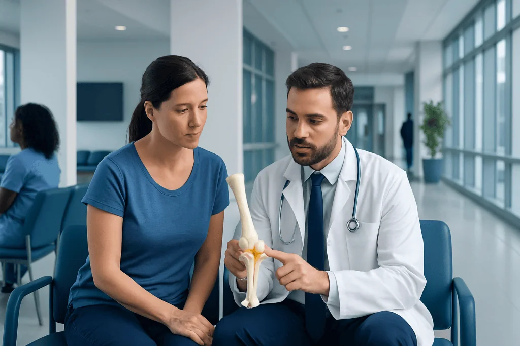
Osteochondritis dissecans usually affects the capitellum, a part of the upper arm bone that meets the radius at the elbow. Surgery is generally considered for unstable lesions, loose fragments, mechanical symptoms, or symptoms that do not improve with rest and rehabilitation.
Key Takeaways
- Osteochondritis dissecans usually affects the capitellum, a part of the upper arm bone that meets the radius at the elbow.
- Surgery is generally considered for unstable lesions, loose fragments, mechanical symptoms, or symptoms that do not improve with rest and rehabilitation.
- Arthroscopy can allow surgeons to assess the joint and perform drilling, fixation, fragment removal, or cartilage-restoration procedures through small incisions.
- Recovery often involves a period of protected movement followed by physiotherapy; return to throwing or impact sport can take several months.
- Results depend on lesion stage, cartilage health, treatment type, rehabilitation, and avoiding an early return to high-load activity.
Osteochondritis dissecans elbow surgery may be recommended when a damaged area of cartilage and underlying bone is unstable, detached, or continues to cause pain and loss of function despite non-surgical care. The procedure is tailored to the lesion’s size, location, stability, and the person’s age, activity goals, and elbow condition.
Overview: what osteochondritis dissecans elbow surgery does
Osteochondritis dissecans elbow surgery treats an area where cartilage and the bone beneath it have been damaged, most often on the capitellum at the outer side of the elbow. The aim is to protect or restore the joint surface, relieve symptoms such as catching or pain, and help preserve elbow movement for daily activities and sport.
Not every person with elbow osteochondritis dissecans needs an operation. Early, stable lesions may heal with stopping aggravating activity, a structured rehabilitation plan, and follow-up imaging. Surgery is more often considered when the fragment is unstable or detached, when there is a loose body in the joint, or when pain and restricted function continue despite appropriate conservative care.
The best procedure differs from person to person. An orthopedic surgeon reviews the lesion’s stability, size, depth, cartilage condition, skeletal maturity, sport demands, and imaging findings before recommending a plan. For broader information about the condition itself, see osteochondritis dissecans.
Symptoms, causes and who may be a candidate
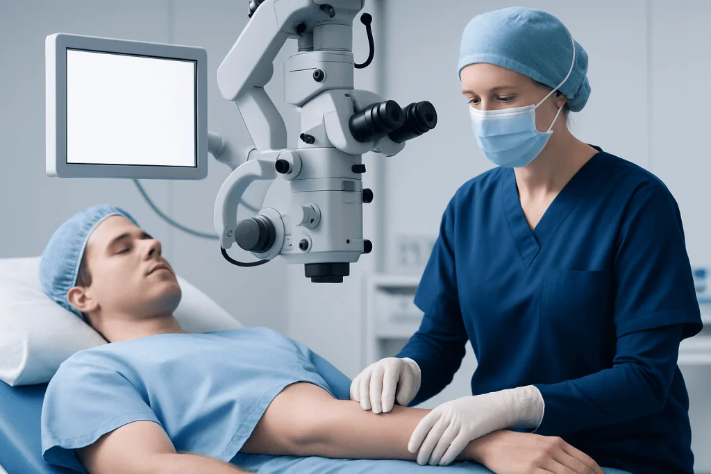
Elbow osteochondritis dissecans commonly develops in adolescents and young adults who repeatedly load the elbow, including overhead athletes and gymnasts. Repetitive compressive and twisting forces between the radius and capitellum are thought to contribute. Some people may also have individual differences in bone blood supply, joint anatomy, growth, or healing capacity.
Symptoms can begin gradually. They may include pain at the outer elbow during activity, swelling after use, stiffness, reduced ability to fully straighten the arm, clicking, catching, locking, or a sense that the elbow is giving way. Mechanical symptoms such as locking can suggest an unstable fragment or loose body and deserve timely medical assessment.
A person may be a surgical candidate when imaging shows an unstable lesion, a detached osteochondral fragment, or loose pieces within the joint. Surgery may also be discussed if a stable lesion has not improved after a period of activity modification and supervised rehabilitation. The decision is individualized; imaging findings alone do not always determine whether an operation is needed.
How osteochondritis dissecans elbow surgery works
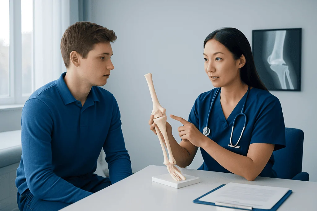
The surgical strategy is chosen to address the condition of both cartilage and underlying bone. In a stable lesion with healthy overlying cartilage, drilling may be used to encourage blood flow and healing in the bone beneath the surface. This may be performed from the back of the bone or through the joint, depending on the lesion and the surgeon’s approach.
If a fragment is partly or fully detached but still repairable, the surgeon may reposition and fix it with small implants. When damaged tissue cannot be repaired, treatment may involve removing unstable material, smoothing the defect, stimulating repair tissue with techniques such as microfracture, or using a cartilage-and-bone graft in selected larger defects.
Many procedures are carried out with elbow arthroscopy, using a camera and fine instruments inserted through small incisions. In some cases, an open incision is needed to safely access, fix, or reconstruct the lesion. The surgeon explains why a particular method is suitable, including how it may influence rehabilitation and return to sport.
Step-by-step: what happens during the procedure
Before surgery, the patient typically has a clinical examination and imaging such as X-rays and magnetic resonance imaging. The care team reviews medical history, medicines, allergies, and anesthesia considerations. The surgeon also discusses the likely procedure, possible alternatives, expected restrictions, and the possibility that the final surgical plan may be adjusted after directly examining the lesion.
On the day of surgery, anesthesia is provided so the patient does not feel the operation. The arm is positioned carefully, and the surgeon examines the elbow through arthroscopy or an open approach. They assess cartilage quality, the stability of the osteochondral fragment, the surrounding joint surface, and whether loose bodies are present.
The necessary treatment is then performed, such as drilling, fixation, removal of loose fragments, microfracture, or grafting. The incisions are closed and covered with dressings. A sling, splint, or brace may be used initially, particularly after fixation or grafting, to protect healing tissues while allowing the rehabilitation plan to begin at the appropriate time.
Recovery timeline and rehabilitation
Recovery begins with protecting the surgical site, controlling swelling, and following the surgeon’s instructions about dressing care, lifting, driving, work, and movement. The first days may involve discomfort, bruising, and stiffness. Pain relief is prescribed or recommended individually, and keeping the hand moving as advised can help limit swelling.
Early rehabilitation often focuses on gradually restoring comfortable elbow movement while protecting any repaired or grafted area. Physiotherapy may later add strengthening for the forearm, upper arm, shoulder blade, and trunk. A progressive program is important because the elbow works as part of the whole throwing and lifting chain.
Timelines vary considerably. Simple arthroscopic procedures may allow earlier motion and functional recovery, while fixation or cartilage-restoration procedures usually need more protection. Many people resume everyday light activities over several weeks, but return to repetitive overhead sport, gymnastics, heavy lifting, or throwing is often measured in months. Clearance should be based on healing, strength, movement, symptoms, and sport-specific testing rather than a date alone.
Rehabilitation after elbow surgery may be coordinated through physical therapy and rehabilitation to help restore safe movement and progressively rebuild strength. Following restrictions carefully is an important part of protecting the repair.
Is elbow surgery hard to recover from?
Elbow surgery can be challenging to recover from because the elbow may become stiff after injury or immobilization, and it is used frequently for everyday tasks. However, a structured plan that balances protection with guided movement can support recovery. The difficulty depends on the type of surgery, the size and stage of the lesion, whether fixation or grafting was required, and the person’s activity needs.
People who play throwing sports or gymnastics often require a longer and more carefully staged return than those whose goals are comfortable daily activities. Rehabilitation is not simply about reducing pain; it also addresses range of motion, strength, coordination, and tolerance of gradual loading.
Contact the surgical team if pain is worsening rather than settling, the hand becomes unusually swollen or numb, the wound develops increasing redness or drainage, or fever occurs. These symptoms do not always mean a serious problem, but they should be assessed promptly.
How long does it take to recover from osteochondritis dissecans surgery?
Recovery from osteochondritis dissecans surgery is individualized. For arthroscopic assessment or limited treatment, daily function may improve over weeks, although full strength and confidence can take longer. Procedures intended to heal or reconstruct cartilage and bone generally require a more cautious recovery because the treated area needs time to become secure.
Many patients follow a phased program over several months. Early phases protect the elbow and restore motion; later phases build strength and introduce task-specific activities. Athletes may need several additional months before a graduated throwing, tumbling, or high-load program is considered safe.
Follow-up visits and, when appropriate, repeat imaging help the surgeon evaluate healing. Returning too quickly to the activity that contributed to the injury can increase the chance of ongoing symptoms or damage, so medical clearance is important even if the elbow begins to feel better.
Benefits, risks and when to seek medical care
Potential benefits of surgery include removing painful loose bodies, reducing catching or locking, improving movement, and addressing an unstable or damaged joint surface. Surgery may also help protect longer-term elbow function, although no procedure can guarantee complete restoration of normal cartilage or a return to a previous level of sport.
Possible risks include infection, bleeding, nerve or blood-vessel injury, stiffness, persistent pain, incomplete healing, failure of fixation or graft tissue, blood clots, anesthesia-related complications, and a need for further treatment. An orthopedic surgeon can explain which risks are most relevant to the proposed technique and the person’s health.
Medical care should be sought for persistent outer-elbow pain, swelling, loss of motion, catching, or locking, especially in a young athlete who repeatedly throws, lifts, or bears weight through the arms. Urgent assessment is appropriate after an injury with deformity, severe pain, loss of sensation, a cold or pale hand, or inability to move the fingers normally.
Acibadem International’s multidisciplinary specialists and JCI-accredited hospitals assess and treat elbow conditions for international patients, with care plans guided by orthopedic evaluation, imaging, surgery when appropriate, and rehabilitation.
Frequently asked questions
How painful is osteochondritis dissecans?
Pain varies with the lesion’s size, stability, and activity level. Early elbow osteochondritis dissecans may cause a mild ache only during throwing, lifting, or weight-bearing through the arm, while an unstable fragment can cause sharper pain, swelling, catching, or locking. Persistent pain should be evaluated rather than pushed through.
What happens during stage 4 of osteochondritis dissecans?
Stage 4 generally describes an advanced lesion in which the osteochondral fragment has become detached, creating a loose body in the joint. Symptoms may include catching, locking, swelling, and loss of motion. Surgery is commonly considered to remove or repair the fragment and address the remaining cartilage-and-bone defect, depending on its condition.
Can elbow osteochondritis dissecans heal without surgery?
Some early, stable lesions can improve with stopping painful activities, temporary activity modification, rehabilitation, and regular follow-up. Healing is more likely when the lesion is stable and identified early, particularly in younger patients. Unstable lesions, loose bodies, and symptoms that continue despite non-surgical care are more likely to need surgical discussion.
When can a person return to sports after elbow OCD surgery?
Return to sport depends on the procedure, healing progress, elbow motion, strength, and the sport’s demands. Low-load activities may resume earlier than throwing, gymnastics, or heavy resistance exercise. The surgeon and rehabilitation team should provide clearance after a graded return plan rather than relying only on time since surgery.
Will elbow osteochondritis dissecans lead to arthritis?
A damaged joint surface can increase the risk of later degenerative changes, especially if an unstable lesion remains untreated or if substantial cartilage loss has occurred. Early assessment and appropriate management aim to preserve joint function, but they cannot eliminate all future risk. Ongoing symptoms after treatment should be reviewed by an orthopedic clinician.
What should patients avoid after osteochondritis dissecans elbow surgery?
Patients should avoid lifting, throwing, pushing, pulling, and weight-bearing through the arm until their surgeon permits progression. They should also avoid returning to sport because pain has temporarily improved, since tissue healing may still be incomplete. Individual restrictions depend on whether the procedure involved drilling, fixation, microfracture, or grafting.
References
- American Academy of Orthopaedic Surgeons
- American Orthopaedic Society for Sports Medicine
- OrthoInfo
- National Institute of Arthritis and Musculoskeletal and Skin Diseases
This article is for general information only and is not a substitute for professional medical advice. Please consult a qualified doctor about your individual situation.
Elbow Surgery in Turkey — costs, top hospitals & a free quote
JCI-accredited · board-certified surgeons · reply within 24h
Add us as a Preferred Source to see more of our trusted health content across Google Search, AI Overviews and Discover.

Orthopedics & Spine
Care for bones, joints and the musculoskeletal system, including joint replacement, sports injuries and trauma.
125 specialists in this unit