

Bone Anchored Hearing Aid MRI Safety: Preparation, Procedure and Results

Never enter an MRI scanner room wearing a bone-anchored hearing aid sound processor. MRI eligibility depends on the exact implant system, including whether it contains a magnet.
Key Takeaways
- Never enter an MRI scanner room wearing a bone-anchored hearing aid sound processor.
- MRI eligibility depends on the exact implant system, including whether it contains a magnet.
- The implant card, device model and manufacturer MRI instructions should be reviewed before the scan is booked.
- Bone-anchored hearing devices and cochlear implants are different systems with different MRI precautions.
- BAHA surgery is commonly performed as a planned outpatient procedure, with pain usually manageable using prescribed or over-the-counter pain relief as advised.
- New pain, redness, drainage or swelling around an implant site should be assessed by a healthcare professional.
Bone anchored hearing aid MRI safety is usually manageable when the radiology team follows the specific instructions for the implanted device. Most external sound processors must be removed before scanning, but MRI conditions for the implanted component vary by model, magnet type and scanner strength.
Bone Anchored Hearing Aid MRI Safety: The Essential Answer
Bone anchored hearing aid MRI safety depends on the exact device a person has. In most cases, the external sound processor must be removed before entering the MRI environment, while the implanted portion may be scanned only under the manufacturer’s stated conditions. These conditions can include limits on MRI strength, body position, scanning region and the use of a protective bandage or magnet-related precautions.
A person should not assume that all bone-anchored hearing devices have the same MRI guidance. Before an MRI is scheduled, they should tell the referring clinician and imaging department about the implant, bring their implant identification card if available, and ask the hearing implant team to confirm the model-specific instructions. This preparation helps the radiology team plan a scan that is both safe and diagnostically useful.
Even when MRI is permitted, the implant may create image distortion, known as artifact, near the ear and skull. This does not necessarily prevent scanning other parts of the body, but it can affect images of nearby structures. The radiologist can advise whether MRI, CT or another imaging method is most appropriate for the clinical question.
How a Bone-Anchored Hearing Aid Works and Who May Benefit
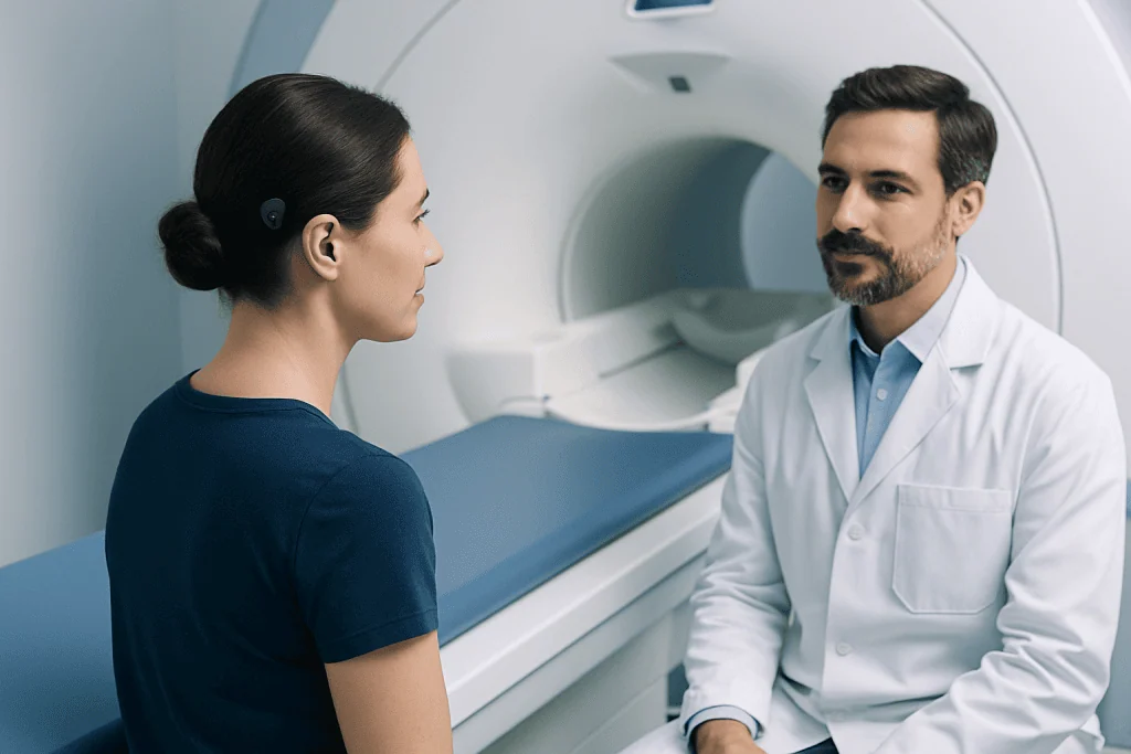
A bone-anchored hearing aid, often called a BAHA, is a hearing system that sends sound vibrations through the skull bone to the inner ear. Rather than relying entirely on the ear canal and middle ear, it uses bone conduction. The sound processor captures sound and converts it into vibrations that reach the cochlea, the hearing organ in the inner ear.
Some systems connect to an implant placed in the bone behind the ear. Others are held against the skin by a magnetic connection, while certain non-surgical options use a headband or adhesive attachment. The design matters because systems with implanted magnets may have additional MRI requirements compared with systems that use a small abutment or have no surgically implanted magnet.
Clinicians may consider bone-conduction systems for people with conductive or mixed hearing loss, chronic ear canal problems, certain congenital ear differences, or single-sided deafness when the opposite ear has usable hearing. Assessment generally includes a hearing test, examination of the ears and discussion of communication needs, anatomy, medical history and expectations. Bone-anchored hearing aid treatment may involve evaluation by ENT specialists, audiologists and, when needed, imaging professionals.
Can You Get an MRI With a Bone-Anchored Hearing Aid?

Many people can have an MRI with a bone-anchored hearing aid, but the answer is device-specific. The external processor is generally MR Unsafe and must be removed before the person enters the MRI scanner room. The implanted fixture, abutment or magnetic component may be MRI conditional, meaning scanning is permitted only when the manufacturer’s detailed conditions are met.
Important details include the brand and model, whether an implant contains a magnet, the magnetic field strength of the MRI scanner, and the area of the body being scanned. Some magnet-containing devices require special precautions, temporary magnet removal or may have restrictions at certain MRI strengths. The implant surgeon, audiology team and radiology department should confirm this information rather than relying on memory or general internet advice.
At the MRI appointment, the patient should report any unusual warmth, pressure, pain or pulling sensation immediately through the scanner communication system. The MRI team will follow a safety checklist, remove the external equipment, position the patient appropriately and use the approved scan settings. If the required device information cannot be verified, the scan may be postponed while the team obtains it or considers a suitable alternative.
BAHA Procedure: Steps, Recovery, Benefits and Risks
When a surgically implanted bone-anchored system is appropriate, the procedure is planned after hearing and medical assessments. It is commonly performed under local anesthesia with sedation or under general anesthesia, depending on the individual, the surgical approach and clinical preferences. The surgeon makes a small incision behind the ear and places a titanium implant in the skull bone; the exact technique differs for abutment-based and magnet-based systems.
In many cases, the implant is allowed time to integrate with the bone before the external processor is fitted and programmed. This healing period varies by device and individual circumstances. Follow-up visits allow the team to check the skin and implant site, manage wound care, and adjust the processor so that sound is comfortable and useful.
Recovery commonly involves short-term tenderness, swelling or bruising around the surgical site. People are usually given guidance on keeping the area clean, protecting it while healing and gradually returning to daily activities. The care team will advise when hair washing, exercise, swimming and use of the processor are appropriate.
Potential benefits include improved access to sound without blocking the ear canal, and better hearing in selected patterns of hearing loss. Risks can include bleeding, infection, skin irritation, delayed healing, numbness, discomfort from pressure, implant failure or a need for revision surgery. An ENT surgeon can explain how these issues apply to the particular device and person.
Is a BAHA Considered a Hearing Aid?
Yes. A BAHA is considered a hearing device or hearing aid, but it works differently from a conventional air-conduction hearing aid. Standard hearing aids amplify sound and direct it through the ear canal, eardrum and middle ear. A BAHA uses bone conduction to deliver sound vibrations to the inner ear.
Because it bypasses part of the outer and middle ear, a bone-anchored device can be helpful when sound cannot travel effectively through those structures or when wearing an in-ear device is not suitable. It does not restore hearing to normal, and benefit varies according to the type and degree of hearing loss, the health of the cochlea and the person’s listening environments.
Bone-anchored devices should also not be confused with cochlear implants. A cochlear implant electronically stimulates the auditory nerve inside the cochlea and is intended for people with more severe sensorineural hearing loss who meet specific criteria. Cochlear implant treatment involves different surgery, programming and MRI safety guidance.
How Painful Is BAHA Surgery?
During BAHA surgery, anesthesia is used so that the person should not feel surgical pain. Afterward, discomfort is often described as soreness, pressure or tenderness behind the ear, especially during the first several days. The level of discomfort differs between individuals and can also depend on the surgical technique and whether a magnet-based or abutment-based system is used.
The surgical team provides individualized instructions for pain relief and wound care. Many people can manage postoperative discomfort with the medicines recommended by their clinician, rest and careful protection of the incision. Increasing pain rather than gradual improvement, or pain accompanied by fever, spreading redness, discharge or marked swelling, should be reported promptly.
Longer-term discomfort is not expected for most people, although skin sensitivity, pressure-related discomfort or irritation can occur. Follow-up with the surgeon and audiologist is important because adjustments to the processor, magnet strength or skin-care routine may improve comfort.
What Happens If You Get an MRI With a Cochlear Implant?
A cochlear implant requires its own MRI safety plan. The external sound processor must be removed before MRI, and the implanted device may be MRI conditional under specific manufacturer instructions. Depending on the implant model and MRI strength, precautions may include a tight head wrap, an MRI kit, temporary removal of the internal magnet by a surgeon, or choosing another imaging method.
If MRI is performed without following the correct instructions, a magnet-containing implant may cause pain or pressure and, in some circumstances, magnet movement or damage to the device. MRI can also create substantial artifact around the implant, limiting images of the nearby brain, ear or facial structures. For these reasons, the imaging center must identify the exact implant before the scan begins.
Someone with a cochlear implant who has already had an MRI and develops persistent pain, swelling, reduced device performance or difficulty attaching the external processor should contact their implant clinic. Assessment may include examination, device testing and imaging if needed. Hearing loss should be evaluated with an individualized plan, particularly when a hearing device is not functioning as expected.
When to Seek Medical Care
Urgent medical assessment is appropriate for severe pain, rapidly increasing swelling, significant bleeding, fever with worsening redness around a recent implant site, pus-like drainage, or new facial weakness. A person who experiences these symptoms after surgery or after an MRI should contact their surgical team or seek urgent care according to the severity of symptoms.
A routine but timely review is sensible for persistent skin irritation, soreness under a magnet, repeated crusting around an abutment, changes in hearing benefit, or a processor that no longer connects securely. Before any MRI, CT scan, surgery or other procedure involving medical equipment, the person should also disclose their hearing implant to the healthcare team.
Acibadem International’s multidisciplinary ENT, audiology and radiology specialists in JCI-accredited hospitals can assess hearing implant needs and coordinate MRI planning for international patients. The safest next step is always to obtain device-specific guidance from the implant team and the radiology service performing the scan.
Frequently asked questions
Do I need to remove my BAHA before an MRI?
The external sound processor should generally be removed before entering the MRI scanner room. Whether the implanted part can remain in place depends on its exact model and the manufacturer’s MRI conditions. The radiology department should verify these details before the scan.
Can a BAHA implant affect MRI images?
Yes. Metal and magnets can cause image artifact, which may obscure structures near the implant, particularly around the ear and side of the head. The effect on a scan of another body area may be limited, but the radiologist will consider this when planning imaging.
Should I bring my implant card to an MRI appointment?
Yes. The implant identification card can help the MRI team identify the device model and locate the correct safety instructions. If the card is unavailable, the implant clinic or manufacturer may be able to provide the necessary information.
Can an MRI damage a bone-anchored hearing aid?
An MRI may create risks if a device is scanned outside its approved conditions, especially if it has an implanted magnet. Following the device-specific MRI instructions and removing the external processor helps reduce avoidable risk. A healthcare team should never proceed without confirming compatibility.
How long does recovery take after BAHA surgery?
Initial wound healing often takes several weeks, while the implant may need additional time to integrate with the bone before the processor is fitted. The exact timeline varies by device, surgical technique and individual healing. The surgeon will provide a personalized schedule for wound checks and activation.
Is a bone-anchored hearing aid the same as a cochlear implant?
No. A bone-anchored hearing aid sends sound vibrations through bone to a functioning inner ear, whereas a cochlear implant directly stimulates the hearing nerve through electrodes placed in the cochlea. They are used for different hearing needs and have different surgical and MRI considerations.
References
- U.S. Food and Drug Administration
- American College of Radiology
- National Institute on Deafness and Other Communication Disorders
- American Academy of Otolaryngology–Head and Neck Surgery
- Device manufacturer MRI safety instructions
This article is for general information only and is not a substitute for professional medical advice. Please consult a qualified doctor about your individual situation.
Explore treatments in Turkey — costs, top hospitals & a free quote
JCI-accredited · board-certified surgeons · reply within 24h
Add us as a Preferred Source to see more of our trusted health content across Google Search, AI Overviews and Discover.

More from the Health Library



Related Specialists

Dr. Muhammet Mustafa Yıldız
Emergency Service
Dr. Doğuş Durmuş
General Surgery
Dr. Ahmad Nejat Ghaffarı
Infectious Diseases & Clinical Microbiology
Dr. Aydın Halefoğlu
Anesthesiology