Sinus CT Scan Cancer: Preparation, Procedure and Results
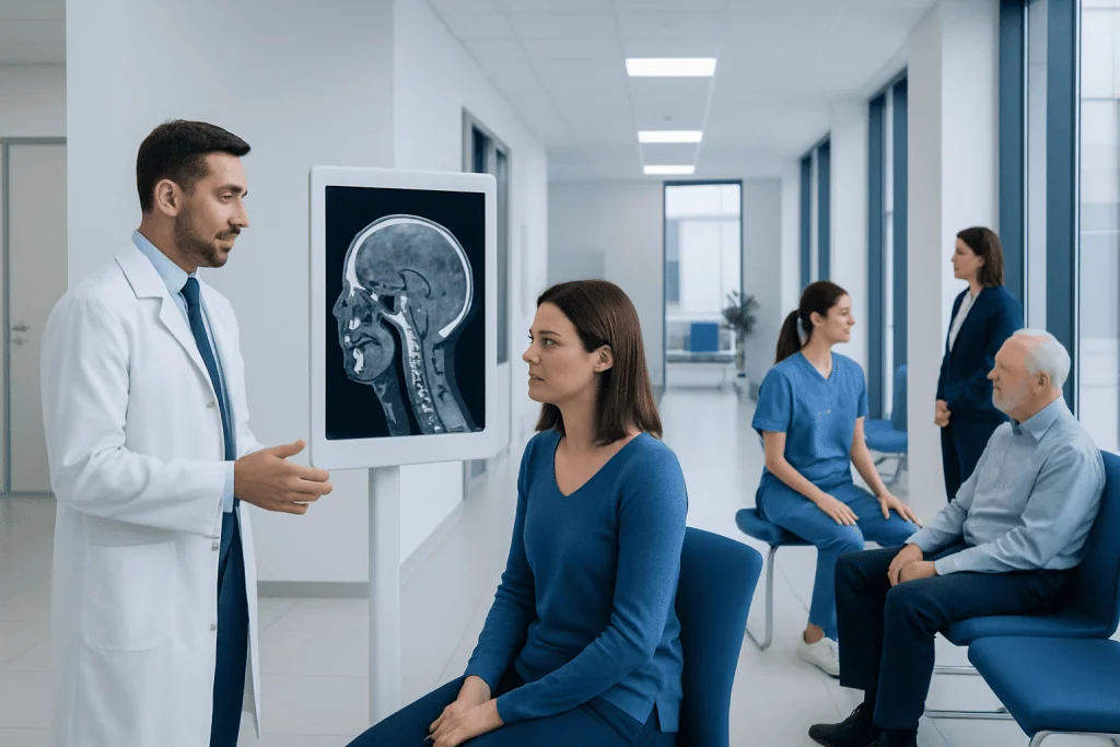
A sinus CT scan is a fast imaging test that helps assess the nasal cavity, sinuses, nearby bones, and surrounding structures. CT can show findings that raise concern for cancer, but a tissue biopsy is needed to make a diagnosis.
Key Takeaways
- A sinus CT scan is a fast imaging test that helps assess the nasal cavity, sinuses, nearby bones, and surrounding structures.
- CT can show findings that raise concern for cancer, but a tissue biopsy is needed to make a diagnosis.
- Most people need little preparation, although contrast dye, pregnancy, kidney disease, and allergies should be discussed beforehand.
- Results may be available within hours to several days, depending on the imaging center and whether urgent review is needed.
- Persistent one-sided nasal blockage, repeated nosebleeds, facial numbness, or eye symptoms should be assessed by a clinician.
A sinus CT scan creates detailed cross-sectional images of the nasal cavity and paranasal sinuses. It can reveal a mass, bone changes, or sinus blockage that may require further testing for cancer, but it cannot confirm cancer on its own; confirmation usually requires a biopsy.
Sinus CT Scan Cancer: What the Scan Can Show
A sinus computed tomography (CT) scan uses X-rays and computer processing to produce detailed images of the nasal passages, paranasal sinuses, facial bones, eye sockets, and nearby soft tissues. It is commonly requested for chronic sinus symptoms, facial injury, surgical planning, or an unexplained abnormality found during an ear, nose, and throat examination.
When cancer is a concern, CT helps clinicians look for a mass, persistent one-sided sinus opacification, destruction or remodeling of bone, extension toward the eye socket or skull base, and enlarged lymph nodes in the neck. These findings can help determine whether more investigation is appropriate and can assist with planning subsequent imaging or treatment.
Many CT findings are caused by non-cancerous conditions, including sinus inflammation, nasal polyps, fungal sinus disease, benign growths, dental disease, or prior injury. A scan therefore provides important anatomical information, but it does not identify the exact cell type of a suspicious area. If needed, an ENT specialist may arrange nasal endoscopy and a biopsy to establish a diagnosis.
How a Sinus CT Scan Works and Who May Need One
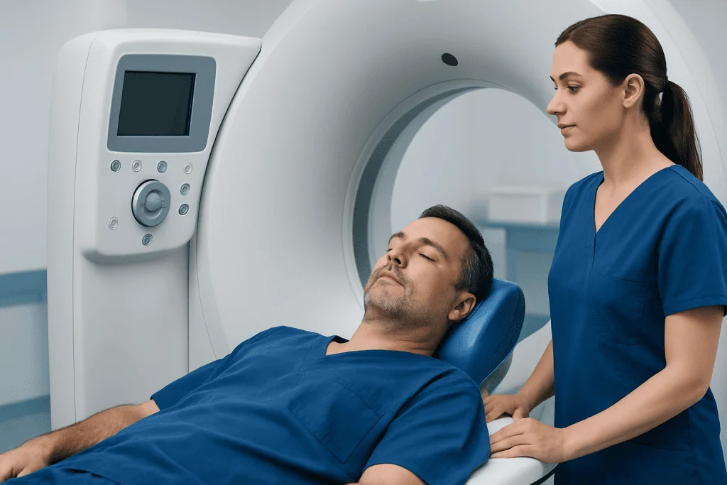
During CT imaging, an X-ray tube rotates around the head while detectors measure how X-rays pass through tissues. A computer then constructs thin image slices, allowing the radiologist to view the sinuses in multiple planes. Compared with a standard X-ray, CT provides much greater detail of bone and air-filled sinus spaces.
A clinician may recommend a sinus CT scan when symptoms have not improved as expected, when there is a suspected structural blockage, or when examination suggests an unusual lesion. It may also be used after a biopsy-proven nasal or sinus cancer diagnosis to assess local extent. Magnetic resonance imaging (MRI) may be added when clinicians need more detail about soft tissues, nerves, the brain, or the eye area.
CT is not routinely needed for ordinary short-term colds or uncomplicated acute sinus symptoms. The decision is based on the person’s symptoms, clinical examination, medical history, and whether imaging results are likely to change care. For patients being evaluated for a possible tumor, care may involve ENT, radiology, pathology, oncology, ophthalmology, and skull-base surgery specialists.
What Should You Not Do Before a Sinus CT Scan?

For a sinus CT scan performed without contrast dye, people can usually eat, drink, and take their usual medicines unless the imaging department provides different instructions. It is helpful to remove metal items around the head and face, such as earrings, hairpins, glasses, removable dental appliances, and hearing aids, because they can create image artifacts.
If contrast dye is planned, the imaging team may ask the patient not to eat for a short period before the examination. Instructions vary by facility and by the person’s health needs, so the appointment guidance should be followed rather than making unnecessary dietary changes independently.
Before the scan, patients should tell the team about pregnancy or a possible pregnancy, kidney disease, diabetes, thyroid problems, previous reactions to iodinated contrast, asthma, and all medicines or supplements. They should not stop prescribed medication unless the clinician specifically advises it. Bringing earlier scans or reports can also help the radiologist compare changes over time.
Step-by-Step: What Happens During the Procedure
On arrival, a radiology staff member confirms the patient’s identity, clinical history, and any relevant safety information. The person may change into a gown and will be asked to lie on their back on a narrow, motorized table. The head is positioned carefully, sometimes with supports to help keep it still.
The table moves through the short, doughnut-shaped CT scanner while images are acquired. The machine can make whirring sounds, but it does not touch the patient and the scan itself is painless. A sinus CT commonly takes only a few minutes, although the full appointment may take longer for registration, positioning, and contrast preparation when applicable.
If intravenous contrast is used, a small cannula is placed in a vein. Some people notice a brief warm sensation, a metallic taste, or a feeling of needing to urinate; these effects generally pass quickly. Staff communicate throughout the scan, and the patient should remain still and follow any simple instructions to ensure clear images.
Would a CT Scan of Sinuses Show Cancer?
A CT scan of the sinuses can show abnormalities that may be consistent with cancer, such as an irregular soft-tissue mass, invasion of nearby structures, bone erosion, or a one-sided sinus blockage that does not look like routine inflammation. It is valuable for showing where an abnormality is located and how it relates to surrounding bones and spaces.
However, CT cannot reliably prove that an abnormality is cancer. Inflammation, infection, polyps, benign tumors, and other conditions can sometimes have overlapping appearances. A radiology report may describe a finding as suspicious or recommend further assessment, but diagnosis requires a sample of tissue examined by a pathologist.
If CT findings or symptoms require further evaluation, an ENT specialist may perform nasal endoscopy and arrange a biopsy. Additional imaging, often MRI and sometimes PET-CT, may be used after diagnosis or when there is a strong clinical concern. This structured approach helps clinicians distinguish cancer from other causes and plan care appropriately.
Recovery, Benefits and Risks of Sinus CT Imaging
There is usually no recovery period after a sinus CT scan without contrast. People can return to normal activities immediately. After contrast, most people can also resume usual activities, and drinking fluids may be encouraged unless a clinician has advised fluid restriction for another health reason.
The main benefit is that CT offers rapid, detailed information about sinus anatomy and possible disease. It can clarify the cause of persistent symptoms, help guide endoscopic assessment or biopsy, and support treatment planning. In people with a confirmed malignancy, imaging is one part of understanding the location and local spread of the disease.
CT uses ionizing radiation, so clinicians aim to use imaging only when it is medically justified and to keep exposure as low as reasonably achievable. Contrast reactions are uncommon and are often mild, but serious reactions can occur rarely. Contrast can also be relevant for people with significant kidney impairment. The radiology team reviews these issues beforehand and can adapt the examination when appropriate.
What Are the Red Flags for Nasal Cancer?
Nasal cavity and paranasal sinus cancers are uncommon, and their early symptoms may resemble allergies or sinusitis. Symptoms that are persistent, progressive, or mainly on one side of the nose deserve medical assessment, particularly when they do not improve with appropriate treatment.
- One-sided nasal blockage that persists or worsens
- Repeated unexplained nosebleeds or blood-stained nasal discharge
- Facial pain, pressure, swelling, numbness, or new dental symptoms
- A visible nasal lump, persistent crusting, or a sore that does not heal
- Double vision, reduced vision, eye prominence, or swelling around an eye
- Persistent headache, hearing changes, or a new lump in the neck
These symptoms do not necessarily mean cancer, and common conditions remain more likely for many people. Nonetheless, prompt assessment by a qualified clinician is important because persistent one-sided symptoms may need examination, endoscopy, or imaging. Information about related diagnoses is available in nasal cancer.
How Long Does It Take to Get Results From a Sinus CT Scan?
The images are produced immediately, but the formal report is prepared after a radiologist reviews the scan. In non-urgent settings, results are often sent to the referring clinician within one to several working days. Timing can differ according to the hospital, the complexity of the scan, and whether prior images need comparison.
If imaging is performed urgently, the radiologist may communicate important findings to the treating team sooner. Patients should ask at the appointment how and when they will receive results, and should arrange follow-up with the clinician who requested the scan. A report may recommend further imaging, ENT review, or biopsy; this wording does not by itself confirm cancer.
When a suspicious lesion is identified, referral pathways are designed to coordinate the next steps. Acibadem International’s multidisciplinary specialists and JCI-accredited hospitals can assess sinus and nasal conditions for international patients, including imaging review, ENT evaluation, biopsy, and treatment planning when needed.
Frequently asked questions
Is a sinus CT scan painful?
A sinus CT scan is painless. The person needs to lie still while the table moves through the scanner, which may feel unfamiliar but usually takes only a few minutes. If contrast is used, there may be a brief warm sensation or metallic taste.
Do I need contrast dye for a sinus CT scan?
Many sinus CT scans are performed without contrast because CT shows sinus anatomy and facial bones well without it. Contrast may be used when a clinician needs more information about a mass, blood vessels, inflammation, or spread into nearby soft tissues. The radiology team will explain the planned approach.
Can sinusitis look like cancer on a CT scan?
Yes. Chronic inflammation, infection, polyps, and fungal sinus disease can sometimes create CT appearances that overlap with those of a tumor. Radiologists assess the pattern of findings, but endoscopy, follow-up imaging, or biopsy may be needed to clarify the cause.
Should I be worried if my report says sinus opacification?
Sinus opacification means that a sinus space that normally contains air appears filled by fluid, swollen lining, mucus, or soft tissue. It is commonly associated with inflammation or infection and is not a cancer diagnosis. The meaning depends on the location, pattern, symptoms, and other features described in the report.
When should someone seek medical care for sinus symptoms?
Medical care is advisable for symptoms that persist despite treatment, recur frequently, or are mainly on one side. Urgent assessment is important for eye swelling, vision changes, severe facial swelling, confusion, a severe worsening headache, or significant persistent bleeding. A clinician can determine whether ENT review or imaging is needed.
What happens if a sinus CT scan finds a suspicious mass?
The referring clinician will usually arrange assessment by an ENT specialist. The next steps may include nasal endoscopy, MRI, and a biopsy to identify the tissue type. If cancer is confirmed, a multidisciplinary team discusses individualized treatment options based on the tumor’s type, location, and extent.
References
- American Cancer Society
- National Cancer Institute
- Radiological Society of North America
- American College of Radiology
- NHS
This article is for general information only and is not a substitute for professional medical advice. Please consult a qualified doctor about your individual situation.
Oncology care in Turkey — second opinion and treatment plan
JCI-accredited · board-certified surgeons · reply within 24h
Add us as a Preferred Source to see more of our trusted health content across Google Search, AI Overviews and Discover.