

Robotic Assisted Mitral Valve Surgery: Procedure, Recovery and Results

Robotic technology allows surgeons to operate through small chest incisions while controlling precise instruments from a console. Mitral valve repair is generally preferred over replacement when a durable repair is feasible.
Key Takeaways
- Robotic technology allows surgeons to operate through small chest incisions while controlling precise instruments from a console.
- Mitral valve repair is generally preferred over replacement when a durable repair is feasible.
- Not every patient or valve condition is appropriate for a robotic approach; anatomy, previous surgery, vascular health and other conditions matter.
- Recovery often begins with walking and breathing exercises in hospital, followed by gradual return to activity over several weeks.
- Possible complications include bleeding, infection, irregular heart rhythm, stroke, kidney problems and the need to convert to an open operation.
Robotic assisted mitral valve surgery is a minimally invasive approach used to repair, and less commonly replace, a diseased mitral valve through small incisions between the ribs. It may offer a smaller incision and a different recovery experience than traditional open-heart surgery, but it is suitable only for selected patients after detailed heart-team assessment.
Overview: What Is Robotic Assisted Mitral Valve Surgery?
Robotic assisted mitral valve surgery is a form of minimally invasive heart surgery used to treat problems affecting the mitral valve, which sits between the left upper and lower chambers of the heart. The surgeon works through several small openings on the right side of the chest rather than making a long incision through the breastbone. A surgical robot does not act independently: the surgeon remains in full control of the camera and instruments throughout the operation.
The procedure is most often performed to repair a leaking mitral valve, known as mitral regurgitation. In selected circumstances, it may be used for mitral stenosis or for valve replacement when repair is not possible. The aim is to restore effective blood flow, relieve symptoms where present, and help protect heart function over time.
For suitable patients, robotic assisted mitral valve surgery can be an alternative to conventional sternotomy, in which the breastbone is divided. However, a small-incision approach is not automatically safer or better for every person. The best technique depends on the valve problem, overall health, imaging findings and the experience of the surgical team.
How the Procedure Works
The mitral valve has two leaflets that open to allow blood to move from the left atrium into the left ventricle and close to prevent backward flow. When the leaflets, supporting cords or valve ring do not work properly, blood may leak backward. This can cause the heart to work harder and may eventually lead to breathlessness, fatigue, abnormal heart rhythms or reduced heart function.
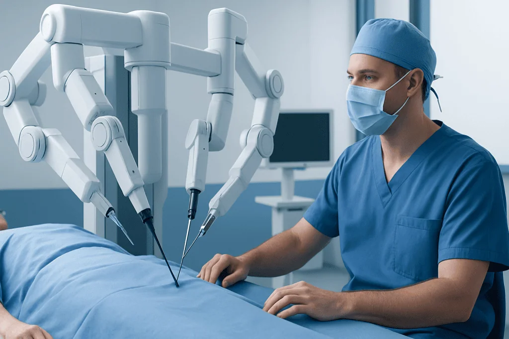
During robotic surgery, the surgeon uses a high-definition three-dimensional camera and wristed instruments introduced through small ports between the ribs. These instruments can make refined movements inside the chest. The surgeon controls them from a nearby console while an experienced bedside surgical team assists directly at the operating table.
Whenever possible, surgeons aim to preserve the patient’s own valve. Repair techniques may include reshaping part of a leaflet, repairing or replacing damaged supporting cords, closing a gap in the leaflets, or placing a supportive ring around the valve opening. If replacement is necessary, the choice between a mechanical and biological valve is individualized and includes discussion of long-term medication needs and valve durability.
Who May Be a Candidate?

Candidacy is determined by a multidisciplinary heart team, commonly involving cardiologists, cardiac surgeons, imaging specialists, anesthesiologists and other clinicians as needed. People with severe mitral regurgitation, particularly degenerative valve disease where a durable repair appears likely, may be considered for robotic repair. Some individuals are evaluated before symptoms become severe if the valve problem is affecting heart size, function or rhythm.
Detailed imaging is essential. Echocardiography assesses valve structure and blood flow, while transesophageal echocardiography may provide a closer view. CT imaging may be used to assess the chest and blood vessels, because access to the heart-lung machine through vessels in the groin is often part of the robotic approach. Coronary artery testing may also be required, depending on age and risk factors.
A robotic procedure may not be the best option for everyone. Previous surgery on the right chest, major disease of the blood vessels, severe lung disease, extensive coronary artery disease requiring bypass surgery, certain complex valve conditions, active infection or urgent instability can make another approach more appropriate. The assessment is designed to identify the safest operation, not simply the least visible incision.
Related conditions can influence treatment planning. For example, mitral valve regurgitation may coexist with atrial fibrillation, tricuspid valve disease or heart failure, and these issues may need to be addressed during the same treatment plan.
Step by Step: What Happens During Surgery?
Before surgery, the patient meets the anesthesia and surgical teams, reviews medications and receives instructions about fasting and medicines that may need to be paused. The operation is performed under general anesthesia, so the patient is asleep and does not feel pain. Monitoring lines are placed to closely track heart function, blood pressure, oxygen levels and other vital signs.
The surgical team makes several small incisions on the right side of the chest for the camera, robotic instruments and access ports. In many cases, the heart-lung machine temporarily takes over circulation and oxygenation while the surgeon works on the valve. Blood vessels in the groin may be used to connect to this machine.
The surgeon carefully examines the mitral valve and performs the planned repair or replacement. An echocardiogram performed during surgery checks that the valve is functioning well before the procedure is completed. If the repair is not satisfactory or a minimally invasive approach cannot be completed safely, the team may need to change the surgical plan, including conversion to a larger incision when necessary.
After the valve is repaired or replaced and the heart is functioning normally, the instruments are removed and the small incisions are closed. Drainage tubes and temporary pacing wires may be used for a short time. Surgery duration varies according to the valve problem, planned repair and whether other procedures are performed at the same time.
Recovery Timeline and Expected Results
After surgery, patients are monitored in an intensive care or high-dependency setting before moving to a regular cardiac ward. The breathing tube is usually removed when it is safe to do so, often within the first day. Pain control, breathing exercises, early sitting and walking, and prevention of blood clots are important parts of initial recovery.
Hospital stay varies based on the individual procedure, heart rhythm, mobility, pain control and any complications. Some people may leave hospital sooner than after a sternotomy, but recovery is not identical for every patient. Small incisions can reduce chest wall discomfort for some people, yet tiredness after heart surgery remains common and should be expected.
At home, patients generally increase walking and daily activity gradually. Driving, returning to work, lifting heavier objects and resuming exercise should follow the specific advice of the surgical team. Cardiac rehabilitation can provide supervised exercise, education and support for a safe return to everyday activities. Follow-up includes wound checks, heart rhythm assessment and echocardiography to evaluate valve function.
When a successful durable repair is achieved, it can improve symptoms and help preserve heart function. Outcomes depend on the cause and severity of valve disease, the condition of the heart before surgery, other health conditions and the quality of the repair. Long-term follow-up with a cardiologist remains important even when the patient feels well.
Benefits, Risks and Important Limitations
Potential benefits of robotic assisted mitral valve surgery include smaller chest incisions, avoidance of dividing the breastbone, less visible scarring and, for some appropriately selected patients, a shorter hospital stay or earlier return to normal movement. The enhanced view and instrument range can also support complex valve repair techniques in experienced hands. These potential advantages should be weighed against the needs of the individual patient.
All heart surgery carries risks. Possible complications include bleeding, infection, reactions to anesthesia, blood clots, stroke, kidney problems, lung complications, irregular heart rhythms, damage to nearby structures and problems with the repaired or replacement valve. Some patients require a blood transfusion, a pacemaker, another procedure or conversion to open surgery. Although serious complications are uncommon in many planned operations, they should be discussed clearly before consent.
Robotic surgery can require specialized equipment, careful preoperative planning and a trained team. The operation may take longer than a conventional approach in some circumstances, particularly during early team experience. A recommended open or minimally invasive non-robotic procedure does not mean that care is inferior; it may be the most appropriate route to a safe, durable result.
Self-care After Surgery and When to Seek Medical Care
Patients should follow their discharge plan closely, including instructions for medicines, wound care, activity, diet and follow-up visits. It is helpful to take short walks as advised, practice breathing exercises, avoid tobacco, and maintain a heart-healthy eating pattern. Family or friends may be useful for practical support during the first period at home, especially with shopping, transport and heavier household tasks.
The surgical team should be contacted promptly for a fever, increasing redness, swelling, drainage or opening at an incision, worsening pain, new palpitations, persistent nausea or difficulty taking prescribed medicines. Patients should also report increasing shortness of breath, rapid weight gain or swelling in the legs, as these may need medical assessment.
Emergency medical care is needed for severe or sudden chest pain, severe breathing difficulty, fainting, signs of stroke such as new facial drooping, weakness or trouble speaking, or uncontrolled bleeding. These symptoms do not always mean a surgical complication, but they require urgent evaluation.
Acibadem International’s multidisciplinary cardiac specialists and JCI-accredited hospitals assess and treat mitral valve disease for international patients, with care plans tailored to clinical findings and individual needs.
Frequently asked questions
Is robotic assisted mitral valve surgery safer than open-heart surgery?
Neither approach is universally safer for every patient. Robotic surgery may offer advantages for appropriately selected people, but its safety depends on the valve condition, overall health, anatomy and the expertise of the treating team. A conventional operation may be the safer and more durable choice in some situations.
How long does robotic mitral valve surgery take?
The length of surgery varies considerably depending on the type of valve problem, whether repair or replacement is needed, and whether another cardiac procedure is performed. Preparation, anesthesia and recovery-room care also add to the total time spent in the operating area. The surgical team can provide a more individualized estimate after reviewing imaging and the operative plan.
Will the mitral valve be repaired or replaced?
Repair is often preferred when the surgeon expects a durable result, especially for certain forms of degenerative mitral regurgitation. Replacement may be necessary if the valve is too damaged, calcified, infected or otherwise unsuitable for repair. The final decision is based on detailed imaging and findings during surgery.
How painful is recovery after robotic mitral valve surgery?
Pain and soreness are expected after any heart operation, but they can usually be managed with a tailored pain-control plan. Discomfort may be felt around the chest incisions, ribs, shoulder or groin access site. Patients should tell their care team if pain is not controlled, as effective pain management supports breathing, movement and recovery.
When can someone return to normal activities after surgery?
Recovery time varies with age, physical condition, the complexity of surgery and whether complications occur. Many people gradually increase walking and light daily activity over the first several weeks, while work, driving and exercise require individual medical clearance. Cardiac rehabilitation and follow-up visits help guide a safe return to usual activities.
Does a repaired mitral valve need lifelong follow-up?
Yes. Even after a successful repair, regular cardiology follow-up and periodic echocardiograms are important to check valve function, heart size and heart rhythm. The schedule depends on the original valve condition, repair type and the patient’s overall cardiac health.
References
- American Heart Association
- American College of Cardiology
- European Society of Cardiology
- Society of Thoracic Surgeons
- National Heart, Lung, and Blood Institute
This article is for general information only and is not a substitute for professional medical advice. Please consult a qualified doctor about your individual situation.
Valve Surgery in Turkey — costs, top hospitals & a free quote
JCI-accredited · board-certified surgeons · reply within 24h
Add us as a Preferred Source to see more of our trusted health content across Google Search, AI Overviews and Discover.
Check your numbers in seconds
BMI, calories, due date, blood pressure and 30+ more clinical calculators — free, instant, doctor-reviewed ranges.

More from the Health Library



Related Specialists

Dr. Duygu Yavuzer Karadeniz
Periodontolgy
Assoc. Prof. Dr. Sevgi Topal
Pediatric Intensive Care Unit
Prof. Dr. Burak Çıtamak
Urology
Hamdi Kandemir
Physical Medicine & Rehabilitation