

MRI Scan: How It Works, Safety, and What Results Can Show
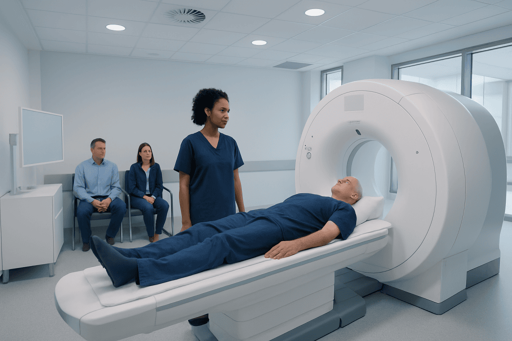
An MRI scan uses magnets and radio waves, not X-rays, to produce detailed images of the inside of the body. Before the scan, patients are carefully screened for implants, metal fragments, kidney problems, pregnancy, and other safety considerations.
Key Takeaways
- An MRI scan uses magnets and radio waves, not X-rays, to produce detailed images of the inside of the body.
- Before the scan, patients are carefully screened for implants, metal fragments, kidney problems, pregnancy, and other safety considerations.
- Some MRI scans use gadolinium-based contrast to make blood vessels, inflammation, tumors, or scar tissue easier to see.
- The scan is painless, but it can be noisy and requires the patient to stay still for clear images.
- MRI results are interpreted by a radiologist and shared with the referring doctor, who explains what the findings mean in the context of symptoms and medical history.
An MRI scan is a detailed imaging test that uses a strong magnetic field and radio waves to create pictures of organs, tissues, joints, and the nervous system. It is non-invasive, does not use ionizing radiation, and is commonly used to help doctors diagnose, monitor, and plan treatment for many conditions.
Overview
An MRI scan, short for magnetic resonance imaging, is a medical imaging test that creates detailed pictures of structures inside the body. It is used in many areas of medicine, including neurology, orthopedics, oncology, cardiology, gastroenterology, and women’s health. MRI is especially helpful for viewing soft tissues such as the brain, spinal cord, muscles, ligaments, cartilage, organs, and blood vessels.
Unlike X-rays or CT scans, MRI does not use ionizing radiation. Instead, it uses a strong magnet, radio waves, and computer processing to produce cross-sectional images. These images can be viewed in different planes, allowing doctors to examine anatomy and disease patterns in detail.
An MRI scan may be requested to investigate symptoms, clarify findings from another test, monitor a known condition, or help guide treatment planning. The test itself is painless and non-invasive, although some people find the scanner noise or enclosed space uncomfortable. With preparation and appropriate safety screening, most patients complete the scan without difficulty.
How an MRI Scan Works
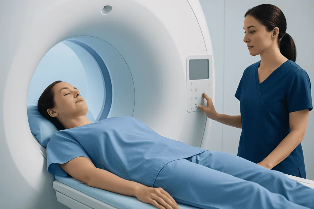
The human body contains a large amount of water, and water molecules contain hydrogen atoms. During an MRI scan, the scanner’s strong magnetic field temporarily aligns these atoms. Short bursts of radio waves then disturb this alignment. As the atoms return to their usual state, they release signals that are detected by the scanner and converted into images by a computer.
Different tissues release signals in different ways. For example, fluid, fat, muscle, scar tissue, inflammation, and tumors may appear differently depending on the MRI sequence used. A sequence is a specific way of collecting MRI information. Radiographers or MRI technologists select sequences based on the body area being examined and the clinical question.
MRI scanners often use specialized coils, which are devices placed around or near the body part being scanned. A head coil, knee coil, breast coil, or spine coil helps improve image quality by receiving signals from the targeted area. The patient does not feel the magnetic field or radio waves, but they may hear loud knocking or tapping sounds as the scanner collects images.
What an MRI Scan Can Show

MRI can show both normal anatomy and changes caused by disease or injury. It is commonly used to assess the brain and spine for conditions such as stroke, multiple sclerosis, tumors, disc problems, nerve compression, inflammation, or infection. In joints and soft tissues, MRI can help evaluate ligament tears, cartilage damage, tendon injuries, bone marrow changes, and muscle conditions.
MRI is also used to examine organs in the chest, abdomen, and pelvis. Depending on the protocol, it can help evaluate the liver, pancreas, kidneys, uterus, ovaries, prostate, heart, blood vessels, and bile ducts. In cancer care, MRI may be used to define the size and location of a tumor, assess nearby structures, plan treatment, or monitor response over time.
It is important to understand that an MRI result is not interpreted in isolation. Some findings may be clearly linked to symptoms, while others may be incidental or age-related. The radiologist describes the imaging findings, and the referring doctor combines this report with the patient’s symptoms, examination, blood tests, and other medical information to decide what the findings mean.
What to Expect Before and During the Scan
Before the scan, patients usually complete a safety questionnaire. This asks about pacemakers, heart valves, aneurysm clips, cochlear implants, neurostimulators, medication pumps, metal fragments, previous surgeries, kidney disease, allergies, and pregnancy. This screening is essential because the MRI magnet is always strong, even when the scanner is not actively taking images.
Patients are typically asked to remove jewelry, watches, hairpins, hearing aids, removable dental appliances, credit cards, and clothing with metal fasteners. In many cases, a hospital gown is provided. If the scan involves the abdomen or pelvis, fasting for a short period may be requested, depending on the protocol and whether contrast is planned.
During the scan, the patient lies on a movable table that slides into the scanner. The technologist communicates through an intercom and can see the patient throughout the examination. Earplugs or headphones are provided because MRI machines make loud rhythmic sounds. Staying still is very important; even small movements can blur images and may require parts of the scan to be repeated.
The length of the scan depends on the area being examined and the number of sequences needed. Some scans are relatively short, while more complex examinations may take longer. If a person is anxious or has claustrophobia, the medical team can discuss options such as relaxation techniques, positioning support, an open or wider-bore scanner when available, or medication prescribed in advance by a doctor.
MRI Contrast: Why It May Be Used
Some MRI scans are performed with contrast material. The most common type is a gadolinium-based contrast agent, which is usually given through a small intravenous line in the arm. Contrast can make certain tissues, blood vessels, inflammation, tumors, infection, or scar tissue more visible, helping the radiologist answer specific diagnostic questions.
Not every MRI needs contrast. The decision depends on the body part, the suspected condition, previous imaging results, and the information the doctor needs. For example, contrast may be useful in many brain, spine, breast, liver, pelvic, vascular, and cancer-related MRI examinations. Other studies, such as many routine joint or spine scans, may be performed without contrast.
Gadolinium-based contrast agents are generally well tolerated. Some people may briefly feel coolness at the injection site or have a metallic taste. Allergic reactions are uncommon but can occur, so patients should tell the team about previous reactions to contrast agents or medicines. People with significantly reduced kidney function require special assessment because certain contrast agents may not be appropriate for them.
Patients who are pregnant, breastfeeding, or trying to become pregnant should inform the imaging team before the scan. MRI without contrast may be considered during pregnancy when medically necessary, but contrast is used only after careful risk-benefit discussion. Breastfeeding guidance can vary depending on the contrast agent and local protocols, so patients should ask their doctor for individualized advice.
Safety, Risks, and Who Needs Extra Screening
MRI is considered a safe and widely used imaging method when proper screening is performed. Because it does not use ionizing radiation, it can be a valuable option when repeated imaging is needed or when soft-tissue detail is important. However, MRI is not suitable for every patient or every medical situation, and the strong magnetic field requires strict safety rules.
People with implanted or internal devices need careful review. Some pacemakers, defibrillators, cochlear implants, brain aneurysm clips, vascular stents, artificial heart valves, neurostimulators, and infusion pumps are MRI-conditional, meaning they may be scanned only under specific conditions. Others may be unsafe. The imaging team may need the device card, model number, operation notes, or confirmation from the treating specialist before proceeding.
Metal fragments in the body can also be important. This is especially relevant for people with a history of metalwork, shrapnel injury, eye injury from metal, or certain older surgical clips. If there is uncertainty, an X-ray may be needed before MRI. Tattoos and permanent makeup may occasionally feel warm during scanning, but serious problems are uncommon when the team is informed and monitoring is in place.
Other possible issues include temporary discomfort from lying still, warmth during some sequences, anxiety in the scanner, or minor bruising from an IV line if contrast is used. The technologist provides a call button or communication method so the patient can alert the team at any time. Patients should always mention pain, unusual heat, dizziness, shortness of breath, or any concern during the examination.
Understanding MRI Results
After the scan, a radiologist reviews the images and prepares a report for the referring doctor. The report typically describes the imaging technique, key findings, impressions, and sometimes recommendations for follow-up. In urgent situations, important findings may be communicated quickly to the clinical team according to hospital procedures.
MRI images can reveal a wide range of findings, from normal results to inflammation, degeneration, injury, blocked or abnormal blood vessels, masses, fluid collections, infection, or changes after surgery. Some findings are specific, while others require correlation with symptoms, laboratory tests, biopsy results, or additional imaging. A normal MRI can also be useful because it may help rule out certain conditions.
Patients should avoid trying to interpret the report without medical guidance. Terms such as lesion, degeneration, cyst, enhancement, or signal change can sound concerning but may have different meanings depending on the body area and clinical context. The referring doctor is best placed to explain whether the findings are significant and what, if any, next steps are needed.
Preparing Well and When to Speak With a Doctor
Good preparation helps the scan go smoothly. Patients should bring prior imaging, reports, implant cards, relevant medical records, and a list of medications if requested. They should follow instructions about eating, drinking, and taking regular medicines. If sedation or anti-anxiety medication is planned, a companion may be needed for travel home, and driving should be avoided until the medication has worn off as advised.
Patients should contact their doctor or imaging center before the appointment if they have an implanted device, possible metal in the body, severe claustrophobia, kidney disease, a history of contrast reaction, pregnancy, or difficulty lying flat. They should also ask questions if they are unsure why the MRI is being performed or whether contrast is planned.
After the scan, most people can return to normal activities immediately unless they received sedation or were told otherwise. If contrast was used, drinking fluids may be advised unless a doctor has restricted fluid intake. Any delayed rash, breathing symptoms, swelling, or unusual reaction after contrast should be reported promptly to a healthcare professional.
For international patients, Acibadem International provides multidisciplinary diagnostic imaging services in JCI-accredited hospitals, including MRI evaluation and specialist follow-up when needed. As with any diagnostic test, the most appropriate imaging plan should be decided by a qualified doctor based on the patient’s individual condition.
Frequently asked questions
Is an MRI scan painful?
An MRI scan is not painful because the scanner does not touch or compress the body in a harmful way. Some patients may feel uncomfortable from lying still, the loud sounds, or the enclosed space. The technologist can communicate with the patient during the scan and provide support if discomfort occurs.
Does an MRI scan use radiation?
No. MRI uses a strong magnetic field and radio waves, not ionizing radiation. This is one reason MRI is often chosen when detailed soft-tissue imaging is needed. However, the best imaging test depends on the medical question, and sometimes CT, ultrasound, or X-ray may be more appropriate.
How long does an MRI scan take?
The time varies depending on the body part and the complexity of the examination. Some MRI scans are completed relatively quickly, while others require more sequences or contrast and take longer. The imaging center can usually provide an estimated duration before the appointment.
Can people with pacemakers or implants have an MRI?
Some people with pacemakers, defibrillators, or other implants can have MRI under specific safety conditions, while others cannot. The device type, model, and MRI compatibility must be checked before the scan. Patients should bring their implant card or device information and inform the imaging team in advance.
Why would MRI contrast be needed?
MRI contrast can help highlight blood vessels, inflammation, tumors, infection, or scar tissue more clearly. It is used only when it is expected to add useful diagnostic information. The team will review kidney function, allergy history, pregnancy status, and other factors before giving contrast.
When will MRI results be available?
A radiologist reviews the images and sends a report to the referring doctor. Timing depends on the hospital workflow, the complexity of the scan, and whether the findings are urgent. The referring doctor will explain the results and discuss whether follow-up, treatment, or additional tests are needed.
References
- American College of Radiology
- Radiological Society of North America
- National Institute of Biomedical Imaging and Bioengineering
- European Society of Radiology
- U.S. Food and Drug Administration
This article is for general information only and is not a substitute for professional medical advice. Please consult a qualified doctor about your individual situation.
Joint and spine care in Turkey — expert assessment & treatment
JCI-accredited · board-certified surgeons · reply within 24h
Add us as a Preferred Source to see more of our trusted health content across Google Search, AI Overviews and Discover.

More from the Health Library



Related Specialists

Prof. Dr. Ercan Karaarslan
Radiology
Prof. Dr. Bora Peynircioğlu
Interventional Radiology
Prof. Dr. Koray Güven
Interventional Radiology
Assoc. Prof. Dr. Deniz Can Aliş
Radiology