PET-CT Scan: Metabolic Imaging for Diagnosis and Treatment Planning
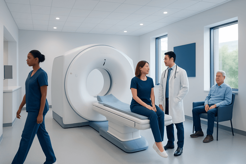
PET-CT combines positron emission tomography and computed tomography in one examination. The scan detects areas of increased or reduced metabolic activity, often using a radioactive glucose tracer called FDG.
Key Takeaways
- PET-CT combines positron emission tomography and computed tomography in one examination.
- The scan detects areas of increased or reduced metabolic activity, often using a radioactive glucose tracer called FDG.
- PET-CT is widely used for cancer staging, treatment response assessment, and radiotherapy or surgical planning.
- Preparation commonly includes fasting, blood sugar checks, and avoiding strenuous exercise before the scan.
- Radiation exposure is carefully controlled, and the benefits usually outweigh the risks when the scan is medically indicated.
- Results should always be interpreted by qualified specialists together with clinical history and other test findings.
A PET-CT scan combines functional and anatomical imaging to help doctors see how tissues and organs are working, not only how they look. It is most often used in cancer care, but it can also support selected heart, brain, and inflammation-related evaluations.
Overview: What Is a PET-CT Scan?
A PET-CT scan is an advanced imaging test that combines two technologies: positron emission tomography, known as PET, and computed tomography, known as CT. PET shows patterns of activity inside the body, such as how cells use glucose or other substances. CT provides detailed images of the body’s structures, including organs, bones, lymph nodes, and soft tissues. When these images are combined, doctors can see both where an abnormality is located and how active it appears.
The most common PET tracer is fluorodeoxyglucose, often called FDG. FDG is a mildly radioactive form of glucose. Because many active cells use glucose for energy, FDG can highlight areas with increased metabolic activity. This is especially useful in oncology, where some tumors and inflamed tissues may take up more FDG than surrounding tissue.
PET-CT does not replace all other imaging tests. Instead, it provides a different type of information. Ultrasound, MRI, CT, laboratory tests, biopsy, and clinical examination may all be needed depending on the medical question. PET-CT is most helpful when the goal is to understand the whole-body distribution and activity of disease, or to plan treatment more precisely.
How PET-CT Works
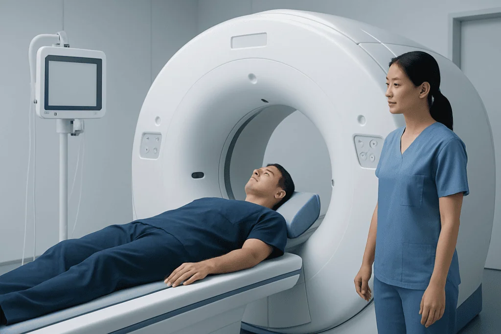
Before the scan, a small amount of radioactive tracer is injected into a vein. The patient then rests quietly while the tracer circulates and is absorbed by tissues. For FDG PET-CT, this uptake period commonly lasts about an hour, although protocols vary by indication and hospital practice. During this time, movement and muscle activity are usually kept to a minimum because active muscles can also absorb FDG.
The PET part of the scan detects signals released by the tracer. A computer converts these signals into images showing areas of tracer uptake. The CT part uses X-rays to create detailed cross-sectional images. Modern PET-CT systems align these two datasets so the metabolic activity can be matched accurately with anatomy.
Different tracers may be used for different medical needs. FDG is the most widely used, but some cancers and conditions are better evaluated with specialized tracers. The choice of tracer depends on the suspected diagnosis, previous test results, treatment history, and the specific question the doctor wants to answer.
Why PET-CT May Be Recommended

PET-CT is commonly recommended when doctors need information about disease activity throughout the body. In cancer care, it can help determine whether a tumor is localized or has spread to lymph nodes or other organs. This process is called staging. Accurate staging helps the care team choose the most appropriate treatment plan, such as surgery, radiotherapy, chemotherapy, targeted therapy, immunotherapy, or a combination of treatments.
PET-CT may also be used after treatment has started or completed. It can help assess whether metabolically active disease has decreased, remained stable, or progressed. In some situations, it is used to investigate whether a finding on CT or MRI represents scar tissue, inflammation, or active tumor. However, PET-CT findings are not interpreted in isolation; biopsy or follow-up imaging may still be required.
Beyond oncology, PET-CT may support selected evaluations in cardiology, neurology, and infectious or inflammatory diseases. For example, it can help assess certain heart muscle viability questions, investigate some causes of fever of unknown origin, or contribute to selected dementia evaluations depending on the tracer and protocol. The test is recommended only when it is expected to provide information that may affect diagnosis or management.
- Common oncology uses include staging, restaging, treatment response assessment, and radiotherapy planning.
- Selected non-cancer uses include infection, inflammation, cardiac viability, and neurological assessments.
- The medical value depends on the clinical question, tracer selection, and expert interpretation.
Preparing for a PET-CT Scan
Preparation is important because it affects image quality. For an FDG PET-CT scan, patients are usually asked to avoid eating for several hours before the appointment. Plain water is often allowed and may be encouraged. Patients should follow the exact instructions from the imaging center, because preparation may differ depending on the tracer, the reason for the scan, and whether contrast material will be used for CT.
Blood sugar control is especially important for FDG PET-CT. High blood glucose levels can interfere with tracer uptake and reduce the clarity of the images. Patients with diabetes should tell the imaging team in advance so medication timing, meals, and appointment scheduling can be coordinated safely. No diabetes medication should be changed without medical advice.
Patients are commonly advised to avoid strenuous exercise for a period before the scan, because active muscles can show increased FDG uptake. Comfortable clothing without metal parts is helpful. Jewelry, belts, hearing aids, removable dental appliances, or other metal items may need to be removed. Patients should also inform staff if they are pregnant, may be pregnant, breastfeeding, have kidney disease, allergies, claustrophobia, or previous reactions to contrast material.
What Happens During the Examination
On arrival, the imaging team reviews the patient’s medical history, current medications, allergies, and the reason for the scan. Blood glucose may be checked for FDG PET-CT. The tracer is then injected through a small intravenous line. After injection, the patient rests in a quiet area while the tracer distributes through the body. This waiting period is a normal part of the examination.
During the scan itself, the patient lies on a table that moves slowly through the PET-CT scanner. The machine is open at both ends and does not usually feel as enclosed as some MRI scanners. The scan is painless, but the patient needs to remain still so the images are clear. The imaging time varies depending on the body area being scanned and the protocol.
Some PET-CT examinations include CT contrast material, either by mouth or through a vein, to improve anatomical detail. If intravenous contrast is planned, the team may ask about kidney function, allergies, asthma, thyroid disease, and previous contrast reactions. After the scan, most patients can return to normal activities unless they have received sedation or have been given specific instructions.
Safety, Radiation, and Possible Limitations
PET-CT involves exposure to a small amount of radiation from both the tracer and the CT scan. Imaging specialists use protocols designed to obtain useful diagnostic information while keeping radiation exposure as low as reasonably achievable. When a PET-CT is recommended, the expected medical benefit should outweigh the potential risk. This balance is considered carefully, especially in children, pregnant patients, and people who need repeated imaging.
The radioactive tracer loses activity over time and leaves the body mainly through urine. Drinking fluids after the scan, if permitted, may help clear the tracer. Patients may be advised to limit close contact with pregnant women or small children for a short time after the examination, depending on local instructions and the tracer used. Breastfeeding patients should ask the imaging team about any temporary precautions.
PET-CT has limitations. Some small lesions may be below the resolution of the scan. Some tumors have low FDG uptake and may not appear strongly on FDG PET-CT. Inflammation, infection, recent surgery, radiotherapy effects, and healing tissues can also show increased uptake and may mimic disease activity. For this reason, PET-CT results are interpreted by nuclear medicine and radiology specialists in the context of the patient’s full medical picture.
Understanding Results and Treatment Planning
PET-CT results are usually reported by a specialist physician trained in nuclear medicine, radiology, or both. The report describes areas of tracer uptake, their location, and whether the pattern is expected, indeterminate, or suspicious. Measurements such as standardized uptake value, often called SUV, may be included, but SUV is only one part of interpretation. It can be influenced by timing, blood glucose, scanner settings, body size, and technical factors.
In cancer care, PET-CT can help the multidisciplinary team plan treatment. It may identify the best biopsy site, guide radiotherapy target areas, support surgical planning, or help decide whether systemic therapy is needed. It can also provide a baseline before treatment so later scans can be compared more accurately. The scan findings are often discussed alongside pathology, blood tests, CT or MRI results, and the patient’s symptoms and goals.
Patients should review their results with the doctor who requested the scan. A PET-CT report may contain technical language, and some findings may need clarification or additional testing. Acibadem International’s multidisciplinary specialists and JCI-accredited hospitals diagnose and treat conditions evaluated with PET-CT for international patients, using imaging results as part of an individualized care plan.
When to Speak With a Doctor
Patients should speak with their doctor if they are unsure why a PET-CT has been recommended or how it may affect treatment decisions. It is reasonable to ask what question the scan is intended to answer, whether other imaging options are available, and what the next steps may be depending on the results. Clear communication can help patients feel more prepared and involved in their care.
Before the appointment, patients should contact the imaging center if they have diabetes, are pregnant or breastfeeding, have kidney problems, have had a reaction to contrast material, or cannot lie still for the expected scan time. These situations do not always prevent PET-CT, but they may require adjusted preparation or additional precautions.
After the scan, patients should follow the discharge instructions provided by the imaging team. They should seek medical advice if they experience symptoms that concern them after receiving contrast material, such as a rash, breathing difficulty, swelling, or persistent dizziness. Serious reactions are uncommon, but timely medical support is important if they occur.
Frequently asked questions
Is a PET-CT scan painful?
The scan itself is not painful. Patients may feel a brief pinch when the intravenous line is placed for the tracer injection. The main challenge for some people is lying still during the imaging period.
How long does a PET-CT appointment take?
The total appointment often takes a few hours because there is a waiting period after the tracer injection before imaging begins. The actual scan time is usually shorter than the total visit. Exact timing depends on the protocol, tracer, and whether contrast-enhanced CT is included.
Can PET-CT detect all cancers?
No imaging test detects all cancers in every situation. Many cancers show increased FDG uptake, but some tumors are slow-growing, very small, or less metabolically active and may not appear clearly. PET-CT is most useful when selected for the right clinical question and interpreted with other medical information.
Is PET-CT safe for people with diabetes?
Many people with diabetes can have PET-CT safely, but preparation needs planning. Blood glucose levels can affect FDG image quality, and medication or meal timing may need adjustment under medical guidance. Patients should inform the imaging center about diabetes when scheduling the scan.
What is the difference between PET-CT and MRI?
PET-CT shows metabolic activity and combines it with CT anatomy, while MRI provides highly detailed soft-tissue images using magnetic fields rather than ionizing radiation. The best test depends on the organ being evaluated and the medical question. In some cases, both tests provide complementary information.
Will a PET-CT result give a definite diagnosis?
PET-CT can provide important evidence, but it may not always give a final diagnosis on its own. Increased tracer uptake can be caused by cancer, inflammation, infection, or healing tissue. A biopsy, laboratory test, follow-up scan, or specialist review may be needed to confirm the cause.
Can patients be around family after a PET-CT scan?
Most patients can resume normal daily activities after the scan. Because the tracer remains in the body for a short time, the imaging team may provide temporary instructions about close contact with pregnant women or young children. Drinking fluids, if allowed, can help the tracer leave the body.
References
- Society of Nuclear Medicine and Molecular Imaging
- American College of Radiology
- European Association of Nuclear Medicine
- National Cancer Institute
- International Atomic Energy Agency
This article is for general information only and is not a substitute for professional medical advice. Please consult a qualified doctor about your individual situation.
Oncology care in Turkey — second opinion and treatment plan
JCI-accredited · board-certified surgeons · reply within 24h
Add us as a Preferred Source to see more of our trusted health content across Google Search, AI Overviews and Discover.