

EMG and Nerve Conduction Studies: Diagnosing Neuromuscular Disease

EMG and nerve conduction studies are often performed together to evaluate nerve signals and muscle activity. The tests can help diagnose conditions such as carpal tunnel syndrome, peripheral neuropathy, radiculopathy, and certain muscle diseases.
Key Takeaways
- EMG and nerve conduction studies are often performed together to evaluate nerve signals and muscle activity.
- The tests can help diagnose conditions such as carpal tunnel syndrome, peripheral neuropathy, radiculopathy, and certain muscle diseases.
- A nerve conduction study uses small electrical pulses, while EMG uses a very thin needle electrode to record muscle activity.
- Most people tolerate the tests well; temporary soreness or tingling may occur afterward.
- Results are interpreted alongside symptoms, physical examination, blood tests, imaging, and medical history.
EMG and nerve conduction studies are diagnostic tests that help doctors understand how well nerves and muscles are working. They are commonly used to evaluate symptoms such as numbness, tingling, weakness, muscle cramps, or pain that may come from nerve or muscle disorders.
Overview
EMG and nerve conduction studies are specialized tests used to examine the electrical function of nerves and muscles. A nerve conduction study measures how quickly and strongly electrical signals travel along a nerve. Electromyography, often shortened to EMG, records the electrical activity produced by muscles at rest and during movement.
These tests are especially helpful when symptoms suggest that a nerve, nerve root, neuromuscular junction, or muscle may not be functioning normally. Common reasons for testing include numbness, tingling, burning pain, muscle weakness, muscle twitching, cramps, or symptoms that radiate from the neck or back into an arm or leg.
Although the tests involve electrical stimulation and, for EMG, a fine needle electrode, they are diagnostic rather than treatment procedures. They do not send electricity through the whole body, and they do not change the underlying condition. Their purpose is to provide objective information that helps a doctor confirm, narrow, or rule out possible causes of symptoms.
What the Tests Measure
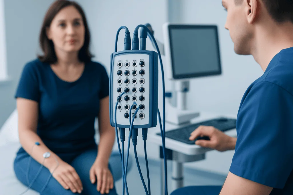
A nerve conduction study focuses on peripheral nerves, which carry signals between the brain, spinal cord, muscles, and skin. During the test, small sensors are placed on the skin, and brief electrical pulses are applied to a nerve. The machine records how fast the signal travels and how large the response is. Slower conduction, reduced signal size, or blocked transmission can point to different types of nerve injury or irritation.
EMG evaluates muscle activity. A doctor inserts a very thin, sterile needle electrode into selected muscles to record electrical patterns. Muscles normally have little electrical activity when relaxed and a predictable pattern when contracted. Changes in these patterns can suggest whether weakness is related to a muscle disorder, a nerve supplying the muscle, or a nerve root leaving the spine.
Together, EMG and nerve conduction studies can provide complementary information. For example, a nerve conduction study may show that a nerve is compressed at the wrist, while EMG can help determine whether muscles supplied by that nerve are affected. In suspected spine-related nerve irritation, EMG may help identify which nerve root is involved and whether the problem appears recent or longer-standing.
Conditions EMG and Nerve Conduction Studies Can Help Diagnose
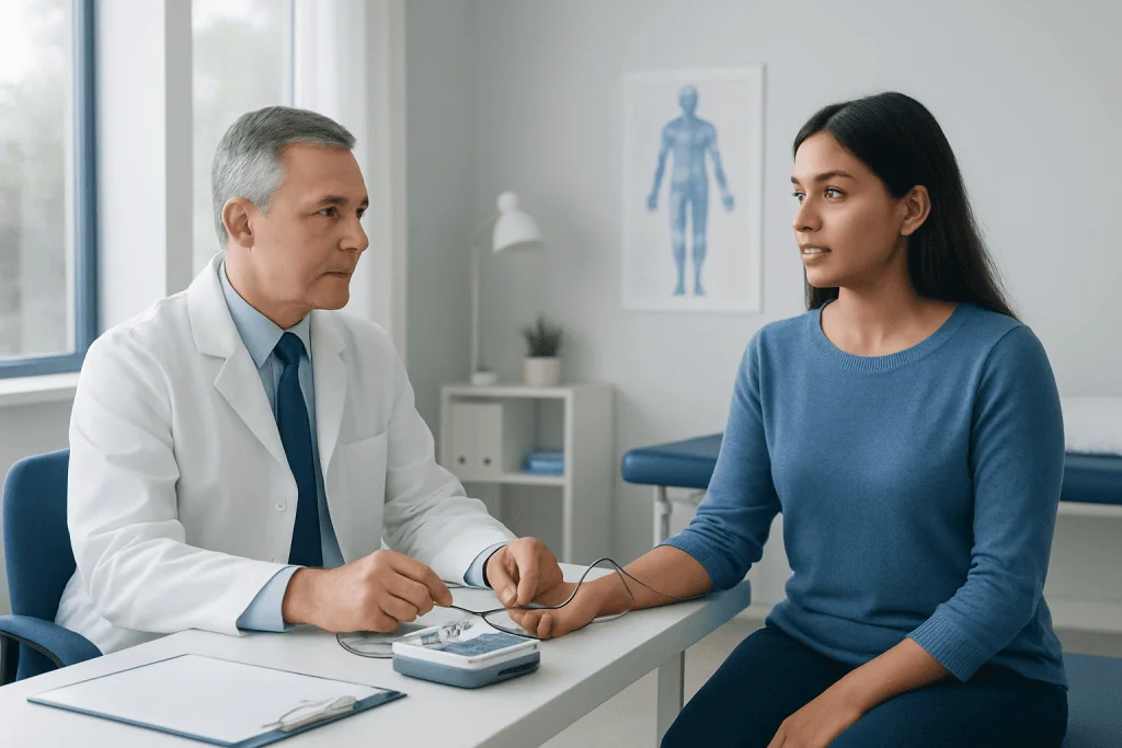
Doctors use these tests for a wide range of neuromuscular concerns. They are commonly used to evaluate entrapment neuropathies, such as carpal tunnel syndrome at the wrist or ulnar nerve compression at the elbow. They can also help assess peripheral neuropathy, a condition in which nerves outside the brain and spinal cord are damaged, often causing numbness, tingling, burning sensations, or balance difficulties.
EMG and nerve conduction studies may be recommended when symptoms suggest radiculopathy, which occurs when a nerve root is irritated or compressed as it exits the spine. This can happen with conditions such as a herniated disc or spinal narrowing. Testing can help distinguish radiculopathy from other causes of arm or leg symptoms, including peripheral nerve compression.
The tests may also support evaluation of certain muscle diseases, motor neuron disorders, neuromuscular junction disorders, and traumatic nerve injuries. They are not the only diagnostic tools used for these conditions, but they often provide important functional information that cannot be seen on an X-ray or standard MRI scan.
- Commonly evaluated symptoms include numbness, tingling, weakness, radiating pain, muscle cramps, and muscle twitching.
- Commonly evaluated conditions include carpal tunnel syndrome, peripheral neuropathy, radiculopathy, plexopathy, and selected myopathies.
- Results help doctors localize the problem, estimate severity, and guide further testing or treatment planning.
How to Prepare for the Test
Preparation is usually simple. Patients are generally asked to wear comfortable, loose clothing that allows access to the area being tested. Skin should be clean and free of lotions, oils, or creams on the day of the examination because these products can interfere with electrode contact during the nerve conduction portion.
The doctor should be informed about implanted devices such as pacemakers or defibrillators, bleeding disorders, use of blood-thinning medicines, or any history of lymphedema. In most cases, testing can still be performed safely, but the medical team may adjust the approach. Patients should also mention infections, skin wounds, or swelling near the area to be tested.
It is helpful for patients to bring previous imaging results, laboratory reports, medication lists, and notes from referring physicians. Because the test is interpreted in context, information about when symptoms began, what improves or worsens them, and whether symptoms are spreading can be as important as the electrical measurements themselves.
What Happens During EMG and Nerve Conduction Studies
The examination is usually performed by a neurologist, physiatrist, or another physician with training in electrodiagnostic medicine, often assisted by a trained technologist. The exact length of the visit depends on how many limbs or muscles need to be studied. Many tests are completed within a single outpatient appointment.
During the nerve conduction study, small recording electrodes are placed on the skin. A brief electrical pulse is delivered to stimulate a nerve. This may feel like a quick tapping, tingling, or mild shock-like sensation. Several nerves may be tested and compared from side to side to look for patterns.
During EMG, the doctor places a fine needle electrode into selected muscles. The patient may be asked to relax the muscle and then gently contract it. The needle does not inject medicine; it records electrical activity. Some people feel brief discomfort or pressure during needle placement, and the sensation usually stops when the needle is removed.
After the test, normal activities can often be resumed unless the doctor gives different instructions. Mild muscle soreness or small bruising can occur at needle sites and usually settles without special treatment. If a patient has specific restrictions because of another medical condition, the testing team will provide individualized guidance.
Understanding the Results
Results are not interpreted as simply normal or abnormal in isolation. The doctor reviews the pattern of findings and compares it with the patient’s symptoms, physical examination, medical history, and other tests. A mild abnormality may be meaningful in one clinical situation and less relevant in another, so interpretation by an experienced physician is important.
Nerve conduction results can show whether a problem affects the nerve’s outer insulating layer, the nerve fibers themselves, or signal transmission at a specific site. EMG findings can suggest whether muscle changes are related to nerve supply, direct muscle disease, or recovery after a previous nerve injury. The pattern can also help determine whether a problem is focal, widespread, recent, or chronic.
Sometimes the tests are normal even when symptoms are real. Very small nerve fibers that carry pain and temperature signals are not well assessed by standard nerve conduction studies. Early disease, intermittent compression, or conditions outside the tested area may also produce normal findings. In such cases, doctors may recommend additional evaluation such as blood tests, imaging, skin biopsy for small fiber neuropathy, or follow-up testing if symptoms change.
Benefits, Limitations, and Safety
The main benefit of EMG and nerve conduction studies is that they assess function rather than structure alone. Imaging tests may show anatomy, while electrodiagnostic testing helps determine whether nerves and muscles are actually transmitting signals properly. This can be particularly useful when symptoms and imaging findings do not fully match.
The tests also have limitations. They do not diagnose every neurological condition, and they may not identify the exact cause of a nerve problem, such as whether neuropathy is due to diabetes, vitamin deficiency, autoimmune disease, infection, or medication exposure. Instead, they help define the type and location of dysfunction, which then guides further investigation.
Safety is generally very good when the tests are performed by trained professionals. The electrical pulses used in nerve conduction studies are brief and localized. EMG needle insertion carries a small risk of temporary soreness, minor bleeding, bruising, or, very rarely, infection. Patients with implanted devices or blood-thinning therapy should tell the doctor in advance so that appropriate precautions can be taken.
When to See a Doctor
A medical evaluation is recommended when numbness, tingling, weakness, or pain persists, worsens, affects walking or hand function, or interferes with daily activities. Evaluation is also important when symptoms follow an injury, radiate from the spine into a limb, or are associated with muscle wasting, repeated falls, or difficulty performing fine movements.
A doctor may recommend EMG and nerve conduction studies after reviewing symptoms and performing a neurological examination. The tests are often part of a broader diagnostic plan that may include blood tests, ultrasound, MRI, or referral to specialists in neurology, orthopedics, endocrinology, rheumatology, or rehabilitation medicine.
Acibadem International’s multidisciplinary specialists and JCI-accredited hospitals diagnose and treat neuromuscular conditions for international patients, using electrodiagnostic testing when it is clinically appropriate. Patients considering testing should consult a qualified physician to understand whether EMG and nerve conduction studies are suitable for their symptoms and medical history.
Frequently asked questions
Are EMG and nerve conduction studies the same test?
They are related but not the same. A nerve conduction study measures how electrical signals travel through nerves, while EMG records electrical activity inside muscles. Doctors often perform them together because the combined information gives a clearer picture of nerve and muscle function.
Do EMG and nerve conduction studies hurt?
Most people tolerate the tests well, although the sensations can be uncomfortable for short moments. Nerve stimulation may feel like a quick tingling or tapping shock, and EMG needle placement may feel like a small pinch or pressure. Any discomfort usually ends quickly after each part of the test.
How long do the tests take?
The length depends on how many nerves and muscles need to be examined. A focused test may take a relatively short time, while evaluation of several limbs or complex symptoms can take longer. The doctor or clinic can give a more specific estimate based on the referral question.
Can EMG diagnose neuropathy?
EMG and nerve conduction studies can help diagnose many types of peripheral neuropathy, especially those affecting larger nerve fibers. They can show whether neuropathy is mainly affecting the nerve covering, the nerve fibers, or both. Some conditions, such as small fiber neuropathy, may require different tests because standard nerve conduction studies can be normal.
Is special preparation needed before the test?
Preparation is usually minimal. Patients are commonly advised to avoid lotions, oils, or creams on the skin on the day of testing and to wear comfortable clothing. The doctor should be told about blood thinners, bleeding disorders, implanted devices, or skin infections near the test area.
When will the results be available?
In many settings, the physician performing the test can review the findings soon after the examination. A formal report may be sent to the referring doctor, who will explain how the results fit with the patient’s symptoms and other tests. Further evaluation or treatment depends on the overall clinical picture.
References
- American Association of Neuromuscular & Electrodiagnostic Medicine
- National Institute of Neurological Disorders and Stroke
- Mayo Clinic
- Cleveland Clinic
- Merck Manual Professional Edition
This article is for general information only and is not a substitute for professional medical advice. Please consult a qualified doctor about your individual situation.
Explore treatments in Turkey — costs, top hospitals & a free quote
JCI-accredited · board-certified surgeons · reply within 24h
Add us as a Preferred Source to see more of our trusted health content across Google Search, AI Overviews and Discover.

More from the Health Library



Related Specialists

Prof. Dr. Ercan Karaarslan
Radiology
Prof. Dr. Bora Peynircioğlu
Interventional Radiology
Prof. Dr. Koray Güven
Interventional Radiology
Assoc. Prof. Dr. Deniz Can Aliş
Radiology