Neurophysiology Tests: EEG, EMG, and Evoked Potentials Explained

Neurophysiology tests record electrical signals from the nervous system and help localize where a problem may be occurring. EEG evaluates brain wave activity and is commonly used when seizures, fainting-like episodes, encephalopathy, or sleep-related events are suspected.
Key Takeaways
- Neurophysiology tests record electrical signals from the nervous system and help localize where a problem may be occurring.
- EEG evaluates brain wave activity and is commonly used when seizures, fainting-like episodes, encephalopathy, or sleep-related events are suspected.
- EMG and nerve conduction studies assess nerves and muscles, helping diagnose neuropathy, nerve compression, radiculopathy, and neuromuscular disorders.
- Evoked potentials measure how well visual, hearing, or sensory pathways carry signals to the brain and spinal cord.
- Most tests are outpatient procedures with minimal risk, and results are interpreted together with the patient’s symptoms, examination, and imaging when needed.
Neurophysiology tests are safe diagnostic procedures that measure electrical activity in the brain, nerves, muscles, and sensory pathways. EEG, EMG, nerve conduction studies, and evoked potentials help doctors understand symptoms such as seizures, numbness, weakness, vision changes, balance problems, and unexplained episodes.
Overview: What Are Neurophysiology Tests?
Neurophysiology tests are a group of diagnostic examinations that record the electrical activity of the nervous system. The brain, spinal cord, nerves, and muscles communicate through tiny electrical signals. By measuring these signals, doctors can evaluate how well different parts of the nervous system are working and whether a symptom is more likely to come from the brain, a nerve pathway, the spinal cord, the neuromuscular junction, or the muscle itself.
The most common neurophysiology tests include electroencephalography, known as EEG; electromyography, known as EMG; nerve conduction studies, often performed together with EMG; and evoked potentials. These tests are different from scans such as MRI or CT. Imaging shows structure, while neurophysiology shows function. In many patients, both types of information are useful and complementary.
Doctors may recommend clinical neurophysiology testing for symptoms such as seizures, loss of awareness, numbness, tingling, weakness, tremor, muscle cramps, vision pathway concerns, hearing pathway concerns, or unexplained sensory changes. The tests are usually performed in a hospital, clinic, or specialized neurophysiology laboratory and are interpreted by trained physicians, often neurologists or clinical neurophysiologists.
EEG: Measuring Brain Wave Activity
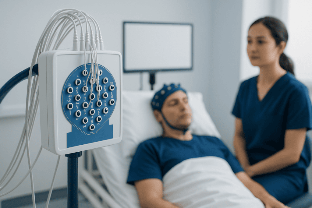
An EEG, or electroencephalogram, records the brain’s electrical activity through small metal sensors placed on the scalp. The sensors do not send electricity into the brain; they only detect natural brain wave patterns. A routine EEG is painless and often takes less than an hour, although longer recordings may be needed when symptoms occur infrequently or mainly during sleep.
EEG is especially helpful when a doctor is evaluating possible epilepsy or seizure-like episodes. It can show patterns that suggest a tendency for seizures, help classify seizure types, or support treatment planning. However, a normal EEG does not always rule out epilepsy, because abnormal activity may not appear during a short recording. For this reason, doctors interpret EEG results together with the medical history, witness descriptions, neurological examination, and sometimes video recordings.
During an EEG, the patient may be asked to rest quietly, open and close the eyes, breathe deeply for a short period, or look at flashing lights. These standard activation methods can make certain brain wave patterns easier to identify. In some cases, sleep-deprived EEG, ambulatory EEG, or video EEG monitoring may be recommended, particularly when episodes need to be captured more clearly. EEG may also be part of an evaluation in neurological sleep medicine when sleep-related events or daytime sleepiness require specialized assessment.
EMG and Nerve Conduction Studies: Testing Nerves and Muscles
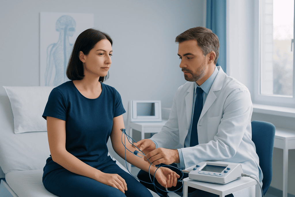
EMG, or electromyography, evaluates the electrical activity of muscles. It is often combined with nerve conduction studies, which measure how quickly and strongly peripheral nerves carry signals. Together, these tests help doctors determine whether symptoms such as numbness, tingling, burning pain, weakness, muscle wasting, or cramps are related to a nerve, nerve root, neuromuscular junction, or muscle problem.
In a nerve conduction study, small surface electrodes are placed on the skin, and a brief electrical pulse is delivered to stimulate a nerve. The response is recorded and analyzed. The sensation may feel like a quick tap or tingling shock, but it is brief and controlled. The test can identify slowed conduction, reduced signal strength, or conduction block, which may occur in conditions such as carpal tunnel syndrome, peripheral neuropathy, ulnar nerve entrapment, or certain inflammatory nerve disorders.
Needle EMG uses a very thin sterile needle electrode inserted into selected muscles to record activity at rest and during gentle contraction. The physician chooses muscles based on the patient’s symptoms and examination findings. EMG may help distinguish between a pinched nerve in the neck or back, a peripheral nerve injury, a motor neuron disorder, a muscle disease, or a disorder affecting communication between nerves and muscles. It is commonly used in the evaluation of neuromuscular diseases and related conditions.
Evoked Potentials: Checking Sensory Pathways
Evoked potential tests measure how the nervous system responds to specific sensory stimulation. The goal is to see whether signals travel normally from the eyes, ears, arms, or legs to the brain and spinal cord. These tests can detect slowed or disrupted conduction along pathways that may not be fully assessed by a routine neurological examination.
The main types include visual evoked potentials, brainstem auditory evoked potentials, and somatosensory evoked potentials. Visual evoked potentials measure the response of the visual pathway, often while the patient looks at a changing checkerboard pattern. Auditory evoked potentials use clicking sounds delivered through earphones to assess hearing pathways and the brainstem. Somatosensory evoked potentials involve mild electrical stimulation of a nerve in the wrist or ankle while responses are recorded from the scalp and spine.
Evoked potentials may be used when doctors are evaluating demyelinating disease, spinal cord pathway problems, optic nerve disorders, or brainstem involvement. They can also be useful when symptoms are subtle or when the doctor needs objective information about a sensory pathway. For patients with vision symptoms linked to the nervous system, evoked potentials may complement specialist assessment in neuro-ophthalmology.
Conditions These Tests May Help Diagnose
Neurophysiology tests do not diagnose every neurological condition on their own. Instead, they provide objective information that helps narrow the diagnosis, confirm a clinical suspicion, determine severity, or guide the next step in care. The same symptom, such as weakness or tingling, can have many possible causes, so the choice of test depends on the pattern of symptoms and examination findings.
EEG is often used for suspected epilepsy, seizure-like episodes, altered awareness, encephalopathy, and some sleep-related events. EMG and nerve conduction studies are commonly used for peripheral neuropathy, nerve entrapment, radiculopathy, plexopathy, motor neuron disease, myopathy, and neuromuscular junction disorders. Evoked potentials may help evaluate optic nerve pathway dysfunction, hearing pathway issues, spinal cord sensory pathway problems, and some demyelinating conditions.
Common reasons for testing may include:
- Episodes of shaking, staring, confusion, or loss of awareness
- Numbness, tingling, burning sensations, or electric-shock-like symptoms
- Weakness in an arm, leg, hand, foot, face, or eye muscles
- Muscle cramps, twitching, wasting, or unusual fatigue with activity
- Vision changes, hearing pathway concerns, balance problems, or sensory changes
- Monitoring of known neurological conditions or response to treatment
Preparation, Procedure, and Safety
Preparation depends on the test type. For EEG, patients are often asked to arrive with clean, dry hair and avoid hair oils, sprays, or gels that can interfere with electrode contact. In some cases, sleep deprivation may be requested, but this should be done only if the clinic gives specific instructions. For EMG and nerve conduction studies, it is usually helpful to keep the skin free of lotion or oil on the day of the test. Patients should inform the medical team about implanted devices, blood-thinning medicines, bleeding disorders, skin infections, or pregnancy when relevant.
During testing, the patient is usually awake and able to communicate with the technologist or physician. EEG and evoked potentials are generally painless, although skin preparation may feel slightly scratchy. Nerve conduction studies can be briefly uncomfortable because of the electrical pulses. Needle EMG may cause a small pinprick sensation and mild muscle soreness afterward, similar to a minor injection or blood test.
Neurophysiology tests are considered low-risk when performed by trained professionals. Serious complications are uncommon. Minor temporary discomfort, small bruising after needle EMG, or skin irritation from electrode paste or tape can occur. Patients who have a pacemaker, implanted defibrillator, deep brain stimulator, spinal cord stimulator, or other implanted device should mention it before testing so that the team can follow appropriate precautions.
Results, Next Steps, and When to See a Doctor
Results are usually reviewed by a specialist who looks at waveforms, timing, signal strength, muscle activity, and patterns that may suggest a specific diagnosis. The report may describe findings as normal, abnormal, or limited by technical or clinical factors. A normal result can still be useful because it may make certain conditions less likely and guide the doctor toward other explanations or tests.
Follow-up depends on the reason for testing. A patient with suspected epilepsy may need medication review, additional EEG monitoring, or brain imaging. A patient with nerve compression may be guided toward splinting, physical therapy, injections, surgery, or other treatments depending on severity. A patient with neuropathy may need blood tests, metabolic assessment, immune evaluation, genetic testing, or rehabilitation planning. The test result is one part of a broader clinical picture.
Medical advice should be sought promptly for new seizures, sudden weakness, sudden vision loss, severe new confusion, loss of bladder or bowel control with back pain, rapidly worsening numbness or weakness, or symptoms after significant injury. Non-urgent consultation is also appropriate for persistent tingling, recurrent unexplained episodes, progressive muscle weakness, or symptoms that interfere with daily activities. Acibadem International’s multidisciplinary specialists and JCI-accredited hospitals diagnose and treat neurological conditions for international patients, including cases that require coordinated neurophysiology, imaging, rehabilitation, and specialist consultation.
Frequently asked questions
Are neurophysiology tests painful?
EEG and most evoked potential tests are painless, although the skin may be gently prepared so electrodes can record clearly. Nerve conduction studies may feel like brief tingling pulses, and needle EMG can feel like small pinpricks with possible mild soreness afterward. The discomfort is usually short-lived and the team can pause if needed.
How long do EEG, EMG, and evoked potential tests take?
A routine EEG often takes about 30 to 60 minutes, while longer or video EEG studies can last several hours or days depending on the purpose. EMG with nerve conduction studies may take 30 to 90 minutes, based on how many nerves and muscles need to be tested. Evoked potentials usually take about 30 to 90 minutes depending on the type and number of pathways examined.
Can a normal EEG rule out epilepsy?
A normal EEG does not always rule out epilepsy because seizure-related electrical patterns may not appear during a short recording. If the history strongly suggests seizures, the doctor may recommend a sleep-deprived EEG, ambulatory EEG, or video EEG monitoring. Diagnosis is based on the full clinical picture, not the EEG alone.
What is the difference between EMG and a nerve conduction study?
A nerve conduction study measures how signals travel through peripheral nerves using surface electrodes and brief electrical stimulation. EMG records electrical activity inside selected muscles using a thin needle electrode. The two tests are often performed together because they provide complementary information about nerves, nerve roots, and muscles.
Do patients need to stop medications before testing?
Patients should not stop prescribed medicines unless the referring doctor or testing center specifically advises it. Some medicines, implanted devices, bleeding risks, or medical conditions may affect test planning, so the medical team should be informed in advance. Clear instructions are usually provided before the appointment.
Are neurophysiology tests safe for children and older adults?
These tests can be performed in children and older adults when clinically appropriate, with adjustments for comfort, cooperation, and medical history. Pediatric testing may use child-friendly explanations and shorter steps when possible. Older adults should tell the team about implanted devices, blood thinners, fragile skin, or mobility limitations before the test.
Will the results be available immediately?
Some preliminary impressions may be discussed after certain EMG and nerve conduction studies, depending on local practice. EEG and evoked potential recordings often require careful review and formal reporting by a specialist. The referring doctor then explains what the results mean in relation to symptoms, examination findings, and any other tests.
References
- American Clinical Neurophysiology Society
- International Federation of Clinical Neurophysiology
- American Association of Neuromuscular and Electrodiagnostic Medicine
- National Institute of Neurological Disorders and Stroke
- American Academy of Neurology
This article is for general information only and is not a substitute for professional medical advice. Please consult a qualified doctor about your individual situation.
Neurophysiology in Turkey — costs, top hospitals & a free quote
JCI-accredited · board-certified surgeons · reply within 24h