Brain Anatomy Explained: Key Structures in Simple Terms

The brain is divided into major regions that work together rather than separately. The cerebrum handles higher functions such as thinking, language, memory, and voluntary movement.
Key Takeaways
- The brain is divided into major regions that work together rather than separately.
- The cerebrum handles higher functions such as thinking, language, memory, and voluntary movement.
- The cerebellum helps coordinate balance, posture, and precise movement.
- The brainstem controls many automatic functions, including breathing, heart rate, and alertness.
- Protective layers, fluid, and blood vessels help support and nourish the brain.
- Changes in speech, strength, balance, vision, or consciousness should be assessed by a doctor promptly.
Brain anatomy can seem complex, but its main structures can be understood in simple, practical terms. Each part of the brain has a distinct role, and together they help control thinking, movement, emotions, memory, and vital body functions.
Overview of the Brain
The brain is the body’s control center. It receives information from the senses, processes that information, and sends signals that help the body move, speak, think, remember, and respond to the world. Although it weighs only a small portion of total body weight, it performs an extraordinary number of tasks every second.
In simple terms, brain anatomy refers to the brain’s physical structures and how they are organized. These structures include large visible regions, smaller deep centers, networks of nerve cells, and protective tissues around them. Each area has specialized jobs, but no part works entirely alone. The brain functions through constant communication between many regions.
The nervous system includes the brain, spinal cord, and nerves throughout the body. The brain acts as the main command center, while the spinal cord and peripheral nerves carry messages back and forth. Understanding the brain’s basic anatomy can make it easier to understand symptoms, injuries, neurological conditions, and the purpose of imaging tests or treatments.
The Cerebrum: The Largest Part of the Brain
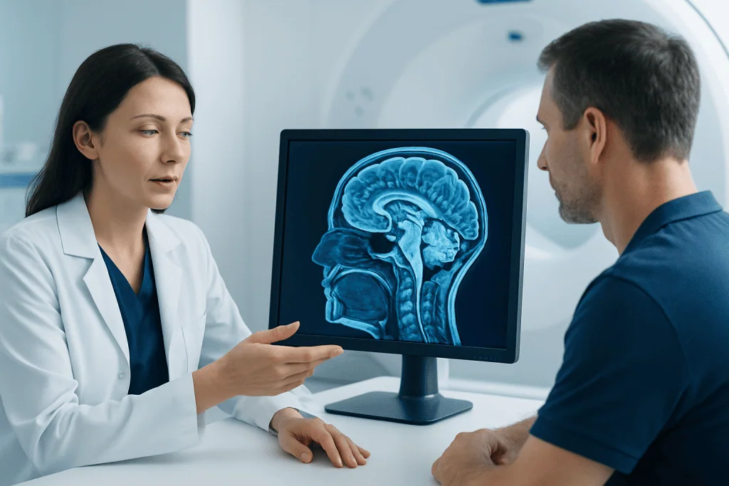
The cerebrum is the largest part of the brain and is the structure most people picture when they think of the brain. It has two halves, called the left and right hemispheres. These hemispheres are connected by a thick band of nerve fibers called the corpus callosum, which allows them to share information quickly.
The outer layer of the cerebrum is called the cerebral cortex. It is folded into ridges and grooves, which increase the surface area and allow more nerve cells to fit into the skull. The cortex is involved in higher functions such as reasoning, planning, language, attention, memory, sensation, and voluntary movement.
Each hemisphere is commonly divided into four main lobes:
- Frontal lobe: supports decision-making, personality, planning, speech production, and voluntary movement.
- Parietal lobe: helps process touch, temperature, pain, and spatial awareness.
- Temporal lobe: is important for hearing, language understanding, and memory.
- Occipital lobe: primarily processes visual information.
Although certain functions are more strongly linked to certain areas, the cerebrum works as an integrated network. For example, speaking involves movement, language, memory, hearing, and attention all at once. This is why symptoms caused by brain disorders can sometimes overlap.
Deep Brain Structures and Their Roles
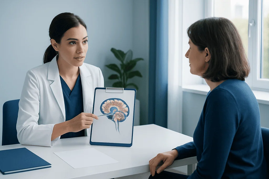
Beneath the outer cortex are deeper brain structures that are essential for emotion, memory, movement, and basic regulation. These areas are smaller and less visible from the outside, but they are central to everyday functioning.
The thalamus acts like a relay center. Many sensory signals, such as touch, pain, and vision-related information, pass through it before reaching the cortex. The hypothalamus helps regulate body temperature, hunger, thirst, sleep-wake cycles, and hormone-related functions. It links the nervous system with the endocrine system through the pituitary gland.
The hippocampus is especially important for forming new memories, while the amygdala plays a key role in emotional responses such as fear and stress. The basal ganglia help regulate movement, posture, and habit-based behaviors. These structures are often involved in neurological movement disorders and other conditions affecting coordination or behavior.
When doctors evaluate symptoms such as memory loss, movement changes, or emotional and behavioral shifts, they often consider these deep structures. In some cases, imaging of the brain may help identify whether a problem involves these regions or other parts of the nervous system.
The Cerebellum and Brainstem
The cerebellum sits at the back and lower part of the brain, underneath the cerebrum. Its main job is to fine-tune movement. It helps the body maintain balance, posture, and coordination, and it supports smooth, precise actions rather than strength itself.
A person uses the cerebellum while walking, reaching for an object, writing, or maintaining balance while turning the head. Problems affecting the cerebellum can cause unsteady walking, poor coordination, tremor, or difficulty with precise hand movements. Speech can also become less coordinated in some cases.
The brainstem connects the brain to the spinal cord and includes the midbrain, pons, and medulla. This area is vital because it helps control automatic functions the body must perform continuously, including breathing, heart rate, blood pressure, swallowing, and alertness. It also contains pathways that carry signals between the brain and the rest of the body.
Because the brainstem is so important, conditions affecting it can be serious and require prompt medical attention. Depending on the situation, doctors may use neurological examinations and MRI scans or other imaging to understand whether the cerebellum, brainstem, or nearby structures are involved.
How the Brain Is Protected and Supplied
The brain is well protected, but it is also delicate. The skull forms a hard outer shield. Beneath the skull are three protective membranes called the meninges: the dura mater, arachnoid mater, and pia mater. These layers help cushion and support the brain.
The brain also floats in cerebrospinal fluid, often called CSF. This clear fluid acts as a shock absorber, helps maintain a stable environment, and assists with waste removal. Spaces within the brain called ventricles produce and circulate this fluid. Problems with CSF flow can lead to pressure changes that may need medical evaluation.
A rich network of blood vessels supplies the brain with oxygen and nutrients. Even brief interruptions in blood flow can affect brain function. This is why symptoms such as sudden weakness, facial drooping, vision changes, or trouble speaking should be treated urgently, as they may suggest a stroke.
The brain is also protected by the blood-brain barrier, a specialized filtering system that helps control which substances can move from the bloodstream into brain tissue. This barrier is important for protection, but it can also make treatment of some brain conditions more complex.
How Brain Cells Communicate
The brain is made up of billions of nerve cells called neurons, along with support cells known as glial cells. Neurons communicate by sending electrical and chemical signals. These messages travel along nerve pathways and allow different brain regions to work together in fractions of a second.
Neurons connect at junctions called synapses. At these points, chemical messengers called neurotransmitters carry signals from one cell to another. Different neurotransmitters are involved in movement, mood, attention, sleep, memory, and many other functions. A healthy brain depends on balanced communication across these networks.
Glial cells support and protect neurons in several ways. They help provide nutrients, maintain the surrounding environment, form myelin to insulate nerve fibers, and participate in immune defense within the nervous system. Although neurons often receive the most attention, glial cells are essential for normal brain function.
When brain cell communication is disrupted, symptoms can vary widely depending on the location and cause. Problems may include seizures, memory difficulties, weakness, numbness, movement changes, or altered thinking. In some cases, symptoms may relate to brain tumors, inflammation, infection, or degenerative neurological disease.
Why Brain Anatomy Matters in Diagnosis and Care
Knowing the basic parts of the brain helps explain why symptoms appear in different ways. For example, difficulty speaking may point to areas that control language, while balance problems may suggest cerebellar involvement. Memory changes may relate to the temporal lobe or deeper memory centers such as the hippocampus. This anatomical understanding guides doctors during neurological examination and testing.
Common tools used to assess the brain include physical and neurological exams, brain imaging, and sometimes specialized tests of electrical activity or blood flow. Depending on the concern, a doctor may recommend brain MRI, CT imaging, or other studies. These tests help identify structural changes, bleeding, blocked blood vessels, inflammation, tumors, or injury.
Treatment depends on the cause rather than anatomy alone. Some problems are managed with medication, rehabilitation, monitoring, or lifestyle changes, while others may need urgent procedures or surgery. For example, treatment pathways can differ greatly between headache disorders, epilepsy, trauma, infection, vascular disease, and tumors.
For patients who need specialist assessment, Acibadem International’s multidisciplinary teams in JCI-accredited hospitals diagnose and treat a wide range of brain and nervous system conditions for international patients, including advanced imaging and neurosurgical care when appropriate.
When to Seek Medical Advice
Many people become interested in brain anatomy after experiencing symptoms themselves or in a loved one. While mild, short-lived complaints may have simple causes, persistent or unusual neurological symptoms should be assessed by a qualified doctor. A medical professional can decide whether the symptoms are likely related to the brain, spinal cord, nerves, or another body system.
Medical advice is especially important for new or worsening headaches, repeated fainting, seizures, confusion, memory decline, weakness, numbness, changes in speech, balance problems, tremor, or visual changes. These symptoms do not always mean a serious condition, but they do deserve proper evaluation.
Emergency care is needed for symptoms that begin suddenly, such as facial drooping, difficulty speaking, severe confusion, loss of consciousness, sudden severe headache, or weakness on one side of the body. These can be signs of a stroke or other urgent neurological event.
Understanding brain anatomy is useful, but it does not replace medical diagnosis. If symptoms are concerning, ongoing, or interfering with daily life, professional assessment offers the safest path toward clarity and appropriate care.
Frequently asked questions
What are the main parts of the brain?
The main parts of the brain are the cerebrum, cerebellum, and brainstem. The cerebrum handles higher thinking, sensation, language, and voluntary movement, while the cerebellum supports coordination and balance, and the brainstem controls automatic life-sustaining functions.
What does the frontal lobe do?
The frontal lobe helps with planning, judgment, personality, problem-solving, and voluntary movement. It also contains areas important for producing speech in many people.
Why is the brainstem so important?
The brainstem helps control breathing, heart rate, blood pressure, swallowing, and alertness. Because it manages many automatic functions essential for life, problems in this area can require urgent medical attention.
What is the difference between the cerebrum and cerebellum?
The cerebrum is the largest brain region and is mainly responsible for conscious thought, language, memory, sensation, and voluntary actions. The cerebellum is smaller and focuses on coordination, posture, and smooth, accurate movement.
How is the brain protected?
The brain is protected by the skull, three membrane layers called the meninges, and cerebrospinal fluid that cushions it. It also has a specialized blood-brain barrier that helps limit harmful substances from entering brain tissue.
Can one part of the brain work completely on its own?
No, most brain functions depend on networks of regions working together. Even simple tasks such as speaking or reaching for an object involve communication between several brain areas.
References
- National Institute of Neurological Disorders and Stroke
- National Institute of Mental Health
- MedlinePlus
- Mayo Clinic
- World Health Organization
This article is for general information only and is not a substitute for professional medical advice. Please consult a qualified doctor about your individual situation.
Explore treatments in Turkey — costs, top hospitals & a free quote
JCI-accredited · board-certified surgeons · reply within 24h
Add us as a Preferred Source to see more of our trusted health content across Google Search, AI Overviews and Discover.
Check your numbers in seconds
BMI, calories, due date, blood pressure and 30+ more clinical calculators — free, instant, doctor-reviewed ranges.