Orthopedic Diagnosis: What to Expect From X-Rays, MRI, and Physical Exams

Orthopedic diagnosis usually begins with a detailed history and physical exam. X-rays are often the first imaging test for fractures, alignment problems, and arthritis.
Key Takeaways
- Orthopedic diagnosis usually begins with a detailed history and physical exam.
- X-rays are often the first imaging test for fractures, alignment problems, and arthritis.
- MRI is especially useful for soft tissues such as ligaments, cartilage, tendons, discs, and muscles.
- Not every patient needs every test; the choice depends on symptoms, age, injury, and exam findings.
- A clear diagnosis helps guide treatment, from rest and physiotherapy to injections or surgery.
- Patients should seek medical care promptly for severe pain, inability to bear weight, deformity, numbness, or symptoms after major trauma.
Orthopedic diagnosis combines a medical history, physical examination, and imaging tests to understand pain, stiffness, weakness, or injury in the bones, joints, muscles, and spine. Knowing what to expect can help patients feel more prepared and take an active role in their care.
Overview
Orthopedic diagnosis is the process doctors use to identify conditions affecting the musculoskeletal system. This includes bones, joints, muscles, ligaments, tendons, and the spine. People may need an orthopedic evaluation for pain, swelling, stiffness, reduced movement, weakness, instability, or an injury after a fall, sports activity, or repetitive strain.
In many cases, diagnosis does not rely on one test alone. Instead, an orthopedic specialist puts together several pieces of information: the patient’s symptoms, medical history, physical examination, and imaging findings. This step-by-step approach helps avoid unnecessary testing and makes it more likely that treatment is matched to the real cause of the problem.
X-rays, MRI scans, and physical exams are among the most common parts of an orthopedic assessment. Each plays a different role. A physical exam shows how the body moves and where symptoms begin. X-rays provide a quick look at bones and joint alignment. MRI gives detailed images of soft tissues that cannot be seen well on standard X-rays.
What Happens During an Orthopedic Physical Exam
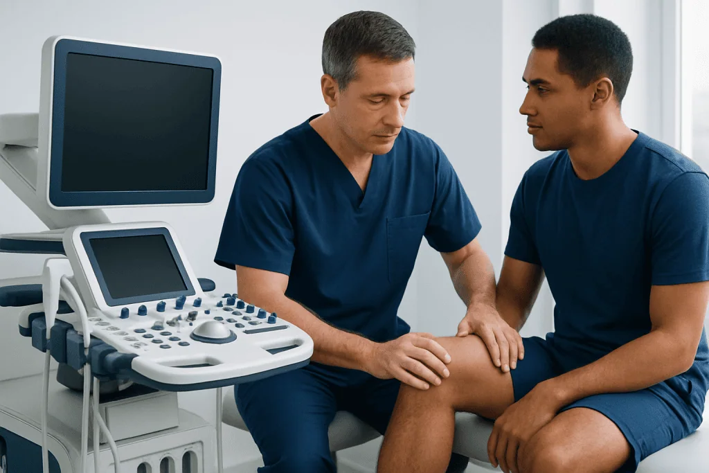
The physical exam is often the foundation of orthopedic diagnosis. The doctor usually begins by asking when the symptoms started, whether there was an injury, what movements make symptoms worse, and whether there are related issues such as tingling, numbness, fever, or night pain. Past surgeries, sports activities, work demands, and underlying conditions such as osteoporosis or arthritis may also be discussed.
Next, the specialist examines the affected area. This may include looking for swelling, bruising, redness, deformity, muscle wasting, or changes in posture. The doctor may gently touch different structures to identify tenderness and compare one side of the body with the other. Range of motion, joint stability, strength, walking pattern, balance, and nerve function may also be checked.
Some patients are surprised that an orthopedic exam can involve nearby body parts. For example, knee pain may be influenced by the hip, foot, or spine. Shoulder symptoms may be related to the neck. Because of this, the exam often looks at how the whole limb or region works together rather than focusing only on one painful spot.
Special maneuvers may help narrow down the cause. These tests can suggest ligament injury, tendon irritation, meniscus damage, nerve compression, or joint instability. While the exam alone may not provide the full answer, it often guides which imaging test, if any, is most useful next.
What X-Rays Show in Orthopedic Diagnosis
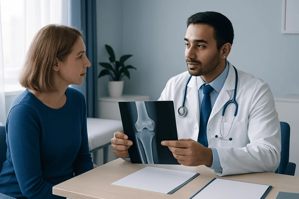
X-rays are usually one of the first imaging tests used in orthopedic diagnosis. They are fast, widely available, and especially helpful for showing bones. An X-ray can reveal fractures, dislocations, changes in bone shape, joint space narrowing, bone spurs, and signs of wear related to osteoarthritis. It can also help assess alignment in the spine, hips, knees, or feet.
For many injuries, an X-ray is the starting point because it helps rule out urgent problems such as a broken bone. However, a normal X-ray does not always mean there is no injury. Soft tissue problems, including ligament tears, tendon injuries, cartilage damage, and some early stress fractures, may not be visible on standard X-rays.
The test itself is simple. The patient is positioned so images can be taken from one or more angles, and it is important to stay still for a few seconds. Sometimes images are done while standing to show how joints line up under body weight. Children and pregnant patients may need special precautions, so it is important to tell the imaging team about any concerns beforehand.
X-rays are often enough to diagnose many common problems, but they may also be followed by other imaging if more detail is needed. In some cases, the findings may point toward conditions such as osteoarthritis or a spine issue that needs further evaluation.
When MRI Is Recommended
MRI, or magnetic resonance imaging, is especially useful when the doctor needs detailed pictures of soft tissues. This includes ligaments, tendons, menisci, cartilage, muscles, spinal discs, and nerves. MRI can help evaluate injuries such as an ACL tear, rotator cuff problem, meniscus injury, stress reaction, or disc-related nerve compression.
Unlike X-rays, MRI does not use ionizing radiation. Instead, it uses a strong magnetic field and radio waves to create images. During the scan, the patient lies on a table that moves into the machine. The test is painless, but it can take longer than an X-ray, and the machine makes loud knocking sounds. Some patients find the space uncomfortable, so they should mention claustrophobia in advance. Metal implants, pacemakers, and some devices may require special review before the scan.
MRI is not always the first test because it is usually used when soft tissue damage is suspected, when symptoms do not match the X-ray findings, or when treatment decisions depend on more detail. For example, MRI can help determine whether persistent knee pain is due to a meniscus tear or whether back pain is related to a disc problem such as herniated disc.
In selected cases, MRI findings may support planning for treatment such as knee replacement, hip replacement, or spine surgery when conservative care has not been enough. The scan is only one part of the decision, and results are always interpreted together with symptoms and the physical exam.
Other Tests That May Be Used
Although X-rays and MRI are common, they are not the only tools in orthopedic diagnosis. Ultrasound may be used to examine tendons, muscles, bursae, and fluid collections in real time. It can be especially helpful for shoulder, elbow, wrist, and ankle problems, and it can also guide some injections or procedures.
CT scans may be recommended when the doctor needs a very detailed view of bone, such as with complex fractures or certain joint and spine conditions. Bone scans and specialized imaging may be used in selected situations, especially if the diagnosis remains unclear after standard tests. Nerve conduction studies and electromyography can help when symptoms suggest nerve compression or muscle disorders.
Lab tests are sometimes part of the workup, particularly if there is concern for infection, inflammation, gout, or autoimmune disease. Blood tests do not diagnose every orthopedic problem, but they can be valuable when symptoms include fever, unexplained swelling, or multiple painful joints.
The important point is that testing is personalized. A person with a recent ankle sprain may need only an exam and perhaps an X-ray, while someone with long-term joint pain, weakness, or neurological symptoms may need a broader evaluation.
How Doctors Put the Results Together
Imaging findings do not always explain symptoms on their own. Many people have age-related changes on scans that are not the true source of pain. For this reason, orthopedic specialists interpret tests in context. A diagnosis is strongest when the patient’s history, exam findings, and imaging all point in the same direction.
For example, an MRI may show a tendon tear, but the doctor will still consider whether that tear matches the patient’s weakness, pain pattern, and movement limits. In the same way, an X-ray may show arthritis, but the specialist will look at whether the degree and location of arthritis fit the patient’s daily symptoms and activity level.
Once a diagnosis is made, the next step is to discuss treatment options. Depending on the condition, this may include rest, activity modification, bracing, medication, physiotherapy, injections, or surgery. If surgery is considered, further imaging or preoperative planning may be needed. In some situations, a patient may be referred for physical therapy and rehabilitation before more invasive treatments are discussed.
Near the end of the diagnostic process, patients should feel comfortable asking questions about what the findings mean, what is still uncertain, and what the next steps are. At Acibadem International, multidisciplinary specialists in JCI-accredited hospitals evaluate international patients with orthopedic conditions and help guide diagnosis and treatment planning.
How Patients Can Prepare and When to Seek Medical Care
Preparing for an orthopedic visit can make the evaluation more efficient. Patients may find it helpful to bring a list of symptoms, past injuries, medications, allergies, and any previous imaging or test reports. Wearing comfortable clothing can make the physical exam easier, especially if the shoulder, knee, hip, or spine needs to be assessed.
Before an MRI, patients should tell the team about any metal implants, surgical hardware, hearing devices, or other medical devices. They should also mention pregnancy, claustrophobia, kidney problems, or prior reactions to contrast if contrast imaging is being considered. Clear communication helps the medical team choose the safest and most appropriate test.
Medical attention should not be delayed if there is severe pain, a visible deformity, inability to move a limb or bear weight, numbness, weakness, loss of bladder or bowel control, signs of infection, or symptoms after significant trauma. These features can suggest a more urgent condition that needs prompt assessment.
Even when symptoms are not urgent, ongoing pain, repeated swelling, joint locking, instability, or reduced function deserve evaluation. Early orthopedic diagnosis can help prevent further injury and support a faster return to daily activities, exercise, and work.
Frequently asked questions
What is the first step in orthopedic diagnosis?
The first step is usually a medical history and physical exam. The doctor asks about symptoms, injury history, daily activities, and then checks movement, strength, tenderness, and stability.
Why might a doctor order an X-ray before an MRI?
X-rays are quick and very good at showing bones, fractures, dislocations, alignment problems, and some signs of arthritis. They are often the best starting point, while MRI is used later if more detail about soft tissues is needed.
Can an MRI show things an X-ray cannot?
Yes. MRI is much better for seeing ligaments, tendons, cartilage, muscles, spinal discs, and nerves. It can help detect injuries and conditions that may not appear on an X-ray.
Is a normal X-ray enough to rule out all orthopedic problems?
No. A normal X-ray does not rule out every injury or condition, especially soft tissue problems or some early stress injuries. If symptoms continue, the doctor may recommend other tests or follow-up imaging.
How long does an orthopedic physical exam take?
The length can vary depending on the problem, but many exams are completed during a routine clinic visit. More complex symptoms, multiple painful areas, or review of previous imaging may take longer.
When should someone seek urgent care for a bone or joint problem?
Urgent care is important for severe pain, major swelling, deformity, inability to bear weight, numbness, weakness, or symptoms after a significant injury. Fever, redness, or sudden loss of bladder or bowel control also need prompt medical evaluation.
References
- American Academy of Orthopaedic Surgeons
- American College of Radiology
- National Institute of Arthritis and Musculoskeletal and Skin Diseases
- Radiological Society of North America
- MedlinePlus
This article is for general information only and is not a substitute for professional medical advice. Please consult a qualified doctor about your individual situation.
Joint and spine care in Turkey — expert assessment & treatment
JCI-accredited · board-certified surgeons · reply within 24h
Add us as a Preferred Source to see more of our trusted health content across Google Search, AI Overviews and Discover.

Orthopedics & Spine
Care for bones, joints and the musculoskeletal system, including joint replacement, sports injuries and trauma.
125 specialists in this unit