

What Connects Bones in a Cartilaginous Joint?

A cartilaginous joint connects bones with cartilage. These joints are designed for support, shock absorption, and limited movement.
Key Takeaways
- A cartilaginous joint connects bones with cartilage.
- These joints are designed for support, shock absorption, and limited movement.
- There are two main types: synchondroses and symphyses.
- Common examples include spinal discs, the pubic symphysis, and rib connections.
- Pain can develop when the cartilage or nearby structures become strained, inflamed, or worn.
In a cartilaginous joint, bones are connected by cartilage rather than a fluid-filled joint space. These joints provide strength, support, and limited movement, especially in areas such as the spine, ribs, and pelvis.
Overview: What Connects Bones in a Cartilaginous Joint?
In a cartilaginous joint, the bones are connected by cartilage. Cartilage is a strong, flexible connective tissue that helps hold bones together while still allowing a small amount of movement in certain parts of the body. Unlike freely movable synovial joints, cartilaginous joints do not have the same type of joint cavity filled with fluid.
These joints play an important structural role. They help support body weight, absorb pressure, and provide stability where too much movement would be harmful. This makes them especially useful in areas that need both firmness and flexibility, such as the spine and pelvis.
There are two main forms of cartilage that may connect bones in these joints. Hyaline cartilage is smoother and more uniform, while fibrocartilage is tougher and better able to resist compression. The type present depends on the joint’s function and location.
Understanding cartilaginous joints can make it easier to see how the skeleton balances strength and movement. These joints are less noticeable than the knee or shoulder, but they are essential for posture, breathing mechanics, and everyday motion.
Types of Cartilaginous Joints
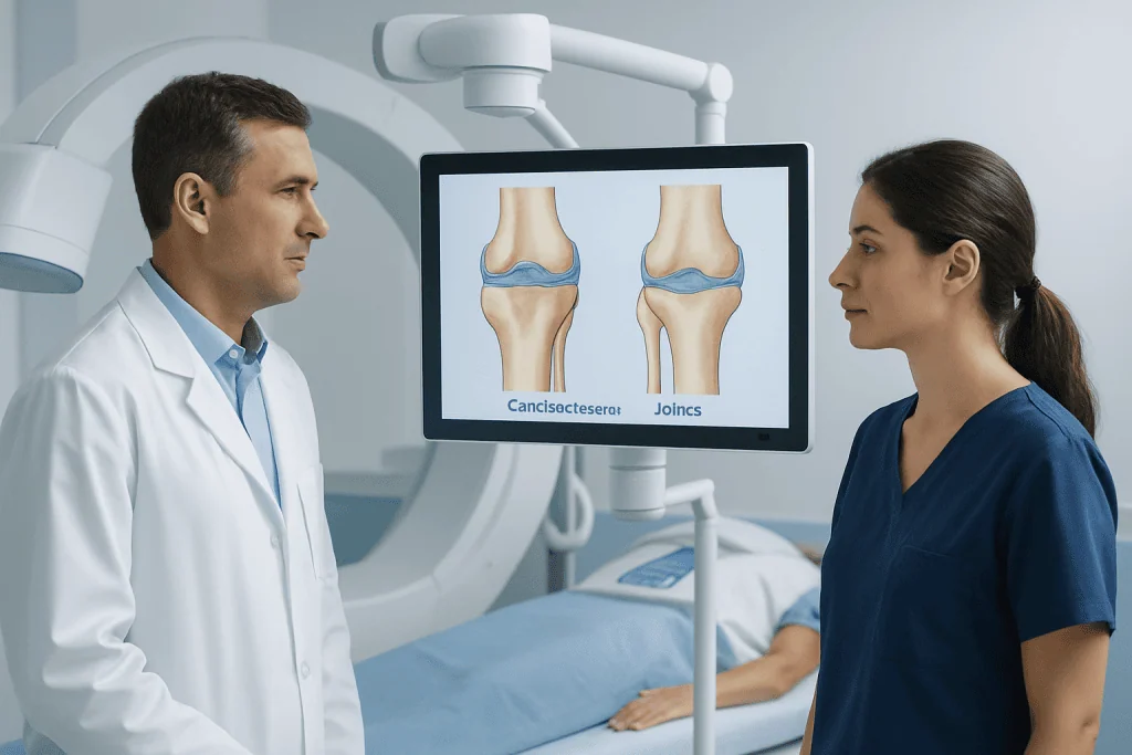
Cartilaginous joints are usually grouped into two types: synchondroses and symphyses. Both connect bones through cartilage, but they differ in structure and how much movement they allow.
Synchondroses are joints in which bones are connected by hyaline cartilage. Many are temporary during growth, such as the growth plates in children, where developing bone is separated by cartilage until maturity. Some synchondroses remain permanent, including certain joints between a rib and the breastbone.
Symphyses are joints in which bones are joined by fibrocartilage. Fibrocartilage is strong and slightly compressible, making it useful where the body must handle repeated stress. These joints allow a little more movement than synchondroses, but still much less than a synovial joint.
- Synchondrosis: bones connected by hyaline cartilage, often for growth or firm support
- Symphysis: bones connected by fibrocartilage, usually for strength and shock absorption
- Function: both types help with stability, load distribution, and controlled movement
Where Cartilaginous Joints Are Found in the Body
Cartilaginous joints are found in several important parts of the skeleton. One of the best-known examples is between the vertebrae of the spine, where intervertebral discs help cushion the bones and allow controlled movement. These discs are made largely of fibrocartilage and are essential for flexibility and shock absorption.
Another example is the pubic symphysis, located at the front of the pelvis. This joint helps unite the two halves of the pelvic bone. It is normally quite stable, but it can become more flexible during pregnancy because of hormonal changes that prepare the body for childbirth.
Some rib connections also involve cartilage. The first rib, for example, connects to the sternum through a synchondrosis. This arrangement provides support while still allowing the chest wall to move in a coordinated way during breathing.
In children and adolescents, the growth plates of long bones are also cartilaginous joints. These areas are vital for bone lengthening during development. Over time, they harden into bone as growth is completed.
What These Joints Do
The main purpose of a cartilaginous joint is to provide a firm connection between bones without making the area completely rigid. This balance allows the skeleton to stay stable while still adapting to normal body movements and forces.
These joints are especially important for shock absorption. In the spine, for example, the cartilage between vertebrae helps reduce the impact of walking, lifting, and twisting. Without this cushioning effect, daily activities would place much more strain on bones and surrounding tissues.
Cartilaginous joints also help distribute pressure. Rather than concentrating force at a single point, the cartilage spreads it more evenly. This supports long-term joint function and protects nearby structures such as ligaments, muscles, and nerves.
Although movement is limited, it is often exactly the amount the body needs. Small motions in the pelvis, ribs, and spine contribute to bending, posture, and breathing. When these joints are healthy, they work quietly in the background and are often unnoticed.
Common Problems That Can Affect Cartilaginous Joints
Even though cartilaginous joints are built for support, they can still become painful or damaged. In the spine, the intervertebral discs may wear down with age or strain. This can contribute to back pain, stiffness, or nerve-related symptoms if nearby nerves are affected. In some cases, doctors may evaluate symptoms related to a herniated disc or other disc problems.
Inflammation may also occur in joints where cartilage connects bones. For example, the rib-sternum area can become tender in conditions such as costochondral irritation. The pubic symphysis may become painful after overuse, sports activity, pregnancy, or childbirth.
Degeneration, injury, poor posture, repetitive stress, and certain inflammatory diseases can all affect these joints. In the spine, long-term wear may be associated with spinal alignment problems or age-related changes that alter how force is distributed.
Symptoms vary by location but may include localized pain, stiffness, tenderness, reduced flexibility, or discomfort with movement. Persistent symptoms should be assessed by a qualified doctor, especially if they interfere with walking, breathing, posture, or daily activity.
How Doctors Diagnose Cartilaginous Joint Problems
Diagnosis usually begins with a medical history and physical examination. A doctor asks where the pain is felt, what movements make it worse, how long it has lasted, and whether there was any injury, recent strain, or underlying medical condition. This helps narrow down whether the problem may involve a cartilaginous joint or surrounding tissues.
During the examination, the doctor may check posture, range of motion, tenderness, nerve function, and signs of inflammation. In the spine or pelvis, they may also assess walking pattern, muscle strength, and flexibility to understand how the area is functioning as a whole.
Imaging may be recommended if symptoms are ongoing, severe, or associated with neurologic findings. Depending on the situation, this may include X-rays, CT scans, or MRI scanning to look at cartilage, discs, bones, and nearby soft tissues in greater detail.
Sometimes additional tests are used to rule out infection, fracture, inflammatory arthritis, or other causes of pain. The goal is to identify the underlying reason for symptoms so treatment can be tailored safely and effectively.
Treatment Options and Self-Care
Treatment depends on the location of the joint, the cause of symptoms, and how much daily life is affected. Many mild or short-term problems improve with rest, activity modification, and supportive care. Doctors often recommend avoiding movements that worsen pain while keeping the body gently active to prevent stiffness.
Physical therapy can be very helpful, especially for spinal or pelvic symptoms. Guided exercises may improve posture, core strength, mobility, and body mechanics. For some people, an individualized physical therapy and rehabilitation plan reduces strain on the affected joint and supports long-term recovery.
When symptoms are related to disc damage or structural spine problems, treatment may involve a broader orthopedic or spine evaluation. Depending on findings, doctors may consider options ranging from medication and exercise to targeted procedures and, in selected cases, spine surgery. If symptoms involve joint degeneration elsewhere, an orthopedic surgery consultation may be appropriate.
Self-care measures can also support comfort and function:
- Use good posture during sitting, standing, and lifting
- Stay physically active with low-impact exercise
- Maintain a healthy weight to reduce pressure on load-bearing areas
- Warm up before sports and repetitive activity
- Seek medical advice if pain persists or worsens
When to See a Doctor
A doctor should be consulted if pain in the back, pelvis, chest wall, or another joint area lasts more than a few days, keeps returning, or limits normal activities. Medical assessment is also important when symptoms follow a fall, sports injury, heavy lifting, or other clear strain.
Urgent evaluation is needed if joint pain is accompanied by numbness, weakness, fever, difficulty breathing, loss of bladder or bowel control, or severe pain that does not improve with rest. These symptoms may point to a more serious problem that needs prompt attention.
People with a history of inflammatory disease, osteoporosis, cancer, or recent infection should also seek medical advice early if they develop new joint or spine pain. A timely diagnosis can help prevent complications and guide the right treatment plan.
For international patients needing specialist evaluation, Acibadem International’s multidisciplinary teams in JCI-accredited hospitals diagnose and treat bone, joint, and spine conditions with individualized care plans.
Frequently asked questions
What connects bones in a cartilaginous joint?
Bones in a cartilaginous joint are connected by cartilage. Depending on the joint, this may be hyaline cartilage or fibrocartilage, both of which provide support and limited movement.
Is a cartilaginous joint movable?
A cartilaginous joint usually allows little or only slight movement. Its main role is stability and cushioning rather than the wide range of motion seen in joints like the shoulder or knee.
What are examples of cartilaginous joints?
Common examples include the intervertebral discs between the spinal bones, the pubic symphysis in the pelvis, and some rib connections to the sternum. Growth plates in children are also a type of cartilaginous joint.
What is the difference between a synchondrosis and a symphysis?
A synchondrosis connects bones with hyaline cartilage and is often very stable. A symphysis connects bones with fibrocartilage, which is stronger against pressure and allows a small amount of movement.
Can cartilaginous joints cause pain?
Yes, they can become painful due to strain, inflammation, degeneration, injury, or nearby structural problems. The exact symptoms depend on the location, such as the spine, pelvis, or chest wall.
How are cartilaginous joint problems treated?
Treatment depends on the cause and may include rest, activity changes, physical therapy, pain management, and imaging or specialist assessment when needed. More advanced treatment is considered if symptoms are persistent or linked to structural damage.
References
- National Library of Medicine
- Cleveland Clinic
- Merck Manual Consumer Version
- American Academy of Orthopaedic Surgeons
- MedlinePlus
This article is for general information only and is not a substitute for professional medical advice. Please consult a qualified doctor about your individual situation.
Joint and spine care in Turkey — expert assessment & treatment
JCI-accredited · board-certified surgeons · reply within 24h
Add us as a Preferred Source to see more of our trusted health content across Google Search, AI Overviews and Discover.

More from the Health Library



Related Specialists

Prof. Dr. Gündüz Tezeren
Orthopedic Surgery & Traumatology
Assoc. Prof. Dr. Mustafa Emir Tavşanlı
Neurology
Dr. Serhat Konar
Anesthesiology
Assoc. Prof. Dr. Ahmet Özveren
Medical Oncology