How to Read a Brain MRI: Basic Terms Patients Often See in Reports
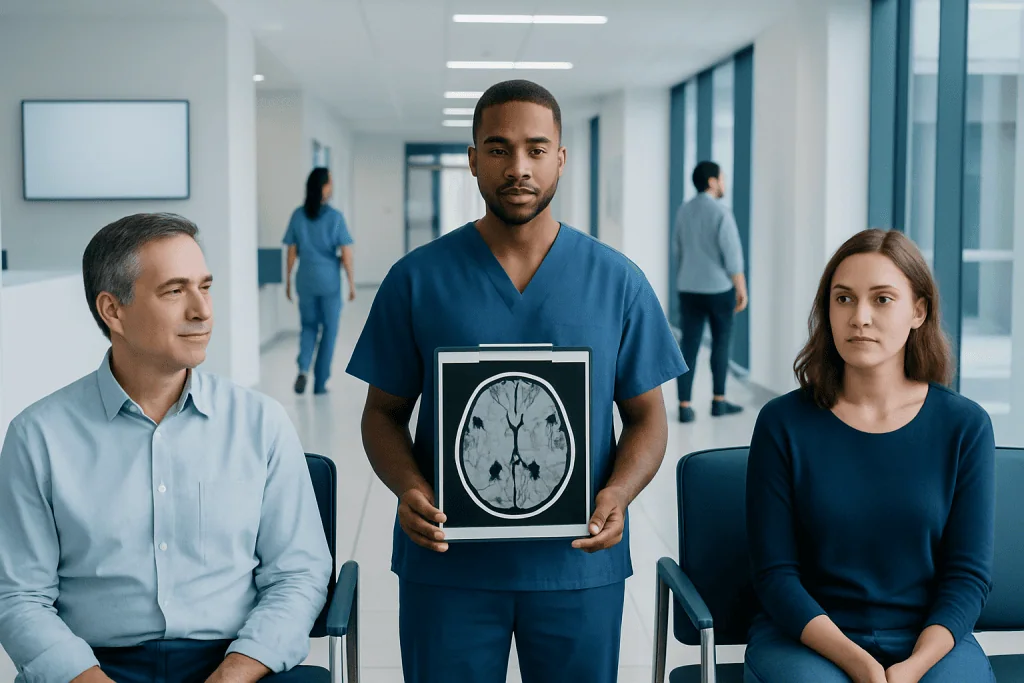
A brain MRI report describes what the radiologist sees, but it does not replace a diagnosis from the treating doctor. Many words in MRI reports are descriptive terms, not final conclusions or signs of serious disease.
Key Takeaways
- A brain MRI report describes what the radiologist sees, but it does not replace a diagnosis from the treating doctor.
- Many words in MRI reports are descriptive terms, not final conclusions or signs of serious disease.
- Common phrases such as lesion, enhancement, white matter changes, and mass effect have specific meanings that need clinical context.
- Normal MRI reports may still mention harmless anatomic variants or age-related changes.
- Patients should review MRI results with a neurologist, neurosurgeon, or referring physician rather than interpreting isolated terms alone.
Brain MRI reports often contain technical words that can sound confusing without medical training. Understanding the most common brain MRI report terms can help patients ask better questions, while remembering that the full meaning depends on symptoms, examination findings, and a doctor’s review.
Overview: What a Brain MRI Report Actually Shows
A brain MRI is an imaging test that uses magnetic fields and radio waves to create detailed pictures of the brain and nearby structures. A radiologist reviews these images and writes a report that describes what is seen. For many patients, the report may be available before a doctor has explained it, which can make routine or unclear wording feel worrying.
It helps to remember that a report is mainly a structured description. It often includes the reason for the scan, the imaging technique used, the main findings, and an impression section summarizing the radiologist’s opinion. Words in the report are chosen to be precise for medical communication, but they are not always easy to understand outside a clinical setting.
Most brain MRI reports contain a mix of normal anatomy, descriptive observations, and comments about whether anything looks urgent or unusual. Some terms may sound serious even when they are mild, nonspecific, or common with age. Others may need prompt medical follow-up. This is why the report should always be interpreted together with symptoms, medical history, and neurological examination.
How a Brain MRI Report Is Organized
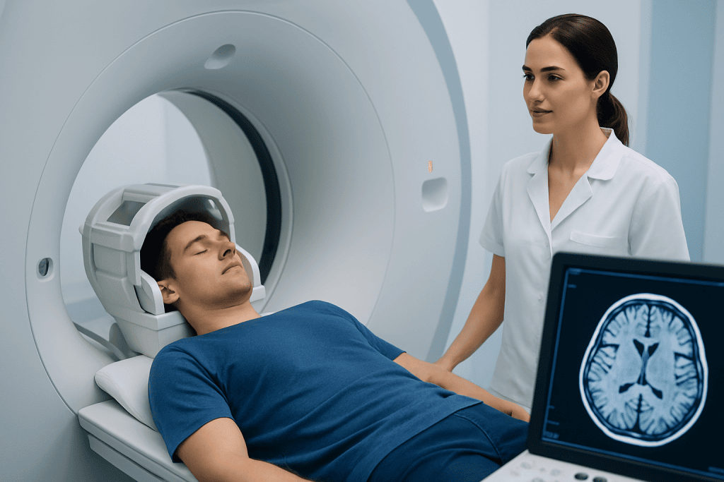
Although wording varies by hospital and radiologist, most reports follow a similar pattern. The first part may list the exam type, such as MRI brain with or without contrast. Contrast refers to a dye-like substance given through a vein to help highlight inflammation, tumors, blood vessels, or breakdown of the blood-brain barrier.
The next section usually describes the technique or sequences used. Patients may see terms such as T1, T2, FLAIR, diffusion, susceptibility, or post-contrast images. These are different ways of looking at brain tissue, fluid, blood products, and abnormal areas. They are technical labels rather than findings by themselves.
Then comes the findings section, where the radiologist describes structures such as the brain tissue, ventricles, midline, blood vessels, sinuses, skull base, and sometimes the orbits. The final impression is often the most important section because it summarizes the key points in plain diagnostic language for the referring doctor.
Patients can use the report as a guide, but the impression should not be read in isolation from the full medical picture. If the wording seems uncertain, this often reflects careful medicine rather than a hidden emergency. Radiologists sometimes use terms like “nonspecific,” “likely,” or “correlate clinically” when findings are not unique to one condition.
Common Normal and Reassuring Terms

Many reports contain phrases that indicate no major abnormality. Terms such as “no acute intracranial abnormality,” “no evidence of infarct,” “no hemorrhage,” or “no mass effect” are generally reassuring. “Acute” means sudden or recent, so this wording often means the MRI does not show a new stroke, active bleeding, or another urgent structural problem.
Patients may also read that the “ventricles are normal in size,” the “midline is preserved,” or “gray-white differentiation is maintained.” These are typical ways of saying that the brain’s structure and symmetry appear normal. The ventricles are fluid-filled spaces in the brain, and the midline refers to the central alignment of brain structures.
Another common phrase is “age-appropriate volume loss” or “mild generalized atrophy.” In older adults, mild shrinkage of brain tissue can be a common age-related finding and may not point to a specific disease on its own. In the same way, “mucosal thickening” in the sinuses may simply indicate mild sinus inflammation rather than a brain problem.
Words such as “incidental finding” can also sound concerning, but they often mean a feature that was noticed unexpectedly and may have little or no clinical importance. Examples can include tiny cysts, mild sinus changes, or harmless anatomic variants. Even so, incidental findings should still be reviewed with a doctor to decide whether any follow-up is needed.
Terms That Often Cause Confusion
One of the most common words in MRI reports is “lesion.” A lesion simply means an area that looks different from the surrounding tissue. It does not automatically mean cancer. Depending on the appearance and location, a lesion could reflect inflammation, a prior injury, changes from small blood vessel disease, infection, a demyelinating process, or a tumor.
Another frequent term is “enhancement.” This means an area becomes brighter after contrast is given, suggesting that the contrast has entered tissue more than expected. Enhancement can be seen with tumors, inflammation, infection, active demyelination, or healing tissue, so it is descriptive rather than diagnostic on its own.
“White matter hyperintensities” or “T2/FLAIR hyperintense foci” are also very common. These phrases describe small bright spots seen on specific MRI sequences. In some people they are related to aging, migraine, high blood pressure, diabetes, smoking, prior inflammation, or other nonspecific changes. In younger patients or in certain patterns, doctors may consider conditions such as multiple sclerosis.
Patients may also see “edema,” which means swelling, “mass effect,” which means a lesion is pushing on nearby structures, or “midline shift,” which means pressure has moved the brain from its usual central position. These terms deserve careful physician review because they can indicate that an abnormality is affecting surrounding tissue.
- Focal: limited to one area
- Diffuse: more widespread
- Chronic: long-standing, not newly developed
- Nonspecific: not unique to a single diagnosis
- Stable: unchanged compared with an earlier scan
What Certain Findings May Refer To
Some report terms point toward broad categories of conditions. “Infarct” usually refers to an area of tissue injury caused by reduced blood flow, as in a stroke. “Acute infarct” suggests a recent event, while “chronic infarct” points to an older, healed area. If a report mentions restricted diffusion, doctors may think about a recent stroke, but the meaning depends on the pattern and timing.
“Hemorrhage” means bleeding. A report might describe microbleeds, subdural collection, intraparenchymal hemorrhage, or subarachnoid blood, each of which has a different significance. “Susceptibility artifact” or “blooming” may suggest old blood products, calcium, or technical effects on a special MRI sequence.
Terms such as “demyelination,” “plaque,” or “periventricular lesions” can be associated with disorders that affect the brain’s insulating nerve covering, including demyelinating disease. Meanwhile, words such as “extra-axial mass,” “meningioma,” “glioma,” or “ring-enhancing lesion” may lead doctors to consider a tumor or another structural lesion, but further tests are often needed before confirming exactly what it is. In some cases, specialists may advise brain tumor treatment planning after full evaluation.
Reports can also mention “hydrocephalus,” meaning enlargement of the fluid spaces in the brain due to altered cerebrospinal fluid flow, or “atrophy,” meaning loss of brain volume. Atrophy can be generalized or focused in certain brain regions and may be seen with aging or in disorders that affect memory or movement, including some neurodegenerative conditions. The report alone usually cannot determine how symptoms will evolve.
Why MRI Terms Need Clinical Context
The same imaging phrase can mean different things in different people. For example, a few white matter spots in an older person with high blood pressure may be interpreted differently from the same pattern in a younger person with numbness, vision changes, or balance symptoms. This is why doctors combine the MRI with a history, examination, and sometimes blood tests, spinal fluid studies, or other imaging.
Comparison with prior scans is also very important. A finding described as “stable” over months or years is often less concerning than one that is new or growing. Radiologists commonly compare current images with older studies to look for changes in size, number, location, or enhancement pattern.
Sometimes the report recommends more testing. This may include a repeat MRI after an interval, MRI with contrast if contrast was not used initially, vascular imaging, or referral for tests such as electroencephalography if symptoms suggest seizures rather than a structural brain problem. In some situations, doctors may recommend a targeted procedure such as brain biopsy or specialist review by neurosurgery when the diagnosis remains uncertain.
For patients, the safest approach is to treat the MRI report as one part of the medical assessment. It can be helpful to write down unfamiliar terms and ask what they mean, whether they are new, whether they explain symptoms, and whether any follow-up is needed.
How Patients Can Read a Brain MRI Report More Confidently
A practical way to read the report is to start with the impression section, then go back to the findings. The impression usually highlights the most clinically important points. Patients can then note unfamiliar words and ask their doctor to explain whether the term is descriptive, incidental, age-related, or something that changes treatment.
It also helps to look for time-related words. “Acute,” “subacute,” and “chronic” suggest when a finding may have developed. “Stable” suggests no important interval change. “Recommend clinical correlation” means the radiologist is asking the treating team to interpret the imaging alongside symptoms and examination rather than implying that something is necessarily wrong.
Patients should be cautious about searching isolated MRI terms online, because many words cover a wide range of harmless and serious possibilities. The meaning of an “enhancing lesion” or “small vessel changes” depends on age, medical history, medications, symptoms, and the exact imaging pattern. Online information may not match an individual case.
For international patients who need specialist review, Acibadem International’s multidisciplinary specialists and JCI-accredited hospitals evaluate brain MRI findings and coordinate diagnosis and treatment when needed. A careful conversation with the treating team is often the clearest way to understand what a report means in real life.
When to Follow Up Promptly After a Brain MRI
Most MRI reports are reviewed in a planned outpatient setting, but some findings or symptoms need faster medical attention. Patients should contact their doctor promptly if the report mentions acute stroke, bleeding, hydrocephalus, significant mass effect, midline shift, or a rapidly growing lesion. These terms may require urgent clinical review even when symptoms seem mild.
Urgent medical care is also important if a person has new weakness, facial droop, trouble speaking, seizure, severe confusion, sudden vision loss, loss of consciousness, or a sudden severe headache. In these situations, symptoms matter as much as the written report. A normal older report should not delay emergency evaluation if new neurological symptoms appear.
Even when findings are not urgent, follow-up still matters. Patients may need repeat imaging, specialist referral, or treatment of underlying risk factors such as high blood pressure, diabetes, migraine, or inflammatory disease. Asking for a plain-language explanation and a clear follow-up plan can make the process much less stressful.
If there is uncertainty, it is reasonable to request that the report be reviewed by the referring doctor, a neurologist, or another appropriate specialist. Second opinions are sometimes helpful, especially for complex imaging findings or when symptoms and MRI results do not seem to match.
Frequently asked questions
Is the impression section the most important part of a brain MRI report?
The impression section is usually the best place to start because it summarizes the main findings and what the radiologist thinks they may mean. However, it should still be interpreted together with the full report, symptoms, and the doctor’s examination.
Does the word “lesion” in a brain MRI report mean cancer?
No. Lesion is a general descriptive term for an area that looks different from normal tissue. It can refer to many causes, including inflammation, old injury, vascular changes, infection, demyelination, or tumor.
What does “nonspecific white matter changes” usually mean?
This phrase describes small MRI signal changes that do not point to one single diagnosis by themselves. They may be related to age, migraine, blood vessel risk factors, prior inflammation, or other common conditions, but the pattern and the patient’s age help guide interpretation.
Should patients worry if a report says “incidental finding”?
Not necessarily. Incidental means something was found unexpectedly while looking for another issue, and many incidental findings are harmless or need only routine follow-up. A doctor can explain whether it matters in a particular case.
Why can an MRI report sound uncertain?
Radiologists often use careful wording when imaging findings are not unique to one condition. Terms like “likely,” “possible,” or “clinical correlation recommended” reflect appropriate medical caution, not poor quality or hidden bad news.
Can a brain MRI be normal even if someone has symptoms?
Yes. Some neurological symptoms can occur without visible structural changes on MRI, especially early in a condition or when the cause is functional, metabolic, electrical, or microscopic. That is why doctors may recommend other tests or follow-up even when the scan looks normal.
References
- Radiological Society of North America
- American College of Radiology
- National Institute of Neurological Disorders and Stroke
- National Institute on Aging
- NHS
This article is for general information only and is not a substitute for professional medical advice. Please consult a qualified doctor about your individual situation.
Explore treatments in Turkey — costs, top hospitals & a free quote
JCI-accredited · board-certified surgeons · reply within 24h
Add us as a Preferred Source to see more of our trusted health content across Google Search, AI Overviews and Discover.
Check your numbers in seconds
BMI, calories, due date, blood pressure and 30+ more clinical calculators — free, instant, doctor-reviewed ranges.