Respiratory System Diagram Explained: From Nose to Lungs

The respiratory system includes the nose, throat, windpipe, bronchi, lungs, and diaphragm. Its main job is to bring oxygen into the body and remove carbon dioxide.
Key Takeaways
- The respiratory system includes the nose, throat, windpipe, bronchi, lungs, and diaphragm.
- Its main job is to bring oxygen into the body and remove carbon dioxide.
- Tiny air sacs called alveoli are where gas exchange happens between the lungs and blood.
- The nose and airways help filter, warm, and moisten inhaled air before it reaches the lungs.
- Healthy habits such as avoiding smoking and treating infections promptly support lung function.
A respiratory system diagram helps show how air travels through the body and how oxygen reaches the blood. Understanding each part—from the nose to the lungs—can make breathing, lung health, and common symptoms easier to understand.
Overview of the Respiratory System
A respiratory system diagram is a simple way to understand how the body breathes. The respiratory system is a connected pathway that carries air from the outside environment into the lungs, where oxygen is absorbed into the bloodstream and carbon dioxide is removed. This process happens automatically throughout the day and night, even during sleep.
When people look at a diagram of the respiratory system, they usually see the upper airways at the top and the lungs lower down in the chest. Each structure has a specific role. Some parts guide air inward, some protect the body from dust and germs, and others help with the actual exchange of gases that keeps organs and tissues working.
The system is often divided into the upper respiratory tract and the lower respiratory tract. The upper part includes the nose, nasal cavity, mouth, throat, and voice box. The lower part includes the trachea, bronchi, bronchioles, and lungs. Together, these structures support breathing, speaking, and even the sense of smell.
The Main Parts: From Nose to Lungs
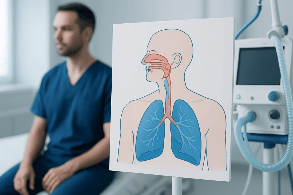
Air usually enters through the nose, although it can also enter through the mouth. The nose is an important first step because it filters, warms, and moistens the air before it travels deeper into the body. Tiny hairs and mucus in the nasal passages help trap dust, allergens, and some germs. From there, air passes through the pharynx, or throat, and then through the larynx, also known as the voice box.
Below the larynx is the trachea, often called the windpipe. The trachea is supported by firm rings of cartilage that help keep it open during breathing. It then divides into two main bronchi, one entering each lung. Inside the lungs, these bronchi branch into smaller tubes called bronchioles, creating a tree-like network that carries air throughout the lung tissue.
At the end of the bronchioles are tiny air sacs called alveoli. These are one of the most important parts of any respiratory system diagram because they are where oxygen moves into the blood and carbon dioxide moves out. Around the lungs are protective membranes called pleura, while below the lungs sits the diaphragm, the main muscle responsible for breathing.
- Nose and nasal cavity: filter, warm, and moisten air
- Pharynx and larynx: guide air and support speaking
- Trachea: carries air toward the lungs
- Bronchi and bronchioles: distribute air inside the lungs
- Alveoli: allow oxygen and carbon dioxide exchange
- Diaphragm: helps the lungs expand and contract
How Breathing Works

Breathing has two main phases: inhalation and exhalation. During inhalation, the diaphragm moves downward and the muscles between the ribs expand the chest. This creates space inside the chest cavity, allowing the lungs to fill with air. During exhalation, these muscles relax, and air flows back out of the lungs.
Once air reaches the alveoli, oxygen passes through very thin walls into tiny blood vessels called capillaries. At the same time, carbon dioxide—a waste gas made by the body’s cells—moves from the blood into the alveoli so it can be exhaled. This exchange is essential for energy production and healthy organ function.
Breathing is controlled by the brain, which constantly monitors oxygen and carbon dioxide levels in the blood. It adjusts the breathing rate automatically during rest, exercise, sleep, or illness. Although breathing usually happens without conscious effort, people may become more aware of it during exercise, stress, or respiratory conditions such as asthma.
What a Respiratory System Diagram Can Show
A clear respiratory system diagram can help patients, students, and families visualize structures that are otherwise hidden inside the body. It often shows the route of air from the nose or mouth, down the throat, through the trachea, and into the branching airways of the lungs. Some diagrams also highlight the diaphragm and the blood vessels around the alveoli to explain gas exchange more fully.
These diagrams can also help explain symptoms. For example, a sore throat affects the upper airway, while wheezing is often linked to narrowing in the bronchi or bronchioles. Mucus buildup may occur anywhere along the airway, and inflammation in the alveoli can affect oxygen transfer. Understanding the location of a problem often makes medical explanations easier to follow.
In clinics and hospitals, respiratory diagrams are commonly used when discussing tests or treatments. They may help explain why a doctor recommends lung function testing, chest imaging, oxygen therapy, or procedures such as bronchoscopy to examine the airways more closely. Visual tools can make complex anatomy feel more manageable and less confusing.
Common Conditions That Affect the Respiratory System
Many conditions can affect different parts of the respiratory system. Common upper airway problems include the common cold, sinus infections, allergies, and throat infections. Lower respiratory problems may include bronchitis, pneumonia, asthma, and chronic obstructive pulmonary disease. Each condition affects breathing in a different way depending on which structures are involved.
For example, asthma often causes inflammation and narrowing of the airways, leading to wheezing, cough, and chest tightness. Pneumonia affects the lung tissue and alveoli, making it harder for oxygen to pass into the blood. Other conditions, such as COPD, may gradually limit airflow and cause long-term shortness of breath.
Symptoms that may suggest a respiratory problem include persistent cough, wheezing, noisy breathing, fever with chest symptoms, breathlessness, or chest discomfort. Some symptoms are mild and temporary, while others need medical attention. When a person has ongoing symptoms or repeated infections, doctors may use examination, imaging, or tests such as pulmonary function tests to assess how well the lungs are working.
How Doctors Assess the Airways and Lungs
Doctors begin with a medical history and physical examination. They usually ask about symptoms such as cough, shortness of breath, chest tightness, sputum, fever, smoking exposure, allergies, workplace irritants, and family history. Listening to the chest with a stethoscope may reveal wheezing, crackles, or reduced airflow.
Depending on the symptoms, additional tests may be recommended. A chest X-ray or CT scan can show the lungs and surrounding structures. Spirometry and other lung function tests measure how much air a person can move in and out and how quickly this happens. Blood tests or pulse oximetry may help assess oxygen levels or signs of infection.
In some cases, specialists may recommend direct airway evaluation. This can be useful if there is unexplained bleeding, a suspected blockage, persistent infection, or a need to collect samples. In larger centers, respiratory care may involve pulmonologists, radiologists, ENT specialists, and critical care teams working together to make a diagnosis and choose the most suitable treatment plan.
Protecting Respiratory Health
Good respiratory health starts with everyday habits. Avoiding smoking and secondhand smoke is one of the most important steps because tobacco smoke irritates and damages the airways and lung tissue. Reducing exposure to dust, fumes, pollution, and workplace chemicals may also help protect the lungs over time.
Preventing infection matters as well. Regular handwashing, staying up to date with recommended vaccines, and seeking treatment when respiratory symptoms worsen can all support recovery and reduce complications. People with chronic lung conditions may benefit from a personalized care plan, regular checkups, and learning how to recognize symptom flare-ups early.
General health also influences breathing. Staying physically active, drinking enough fluids, sleeping well, and managing allergies or acid reflux may all support respiratory comfort. For some people with more complex symptoms, doctors may suggest targeted treatments such as oxygen therapy or pulmonary rehabilitation as part of ongoing care.
When to Seek Medical Advice
Many respiratory symptoms improve with rest, fluids, and time, especially when caused by mild viral infections. However, medical advice is important if symptoms are severe, do not improve, or return often. A doctor should assess unexplained shortness of breath, a cough lasting several weeks, wheezing, coughing up blood, chest pain with breathing, or repeated chest infections.
Urgent medical care is needed if breathing becomes difficult, lips or fingertips look bluish, confusion develops, or a person cannot speak normally because of breathlessness. Young children, older adults, and people with chronic illnesses may need earlier assessment because respiratory problems can worsen more quickly in these groups.
Understanding a respiratory system diagram can help people describe their symptoms and follow medical explanations more easily, but it does not replace professional evaluation. If symptoms suggest a lung or airway problem, a qualified doctor can guide the next steps. For international patients who need evaluation or treatment, Acibadem International’s multidisciplinary specialists in JCI-accredited hospitals provide diagnosis and care for respiratory conditions.
Frequently asked questions
What is the respiratory system?
The respiratory system is the group of organs and airways that allows the body to breathe. Its main job is to bring oxygen into the body and remove carbon dioxide.
What are the main parts shown in a respiratory system diagram?
Most diagrams show the nose, mouth, pharynx, larynx, trachea, bronchi, bronchioles, lungs, alveoli, and diaphragm. Together, these parts move air and support gas exchange.
Where does oxygen enter the blood?
Oxygen enters the blood in the alveoli, which are tiny air sacs inside the lungs. Their thin walls lie next to small blood vessels, allowing oxygen and carbon dioxide to move between air and blood.
Why is the nose important in breathing?
The nose helps filter, warm, and moisten the air before it reaches the lungs. This makes breathing more efficient and helps protect the lower airways from dust and irritation.
What symptoms may suggest a respiratory problem?
Common signs include cough, wheezing, shortness of breath, chest tightness, noisy breathing, or repeated respiratory infections. If symptoms are persistent, severe, or worsening, a doctor should evaluate them.
Can a respiratory system diagram help patients understand disease?
Yes, diagrams can make it easier to see where a problem is happening, such as in the throat, bronchi, or lungs. This often helps patients understand symptoms, tests, and treatment options more clearly.
References
- World Health Organization
- National Heart, Lung, and Blood Institute
- American Lung Association
- MedlinePlus
- NHS
This article is for general information only and is not a substitute for professional medical advice. Please consult a qualified doctor about your individual situation.
Explore treatments in Turkey — costs, top hospitals & a free quote
JCI-accredited · board-certified surgeons · reply within 24h
Add us as a Preferred Source to see more of our trusted health content across Google Search, AI Overviews and Discover.