

R/O Mass on a Scan: What It Means and What Usually Happens Next
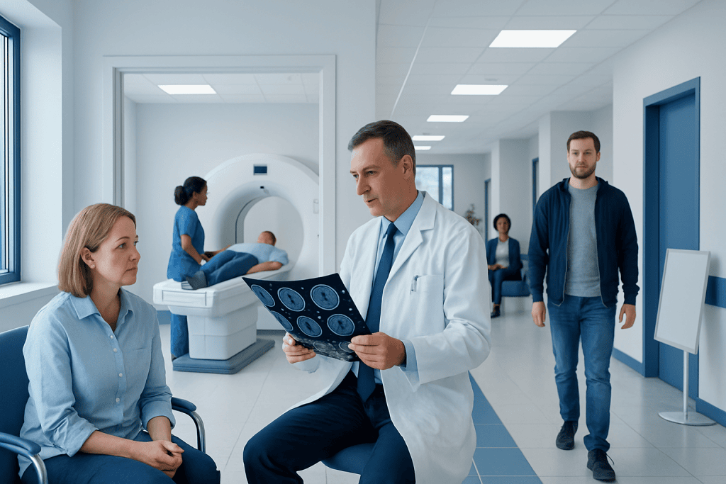
“R/O” stands for “rule out,” meaning a finding needs clarification, not that a diagnosis has been made. A mass on imaging can have many causes, including cysts, inflammation, scar tissue, benign tumors, or cancer.
Key Takeaways
- “R/O” stands for “rule out,” meaning a finding needs clarification, not that a diagnosis has been made.
- A mass on imaging can have many causes, including cysts, inflammation, scar tissue, benign tumors, or cancer.
- Most people need follow-up imaging, a specialist review, or additional tests before doctors can say what the finding means.
- The report wording reflects imaging uncertainty; the final interpretation depends on the whole clinical picture.
- Prompt follow-up matters, but many masses turn out not to be cancer.
Seeing “R/O mass” on a scan report can be unsettling, but it usually means the radiologist wants to rule out an abnormal growth rather than confirm one. The next step is typically more focused imaging, comparison with past scans, or sometimes a biopsy to clarify what the finding is.
Overview: What “R/O Mass” Means on a Scan
When a scan report says “R/O mass,” it usually means “rule out mass.” This is a common medical shorthand used by radiologists and other doctors when an image shows an area that is not fully explained and may need further evaluation. It is not a final diagnosis. Instead, it signals that the doctor wants to consider whether the area could represent a mass and, if so, what type.
In imaging, the word “mass” simply describes an abnormal lump, growth, or area of tissue that looks different from surrounding structures. A mass may be solid or fluid-filled, large or small, clearly harmless or uncertain. Some masses are benign, such as cysts, fibroids, lipomas, or enlarged lymph nodes due to infection. Others may require testing to exclude cancer or another significant condition.
Radiology reports often use cautious language because scans have limits. The appearance of an area can be influenced by scan quality, body position, contrast use, movement, nearby structures, and whether there are older images for comparison. For this reason, terms such as “possible,” “suspicious for,” or “cannot exclude” are common and do not mean that the worst-case explanation is likely.
Why a Mass May Appear on Imaging
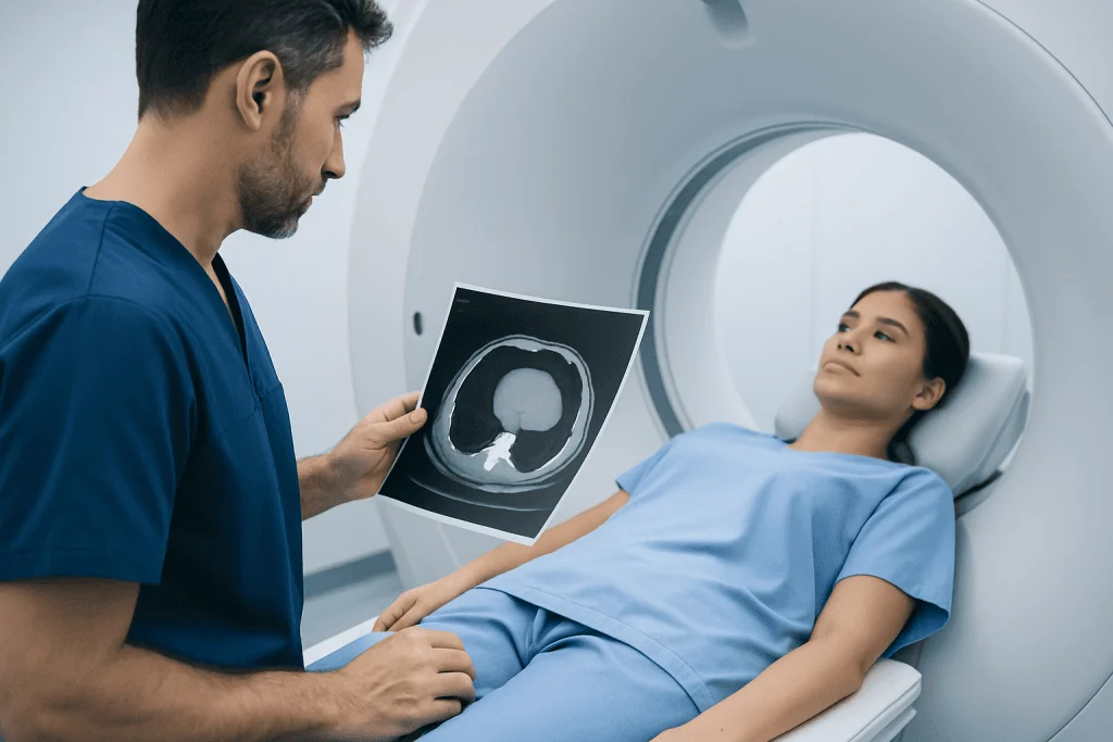
A scan may suggest a mass for many reasons. Sometimes there truly is a growth, but not all growths are dangerous. Benign causes include simple cysts, fatty growths, scar tissue, fibroids, inflammation, abscesses, swollen glands, or normal structures that look unusual from a certain angle. In other situations, the radiologist may see an area of thickening, a shadow, or a small nodule that deserves a closer look.
The type of scan also matters. An X-ray gives limited detail compared with ultrasound, CT, or MRI. A CT scan can show shape, density, and how a structure relates to nearby tissues. MRI often gives more information about soft tissue. Ultrasound can help determine whether a finding is solid or fluid-filled. If the report raises concern after one test, another imaging method may be used to learn more.
Location is important as well. A “mass” in the lung, liver, kidney, breast, thyroid, ovary, brain, or lymph nodes may be approached differently because each organ has its own common benign and malignant conditions. For example, a scan might prompt evaluation for a lung mass, a breast lump, or a brain tumor depending on where the finding appears and how it looks.
Symptoms and What They Do or Do Not Mean
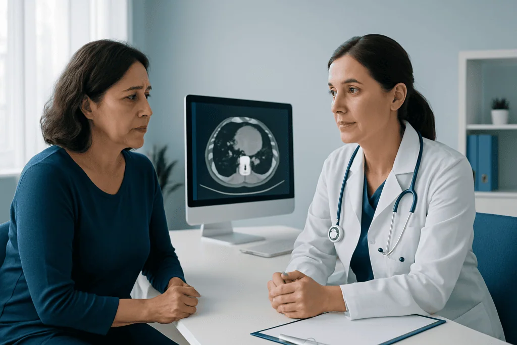
Some people with a reported mass have no symptoms at all. Many findings are discovered incidentally during scans done for another reason, such as back pain, cough, kidney stones, or a routine health check. Incidental findings are common, especially as imaging becomes more detailed. An unexpected report can be stressful, but lack of symptoms is not unusual.
When symptoms are present, they vary widely based on the part of the body involved. Possible symptoms may include pain, swelling, a visible lump, unexplained weight loss, fatigue, cough, bleeding, changes in bowel or bladder habits, headaches, or difficulty swallowing. However, these symptoms are not specific and can occur with many noncancerous conditions too.
Symptoms alone cannot show whether a mass is benign or malignant. Likewise, the absence of symptoms does not always mean a finding is unimportant. Doctors interpret the report alongside age, medical history, family history, smoking history, recent infections, prior surgeries, and physical examination findings. This broader context helps decide how quickly the mass should be investigated.
How Doctors Evaluate an R/O Mass Finding
The first step is usually careful review of the radiology report and the actual images. The doctor may compare the scan with prior studies to see whether the area is new, stable, or changing over time. Stability over months or years can be reassuring for some types of masses, while growth may suggest the need for more testing.
Additional imaging is often the next step. Depending on the body area, this may include a dedicated ultrasound, a contrast-enhanced CT, an MRI scan, a PET-CT scan, or repeat imaging after a set interval. These tests can help define size, shape, borders, blood supply, and whether the area involves nearby structures. In many cases, more detailed imaging provides enough information to show that the finding is likely benign.
If uncertainty remains, doctors may order blood tests or recommend referral to a specialist such as an oncologist, pulmonologist, breast specialist, gynecologist, urologist, or surgeon. The exact path depends on where the mass is located and what the scan suggests. Clear communication with the referring doctor is important so the next step matches the degree of concern described in the report.
When a Biopsy Is Needed
A biopsy is not always necessary, but it is sometimes the best way to make a definite diagnosis. In a biopsy, a doctor removes a small sample of tissue so it can be examined under a microscope. This helps determine whether the mass is benign, inflammatory, infectious, precancerous, or cancerous. Imaging can strongly suggest a diagnosis, but tissue testing may be needed when treatment decisions depend on certainty.
Biopsies can be done in different ways. Some are performed with a needle using ultrasound or CT guidance, while others require endoscopy or a minor surgical procedure. The choice depends on where the mass is located, how easy it is to reach, and whether there are risks such as bleeding or injury to nearby structures. Doctors weigh these factors carefully before recommending the procedure.
For selected cases, the medical team may suggest biopsy after imaging review. If cancer is confirmed, further tests may be needed to determine the exact type and stage before treatment planning begins. At that point, some patients also undergo oncology evaluation and cancer treatment planning with a multidisciplinary team.
Possible Outcomes After Follow-Up Testing
There are several common outcomes after an R/O mass finding. The area may turn out to be a normal anatomical variant or imaging artifact, meaning it only looked suspicious because of technical factors or the angle of the scan. It may also be identified as a benign condition, such as a cyst, lipoma, hemangioma, fibroid, or reactive lymph node, requiring only observation or no treatment at all.
In other cases, the mass may need treatment even if it is not cancer. Examples include abscesses, large benign tumors causing pressure symptoms, or lesions that affect organ function. Treatment may involve medication, drainage, surgery, or follow-up surveillance depending on the diagnosis and the patient’s symptoms.
Sometimes the tests show a cancerous or precancerous lesion. If that happens, the next steps usually include staging, specialist consultation, and a personalized treatment plan. Modern care often involves a combination of surgery, medication, radiation therapy, or close monitoring, depending on the tumor type and stage. Although this possibility is the reason follow-up matters, it is important to remember that “R/O mass” alone does not mean cancer.
What Patients Can Do While Waiting
Waiting for clarification can be emotionally difficult. A helpful first step is to ask the ordering doctor to explain the report in plain language: where the mass is, how concerning it appears, and what test is recommended next. Patients may also ask whether the finding was incidental, whether older scans are available for comparison, and how soon follow-up should be arranged.
Keeping medical information organized can make the process smoother. It helps to gather copies of the report, prior imaging, medication lists, and any relevant family history. If symptoms are present, writing down when they began, how often they occur, and whether they are changing can help the doctor assess the situation more accurately.
It is usually best to avoid assuming the worst based only on report wording or online searches. Reliable guidance comes from a qualified clinician who can interpret the scan in context. Near the end of the evaluation process, some patients seek care at specialized centers; Acibadem International’s multidisciplinary specialists in JCI-accredited hospitals diagnose and treat patients with suspicious imaging findings and confirmed cancers from many countries.
When to See a Doctor Urgently
Most R/O mass findings are addressed through scheduled follow-up rather than emergency care, but some situations deserve prompt medical attention. A person should contact a doctor quickly if there is severe pain, new neurological symptoms, shortness of breath, significant bleeding, rapidly worsening swelling, persistent vomiting, or signs of serious infection such as high fever and chills.
Even without urgent symptoms, it is wise not to ignore the report. Delayed follow-up can make diagnosis slower and add unnecessary stress. If the radiology report recommends a timeline, such as repeat imaging in a few weeks or specialist review within a certain period, that guidance should be followed whenever possible.
If communication is unclear, patients should ask for a written plan. Knowing the next step, expected timing, and which doctor is coordinating care can reduce uncertainty. In general, timely evaluation is important, but there is usually enough time to ask questions, understand options, and make informed decisions with the healthcare team.
Frequently asked questions
Does “R/O mass” mean cancer?
No. “R/O” means “rule out,” so the report is saying that a possible mass needs further evaluation. Many findings that are described this way turn out to be benign or even related to normal anatomy.
Why would a radiologist use uncertain wording on a scan report?
Radiologists often use cautious language when an image is not fully specific. A scan may show an abnormal area, but the exact cause may not be clear without more detailed imaging, comparison with older scans, or tissue testing.
What usually happens after a scan says R/O mass?
The next step is commonly a review by the referring doctor, often followed by repeat or more targeted imaging. Depending on the location and appearance, some people also need blood tests, a specialist appointment, or a biopsy.
Can a mass on a scan be harmless?
Yes. Many masses are benign, including cysts, fibroids, lipomas, scar tissue, and enlarged lymph nodes from infection or inflammation. The purpose of follow-up is to distinguish harmless findings from those that need treatment.
How fast should follow-up happen?
The timing depends on how suspicious the finding appears, where it is located, and whether symptoms are present. Some findings need prompt specialist review, while others are safely rechecked after a set interval. The ordering doctor can explain the recommended timeline.
Will everyone with an R/O mass need a biopsy?
No. If additional imaging strongly suggests a benign diagnosis, a biopsy may not be necessary. Biopsy is usually reserved for cases where the diagnosis remains uncertain or where treatment decisions require tissue confirmation.
References
- American Cancer Society
- National Cancer Institute
- Radiological Society of North America
- American College of Radiology
- National Health Service
This article is for general information only and is not a substitute for professional medical advice. Please consult a qualified doctor about your individual situation.
Oncology care in Turkey — second opinion and treatment plan
JCI-accredited · board-certified surgeons · reply within 24h
Add us as a Preferred Source to see more of our trusted health content across Google Search, AI Overviews and Discover.

More from the Health Library



Related Specialists

Dr. Burak Sercan Erçin
Aesthetic Plastic & Reconstructive Surgery
Dr. G. Pınar Özeren
Gynecology & Obstetrics
Dt. Canan Duman
Dentistry
Dr. Hasan Cem Güneyli
Anesthesiology