Brain AVM: Seizures, Headaches, and When Imaging Finds an Abnormal Tangle of Vessels

A brain AVM is a tangle of abnormal blood vessels that disrupts normal blood flow in the brain. Symptoms can include seizures, headaches, weakness, vision or speech changes, or sudden bleeding, but some AVMs cause no symptoms.
Key Takeaways
- A brain AVM is a tangle of abnormal blood vessels that disrupts normal blood flow in the brain.
- Symptoms can include seizures, headaches, weakness, vision or speech changes, or sudden bleeding, but some AVMs cause no symptoms.
- Diagnosis usually involves brain imaging such as CT, MRI, and specialized blood vessel imaging.
- Treatment depends on the AVM’s size, location, bleeding risk, symptoms, and the person’s overall health.
- Ongoing follow-up with a neurologist, neurosurgeon, or interventional neuroradiology team is important for safe, individualized care.
A brain AVM is an abnormal connection between arteries and veins in the brain. Some cause seizures, headaches, or bleeding, while others are found unexpectedly on brain imaging done for another reason.
Overview
A brain arteriovenous malformation, often called a brain AVM, is an abnormal tangle of blood vessels in the brain. In a typical circulation pattern, arteries carry oxygen-rich blood from the heart to the brain, and veins return blood back toward the heart after it has passed through tiny capillaries. In an AVM, arteries connect to veins more directly, without the usual capillary network in between.
This unusual vessel pattern can change blood flow and pressure in the surrounding brain tissue. Over time, some AVMs may irritate nearby brain areas and lead to symptoms such as seizures or headaches. In other cases, the main concern is bleeding, because the involved vessels may be more fragile than normal.
Not every brain AVM causes symptoms. Some are discovered by chance when imaging is performed for headaches, a minor injury, dizziness, or another unrelated concern. Once found, a brain AVM should be evaluated carefully by specialists who can estimate its risks and discuss whether monitoring or treatment is the better option.
Symptoms
Symptoms of a brain AVM vary widely. Some people have no symptoms at all, while others first come to medical attention after a seizure, a severe headache, or sudden neurological changes. The exact symptoms often depend on the AVM’s size and where it is located in the brain.
Possible symptoms may include:
- Seizures
- Headaches, which may be ongoing or intermittent
- Weakness or numbness in part of the body
- Problems with speech or understanding language
- Vision changes
- Dizziness or balance problems
- Difficulty with memory, concentration, or other thinking skills
If a brain AVM bleeds, symptoms may begin suddenly and can be more serious. A person may develop a sudden severe headache, vomiting, confusion, weakness, trouble speaking, or loss of consciousness. Bleeding in the brain is a medical emergency and needs immediate assessment.
Headaches and seizures can have many causes, and a brain AVM is only one possibility. Related conditions that may also affect brain blood vessels include brain aneurysm and stroke, which are evaluated differently and may require urgent care.
Causes and Risk Factors

The exact cause of most brain AVMs is not fully understood. They are generally believed to form before birth, during the development of the blood vessels, although they may not be detected until childhood or adulthood. In most people, a brain AVM is not caused by anything they did or did not do.
Brain AVMs are usually considered rare. Most occur sporadically, meaning they happen without a clear inherited pattern. In a small number of cases, AVMs can be associated with genetic conditions that affect blood vessel formation, but this is not the situation for most patients.
Doctors look at several factors when considering future risks. These can include whether the AVM has bled before, its size, how deep it is in the brain, and whether it drains through deep veins. The patient’s age, symptoms, and general health also help shape decisions about treatment or observation.
It can be reassuring to know that many people with brain AVMs are diagnosed before a major complication happens. Careful evaluation helps the medical team estimate risk and choose the safest path forward.
How Brain AVM Is Diagnosed
Diagnosis usually begins with a medical history and neurological examination. The doctor asks about headaches, seizures, weakness, vision changes, and any sudden episodes that might suggest prior bleeding. If a brain AVM is suspected, imaging tests are used to confirm the diagnosis and map the blood vessels in detail.
A CT scan is often used urgently if bleeding is suspected, because it can quickly show blood in or around the brain. MRI provides more detailed images of brain tissue and can help define the AVM and its effects on surrounding structures. Doctors may also use specialized scans to better understand blood flow.
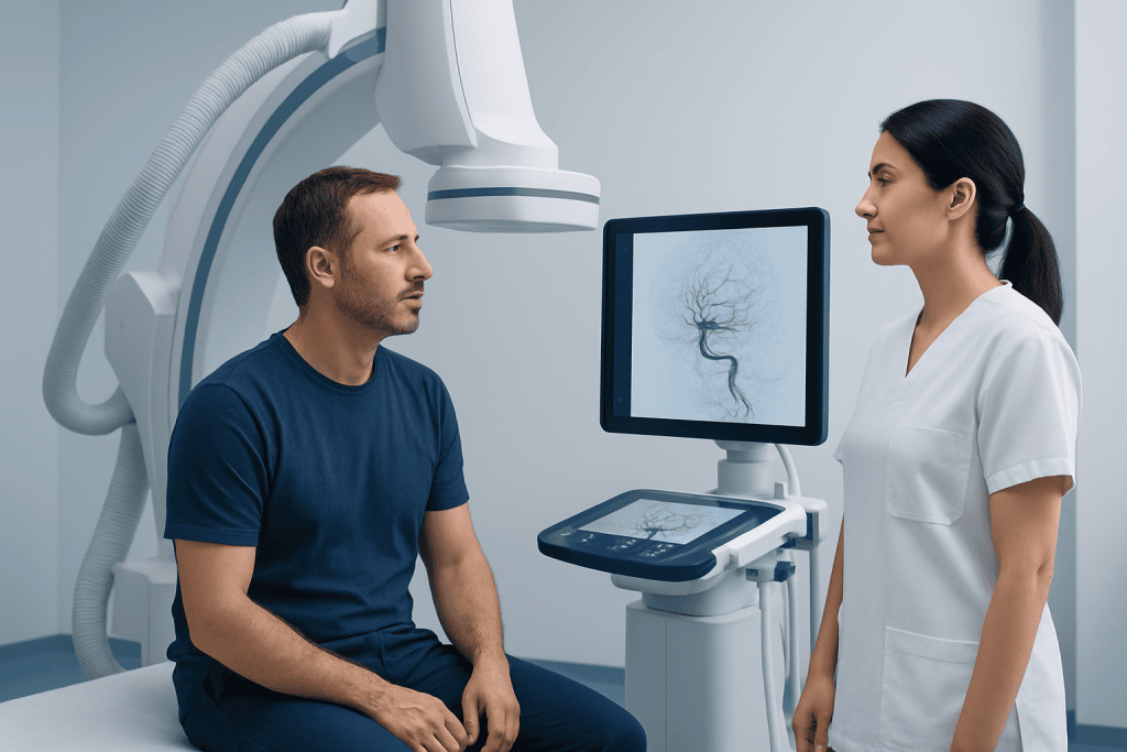
Cerebral angiography, also called a brain angiogram, is often the most detailed test for planning treatment. During this procedure, contrast dye is injected into blood vessels so the care team can see the feeding arteries, the abnormal tangle, and the draining veins. This information is especially important if the AVM might be treated with surgery, radiosurgery, or endovascular techniques such as interventional neuroradiology procedures.
In some cases, an AVM found on imaging is small and not causing symptoms. Even then, follow-up with experienced specialists matters, because treatment decisions depend on a careful balance of the AVM’s natural risk versus the risks of intervention.
Treatment Options
Treatment for a brain AVM is highly individualized. Some AVMs are best monitored over time, especially if they are in a sensitive area of the brain or if the risks of treatment may outweigh the risks of the AVM itself. Others are treated to reduce the chance of bleeding or to control symptoms such as seizures.
The main treatment approaches include microsurgery, endovascular embolization, and stereotactic radiosurgery. Microsurgery aims to remove the AVM completely, often through neurosurgery. Endovascular embolization involves guiding a small catheter through blood vessels and placing materials that reduce blood flow into the AVM; this may be used alone in selected cases or as part of a broader treatment plan. Stereotactic radiosurgery delivers focused radiation to help close off the abnormal vessels over time and may be considered for certain AVMs that are difficult to reach surgically.
Many patients also need treatment for symptoms. Anti-seizure medicines may help control seizures, and supportive care may be needed after a bleed or after a procedure. If a person has had a hemorrhage, treatment decisions are often more urgent because prior bleeding can affect future risk.
The best plan is usually made by a multidisciplinary team that may include neurology, neurosurgery, and interventional radiology or interventional neuroradiology specialists. Near the end of the care journey, some international patients may choose centers such as Acibadem International, where multidisciplinary specialists in JCI-accredited hospitals evaluate and treat brain AVMs with individualized planning.
Prevention and Self-Care
Because most brain AVMs are thought to develop before birth, there is no known way to prevent them. Self-care therefore focuses on reducing complications, recognizing warning signs, and staying engaged with follow-up care. A person with a diagnosed AVM should understand their treatment plan and know when to seek urgent help.
If seizures have occurred, taking prescribed medication consistently and following safety advice is important. Depending on local laws and the doctor’s guidance, activities such as driving, swimming alone, climbing heights, or operating machinery may need temporary restrictions until seizures are well controlled. Good sleep, regular meals, and avoiding missed medications may also help reduce seizure triggers.
General brain and vascular health still matter. It is sensible to manage blood pressure, avoid smoking, limit excess alcohol, and discuss all medicines with a clinician, including blood thinners or supplements that might affect bleeding risk. Patients should not stop or start medication on their own without medical advice.
Emotional support can also be helpful. Learning that there is an abnormal blood vessel tangle in the brain can feel overwhelming, especially if it was found unexpectedly. Clear information, specialist follow-up, and support from family or counseling services can help people feel more confident and prepared.
When to See a Doctor
A person should arrange medical evaluation if they have unexplained seizures, recurrent severe headaches, new weakness, changes in speech, or other neurological symptoms. Even if symptoms seem mild or improve, they can signal a condition that needs specialist assessment and brain imaging.
Emergency care is needed for signs that could suggest bleeding in the brain. These include a sudden severe headache unlike usual headaches, fainting, confusion, vomiting, a seizure in someone without a known seizure disorder, sudden weakness, facial drooping, or trouble speaking. Acting quickly can be very important.
People who have already been diagnosed with a brain AVM should keep scheduled follow-up visits and ask questions if anything changes. A new symptom does not always mean the AVM has bled, but it should not be ignored. Timely review helps doctors decide whether repeat imaging or a change in treatment is needed.
Frequently asked questions
Is a brain AVM always dangerous?
Not always. Some brain AVMs never cause symptoms and are found incidentally on imaging. However, they can carry risks such as bleeding or seizures, so each case should be assessed individually by specialists.
Can a brain AVM cause headaches?
Yes, a brain AVM can be associated with headaches, although headaches are common and have many possible causes. The pattern of headache does not always reveal whether an AVM is present, so imaging may be needed if a doctor is concerned.
What is the difference between a brain AVM and an aneurysm?
A brain AVM is a tangle of abnormal connections between arteries and veins. An aneurysm is a weakened, bulging area in a blood vessel wall. Both involve blood vessels in the brain, but they are different conditions with different treatment considerations.
Does every brain AVM need treatment?
No. Some AVMs are monitored rather than treated, especially if they are small, symptom-free, or located in a high-risk area for intervention. The decision depends on factors such as age, symptoms, prior bleeding, and the AVM’s anatomy.
Can a brain AVM be cured?
In some cases, yes. If the AVM can be completely removed or successfully closed off, treatment may be considered curative. However, not every AVM is suitable for the same approach, and some require staged treatment or long-term monitoring.
What tests are used to diagnose a brain AVM?
Doctors commonly use CT scans, MRI, and cerebral angiography. CT is especially useful in emergencies to check for bleeding, while MRI and angiography provide more detailed information about the AVM’s size, location, and blood flow.
References
- National Institute of Neurological Disorders and Stroke
- American Stroke Association
- National Organization for Rare Disorders
- Radiological Society of North America
- National Health Service
This article is for general information only and is not a substitute for professional medical advice. Please consult a qualified doctor about your individual situation.
Explore treatments in Turkey — costs, top hospitals & a free quote
JCI-accredited · board-certified surgeons · reply within 24h
Add us as a Preferred Source to see more of our trusted health content across Google Search, AI Overviews and Discover.