Digital PET vs Standard PET: What Patients Should Know

Both digital PET and standard PET use radioactive tracers to show metabolic activity inside the body. Digital PET uses newer detector technology that can improve image quality and timing performance.
Key Takeaways
- Both digital PET and standard PET use radioactive tracers to show metabolic activity inside the body.
- Digital PET uses newer detector technology that can improve image quality and timing performance.
- Clearer images may help doctors detect small abnormalities and interpret scans with greater confidence.
- The patient experience is often similar for both types of PET scans, including preparation and tracer injection.
- The best choice depends on the medical question, the body area being studied, and the imaging center’s expertise.
Digital PET and standard PET are both imaging methods that help doctors see how tissues and organs are functioning. The main difference is the detector technology: digital PET can provide clearer images and may improve lesion detection, especially in certain clinical situations.
Overview: What Digital PET and Standard PET Mean
Positron emission tomography, or PET, is an imaging test that helps doctors evaluate how the body is working rather than only how it looks. After a small amount of radioactive tracer is given, the scanner detects signals from the tracer and creates images that show patterns of metabolism or other biologic activity. PET is often used in oncology, neurology, and cardiology because many diseases cause measurable changes in tissue function.
Standard PET and digital PET are based on the same medical principle. Both scans detect positrons emitted by the tracer and convert that information into images. The difference lies in the hardware that captures the signals. Traditional systems use older analog detector technology, while digital PET systems use newer digital detectors and electronics designed to measure the incoming signals more precisely.
For patients, this usually means that the reason for the scan, the tracer injection, and the general steps of the appointment are quite similar. The main advantage of digital PET is not that it is a completely different test, but that it may produce sharper images, better contrast, and improved detection of small or faint abnormalities in some cases.
How PET Scans Work
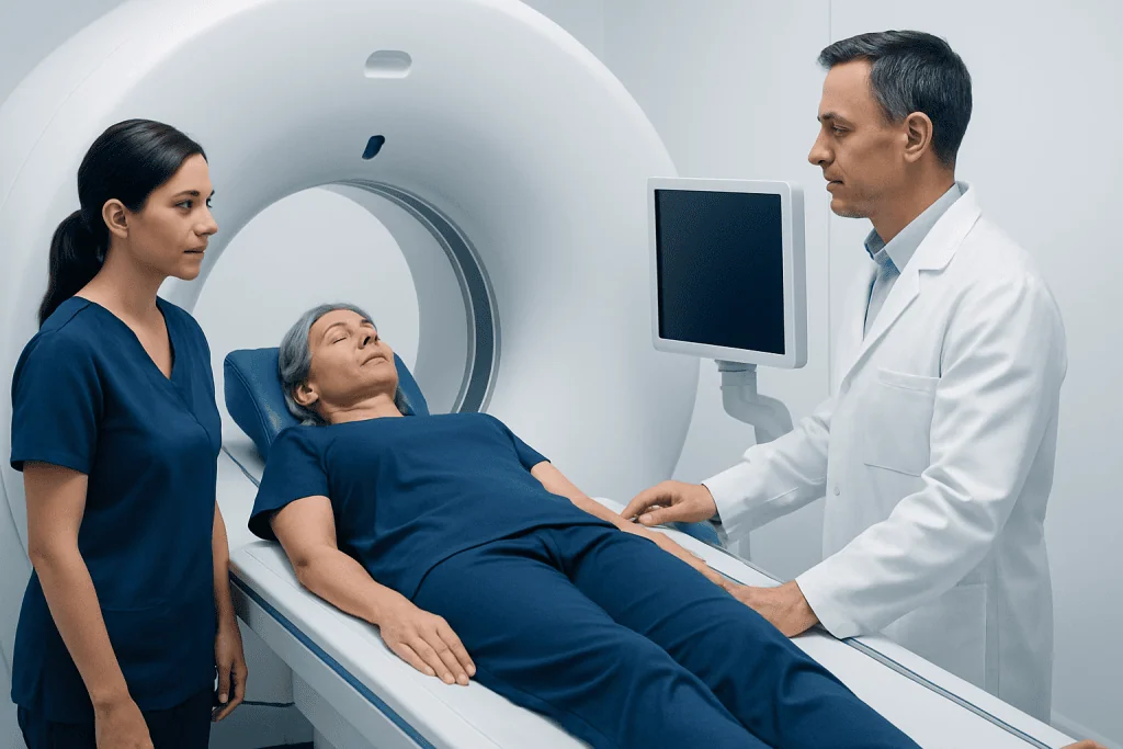
A PET scan begins with a radioactive tracer, most commonly a glucose-like substance that travels through the bloodstream and is taken up by tissues. Cells that are more metabolically active, such as some cancer cells, inflamed tissue, or parts of the brain with high activity, may absorb more tracer. The PET scanner then records this activity and reconstructs it into images.
In many hospitals, PET is combined with CT, and in some centers with MRI, to provide both functional and structural information in one session. This can help doctors match areas of high tracer uptake with precise anatomy. In practice, PET is often used to help diagnose disease, stage cancer, assess treatment response, or monitor for recurrence.
Digital PET and standard PET both perform these tasks. The difference is in how accurately and efficiently the scanner captures the light signals produced inside the detector crystals. More advanced signal capture can improve image resolution and timing, which may help reduce image noise and make subtle findings easier to see.
The Main Differences Between Digital PET and Standard PET
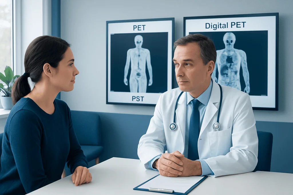
The most important difference in digital PET vs standard PET is detector design. Standard PET systems typically rely on photomultiplier tube-based technology, while digital PET systems use digital photon-counting or silicon photomultiplier-based detectors. These newer systems can measure signals more directly and with greater precision.
For patients, this technical change may translate into practical benefits. Digital PET may provide:
- Higher image resolution
- Better contrast between normal and abnormal tissue
- Improved sensitivity for detecting small lesions
- More accurate timing information, which can support better image reconstruction
- Potentially shorter scan times or lower tracer doses in selected protocols, depending on the center and clinical need
It is important to keep these benefits in context. A digital PET scanner does not automatically replace good medical judgment, and standard PET remains a valuable, widely used diagnostic tool. In many cases, standard PET provides excellent clinical information. Digital PET may be especially helpful when doctors are looking for very small findings, need higher confidence in image interpretation, or want to optimize image quality in challenging cases.
Image quality can also be influenced by factors beyond the scanner itself, including the type of tracer used, the patient’s body size, movement during the scan, preparation before the test, and the experience of the imaging team. For that reason, the quality of the center and the appropriateness of the scan matter as much as the technology.
When Digital PET May Be Especially Helpful
Digital PET may be particularly useful in cancer care, where detecting a small lesion or assessing faint tracer uptake can change treatment decisions. It can support staging, restaging, and treatment monitoring in several tumor types. In this setting, PET is often part of a broader diagnostic pathway that may also include PET-CT imaging, biopsy, laboratory tests, and other scans.
Digital PET can also be useful in neurologic imaging, such as evaluating certain brain disorders, and in cardiac imaging, where high-quality functional information can support diagnosis. For example, better timing performance may improve how clearly the scan distinguishes true tissue activity from background noise.
Even so, not every patient needs digital PET. If a standard PET scan can answer the clinical question clearly, it may be fully appropriate. The decision depends on why the scan is being ordered, what condition is being investigated, and whether the result could affect treatment planning. Patients being assessed for conditions such as lung cancer or breast cancer may hear their doctor discuss which imaging platform is most suitable for their case.
What Patients Can Expect Before, During, and After the Scan
The patient experience with digital PET is usually very similar to that of standard PET. Before the appointment, the imaging team may give instructions about eating, drinking, exercise, and medications. For many common PET tracers, fasting for a period before the test is recommended. People with diabetes may receive specific instructions because blood sugar levels can affect tracer uptake.
At the imaging center, the tracer is injected through a vein, and there is usually a waiting period to allow it to circulate in the body. During the scan, the patient lies still on the scanner table while images are acquired. The scan itself is generally painless, although staying still may be uncomfortable for some people. If PET is combined with CT, the CT portion is completed as part of the same visit. In some settings, this may be discussed as CT imaging used alongside PET.
After the scan, most patients can return to normal activities unless told otherwise. Drinking fluids may help clear the tracer from the body. The amount of radiation involved is carefully considered by the medical team, and the test is ordered when the expected benefit outweighs the risk. Patients who are pregnant, may be pregnant, or are breastfeeding should tell their doctor and the imaging staff before the appointment.
People often assume that a newer scanner feels different, but in many cases the appointment flow is nearly the same. The main difference is in the images produced and how the information may help the radiologist or nuclear medicine specialist interpret the findings.
How Doctors Decide Which PET Technology to Use
Choosing between digital PET and standard PET is usually a clinical decision rather than a patient preference alone. Doctors consider the goal of the scan, the type of disease suspected or already diagnosed, the urgency of the situation, and whether very high image detail may influence treatment decisions. Availability also matters, since not every imaging center has digital PET systems.
In oncology, doctors may prefer the most sensitive imaging available when they need to look for very small metastases, evaluate possible recurrence, or assess how well treatment is working. In some cases, additional imaging or procedures such as MRI scanning may complement PET findings, especially when detailed soft tissue evaluation is needed.
At the same time, standard PET remains clinically effective and is supported by many years of evidence and experience. Patients should not assume that a standard PET scan is inadequate. What matters most is whether the chosen test can answer the medical question accurately enough to guide care. Results are also interpreted in the context of symptoms, examination findings, pathology reports, and other tests.
Benefits, Limits, and Questions to Ask
Digital PET offers meaningful technical advantages, but it still has limitations. PET findings can sometimes be nonspecific, because increased tracer uptake may occur in cancer, inflammation, infection, or healing tissue. A clearer image does not always eliminate the need for biopsy or follow-up imaging. Also, not every abnormality seen on PET is dangerous, and not every disease is best evaluated with PET.
Patients can feel more prepared by asking a few practical questions before the scan:
- Why is this PET scan being recommended?
- Will the scan be digital PET or standard PET, and does that matter in this case?
- How should the patient prepare, especially regarding food, exercise, and medications?
- Will PET be combined with CT or another imaging test?
- How and when will the results be explained?
A thoughtful discussion with the doctor can help patients understand the purpose of the test and what information it can realistically provide. Near the end of the care journey, some patients also seek evaluation at centers with advanced imaging platforms. Acibadem International’s multidisciplinary specialists and JCI-accredited hospitals diagnose and treat international patients using modern imaging and coordinated clinical care.
The overall message is reassuring: both digital PET and standard PET are established tools, and each can be valuable when chosen for the right reason. The most appropriate scan is the one that best answers the patient’s specific medical question safely and clearly.
Frequently asked questions
Is digital PET more accurate than standard PET?
Digital PET can improve image quality and may detect small or low-contrast abnormalities more clearly in some situations. However, accuracy also depends on the tracer used, the condition being evaluated, the patient’s preparation, and the expertise of the imaging team.
Does a digital PET scan expose the patient to more radiation?
The type of detector does not automatically mean more radiation. Radiation exposure depends mainly on the tracer, the CT component if used, and the imaging protocol. In some settings, digital PET may support efficient imaging without increasing dose.
Will the scan feel different if it is digital PET instead of standard PET?
For most patients, the experience feels very similar. The preparation, tracer injection, waiting time, and need to lie still during imaging are generally the same. The difference is mainly in the scanner technology and the images it produces.
Why would a doctor still order a standard PET scan if digital PET is available?
Standard PET is still a reliable and widely used imaging test that can answer many clinical questions well. If it provides the information needed for diagnosis or treatment planning, it may be completely appropriate. Availability, urgency, and the specific medical question also influence the choice.
Is digital PET only used for cancer?
No. Although PET is commonly associated with cancer care, it can also be used in neurology and cardiology. The usefulness of digital PET depends on the condition being evaluated and whether higher image detail may add value.
Should patients ask specifically for digital PET?
Patients can certainly ask what type of PET scanner will be used and whether it matters in their case. Still, the best approach is to discuss the goal of the scan with the doctor, since the most important issue is whether the chosen test will answer the clinical question clearly and safely.
References
- World Health Organization
- Society of Nuclear Medicine and Molecular Imaging
- Radiological Society of North America
- American College of Radiology
- National Cancer Institute
This article is for general information only and is not a substitute for professional medical advice. Please consult a qualified doctor about your individual situation.
Medical weight-loss options in Turkey — costs & eligibility
JCI-accredited · board-certified surgeons · reply within 24h
Add us as a Preferred Source to see more of our trusted health content across Google Search, AI Overviews and Discover.