Pet scan vs ct scan: How to Tell the Difference and When It Matters

A CT scan creates detailed pictures of organs, bones, and tissues using X-rays. A PET scan shows metabolic activity and can highlight areas that are more or less active than expected.
Key Takeaways
- A CT scan creates detailed pictures of organs, bones, and tissues using X-rays.
- A PET scan shows metabolic activity and can highlight areas that are more or less active than expected.
- PET and CT are often combined in one exam to match function with anatomy.
- The best test depends on the reason for imaging, not on one test being universally better.
- Preparation, contrast use, and radiation exposure differ between PET and CT scans.
- A doctor should interpret results in the context of symptoms, examination, and other tests.
PET scan vs CT scan is mainly a question of function versus structure: a CT scan shows detailed anatomy, while a PET scan shows how tissues are working metabolically. Doctors often use one or both depending on the clinical question, such as finding a cause of symptoms, evaluating cancer, or checking treatment response.
Overview: PET scan vs CT scan at a glance
If someone is comparing pet scan vs ct scan, the shortest answer is this: a CT scan shows what the body looks like, and a PET scan shows how parts of the body are functioning. CT is especially useful for anatomy, such as the size, shape, and location of a structure. PET is especially useful for spotting changes in tissue activity that may not yet be obvious on a standard structural scan.
Doctors do not usually choose between them in a vacuum. They choose the test that best answers a specific clinical question. In some situations, a CT scan alone is enough. In others, a PET scan adds important information, and many patients have a combined PET-CT study so both structure and function can be assessed together.
For quick comparison, this side-by-side guide helps clarify how the two tests differ:
- CT scan: Uses X-rays to create cross-sectional images of the body.
- PET scan: Uses a small amount of radioactive tracer to show metabolic or biochemical activity.
- Best for CT: Anatomy, injuries, bleeding, infections, organ detail, and many routine diagnostic questions.
- Best for PET: Cancer staging and follow-up, selected brain disorders, and certain heart-related assessments.
- Time: CT is usually quicker; PET usually takes longer because of tracer uptake time.
- Preparation: PET often requires more specific preparation, especially regarding food and blood sugar.
- Output: CT shows structure; PET highlights active or inactive areas within tissues.
- Combined exam: PET-CT helps doctors match abnormal activity to an exact location in the body.
How clinicians tell them apart and when each matters
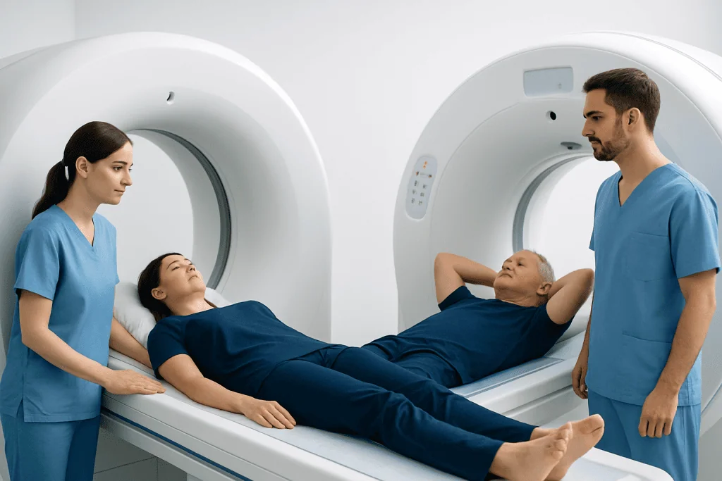
In practice, clinicians distinguish PET and CT by the type of answer each test provides. A CT scan helps answer questions like: Is there a mass? Is an organ enlarged? Is there bleeding, swelling, a fracture, a blocked passage, or a visible change in tissue structure? It is often one of the first imaging tests because it is widely used, fast, and highly effective for many urgent and non-urgent problems.
A PET scan is chosen when the question is more about tissue behavior than appearance. For example, a doctor may want to know whether a suspicious area is metabolically active, whether cancer has spread, or whether treatment is working. PET can sometimes detect changes before clear structural changes appear on CT alone.
That said, PET is not simply a “better” scan. It is more specialized. Some abnormalities show up well on CT and may not require PET at all. In other cases, PET findings can be nonspecific and still need correlation with CT, MRI, laboratory tests, or biopsy.
The distinction matters most when results will change next steps. A CT scan may be enough to explain symptoms such as abdominal pain, kidney stones, or trauma-related concerns. A PET scan may matter more when there is a need to assess cancer activity, search for disease in multiple areas, or clarify whether tissue seen on a structural scan is likely active disease or scarring.
How a CT scan works and what it is used for
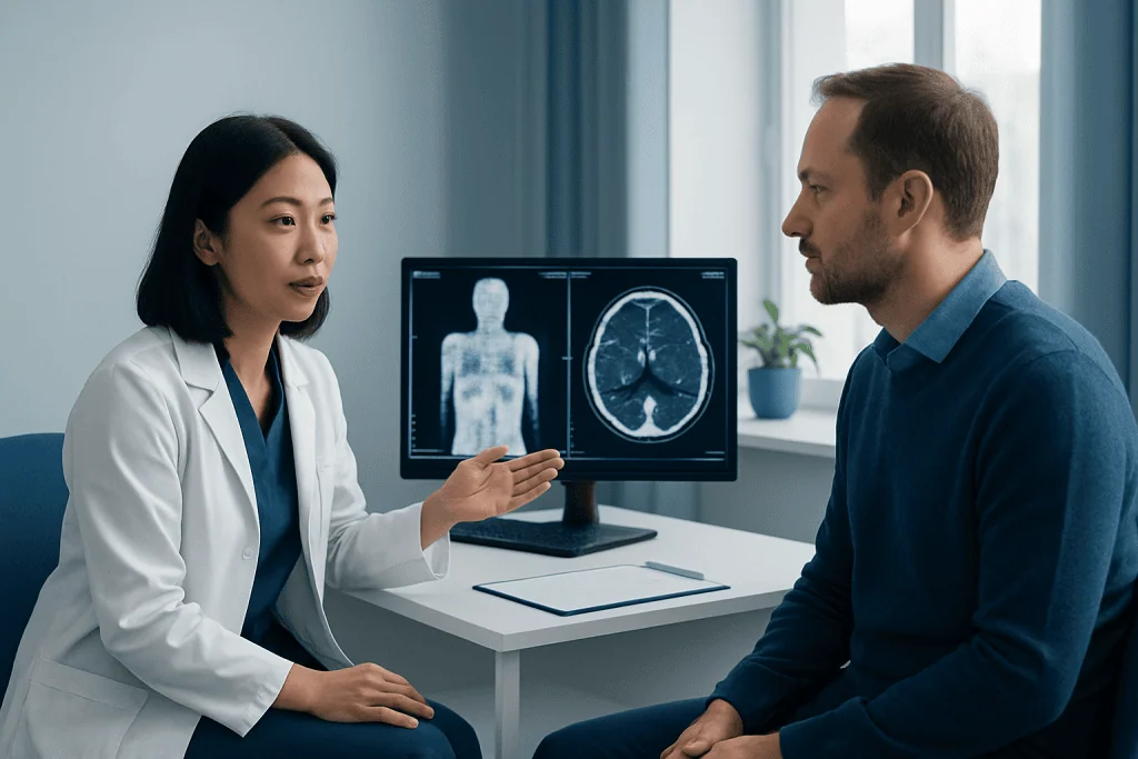
A CT, or computed tomography, scan uses multiple X-ray images processed by a computer to create detailed cross-sectional views of the body. It can image the head, chest, abdomen, pelvis, blood vessels, bones, and soft tissues. In many settings, it is a first-line imaging test because it is fast and provides clear anatomical detail.
CT is commonly used to evaluate injuries, infections, abdominal and pelvic symptoms, chest symptoms, suspected stroke or bleeding, and many other conditions. It also plays a major role in cancer care, often helping identify the location and size of a tumor and guiding procedures or follow-up. When a doctor is investigating a possible lung cancer or another structural abnormality, CT is often one of the earliest imaging tools used.
Some CT scans are done without contrast, while others use contrast material to make blood vessels or organs easier to evaluate. Contrast may be swallowed or given through a vein, depending on the body area being studied. The need for contrast depends on the medical question, kidney function, allergy history, and the type of anatomy that needs to be seen.
CT does involve radiation exposure, but the amount varies by the body part and protocol. Doctors order it when the expected benefit outweighs the risk. For many patients, CT offers a rapid and reliable way to answer an important diagnostic question.
How a PET scan works and what it is used for
A PET, or positron emission tomography, scan shows how tissues are functioning by tracking a small amount of radioactive tracer. The most commonly used tracer highlights glucose use, which can help reveal tissues with unusually high or low metabolic activity. Because many diseases alter metabolism, PET can provide information that structural imaging alone may not capture.
PET is widely used in oncology. It can help with detecting metabolically active disease, checking whether cancer has spread, evaluating response to treatment, and sometimes distinguishing active disease from scar tissue after therapy. It may be part of the work-up for conditions such as lymphoma or other cancers where whole-body metabolic information is helpful.
PET can also be used in selected cardiology and neurology situations. For example, it may help assess heart muscle viability or evaluate certain brain disorders. However, PET is not used for every symptom or every condition. It is a targeted test, ordered when metabolic information is likely to improve diagnosis or management.
Most PET scans today are performed as PET-CT. The CT portion helps localize the PET findings precisely, which makes interpretation more accurate. When cancer care is involved, this combined approach can be especially useful alongside treatments such as medical oncology or radiation oncology when doctors are planning or monitoring care.
What to expect before, during, and after each scan
The experience of having a CT scan is usually straightforward. The patient lies on a table that moves through the scanner. The test is often brief, though the total visit may be longer if contrast is needed or if preparation is required. Some people notice a warm sensation or metallic taste if IV contrast is used, but this is typically temporary.
A PET scan usually takes more time. After the tracer is injected, there is generally a waiting period so the body can absorb it properly before imaging begins. Patients are often asked to avoid strenuous exercise beforehand and to follow specific instructions about eating. Blood sugar control can be particularly important, especially for people with diabetes, because it can affect image quality.
For both tests, it helps to wear comfortable clothing and bring information about medications, allergies, pregnancy, breastfeeding, kidney problems, or implanted devices if relevant. Most scans are painless, although lying still can be uncomfortable for some people. Those who feel claustrophobic should tell the imaging team in advance, since support may be available.
After imaging, many people can return to normal activities right away unless they were given special instructions. Results are interpreted by a radiologist or nuclear medicine specialist and then discussed by the referring doctor. The report is most useful when combined with symptoms, examination findings, and any previous imaging.
How doctors decide which scan to order
The choice between PET and CT depends on the suspected condition and the clinical goal. If the main question is structural, CT is often the better first step. If the main question is whether tissue is active, aggressive, inflamed, or responding to treatment, PET may be more informative. Sometimes the right answer is sequential testing: CT first, then PET only if needed.
In cancer care, CT may help identify a suspicious lesion, while PET may help stage disease or evaluate treatment response. For example, if a tumor is visible on CT, a PET-CT may clarify whether there are other active areas elsewhere in the body. In some cases, tissue sampling is still necessary, and imaging helps guide biopsy rather than replacing it.
Doctors also consider practical and safety factors. CT is often preferred in urgent situations because it is fast and widely available. PET may be less useful in some settings, and not every abnormality is best assessed metabolically. Recent surgery, infection, or inflammation can also affect PET findings and sometimes make interpretation more complex.
The best next step after either scan may include no further testing, repeat imaging later, another type of scan such as MRI or ultrasound, or referral to a specialist. When symptoms are ongoing or results are unclear, asking what specific question the scan is meant to answer can help patients better understand the plan.
Limits, risks, and common misunderstandings
One common misunderstanding in pet scan vs ct scan comparisons is assuming that one test replaces the other. In reality, they provide different kinds of information. A normal CT does not always rule out disease, and a PET finding does not automatically mean cancer. Imaging results need clinical context.
Both PET and CT involve radiation, though the amount differs depending on the scan type and protocol. Imaging is ordered thoughtfully, especially for children, pregnant patients, and people who may need repeated scans. Contrast material used in some CT studies may not be suitable for everyone, particularly people with certain allergies or kidney concerns.
PET scans can also show increased uptake in non-cancerous conditions such as inflammation or infection. CT scans may reveal incidental findings that are harmless but still need clarification. This can understandably cause anxiety, but many findings turn out to be benign or manageable once fully assessed.
If a person has been told they need advanced imaging, it does not automatically mean a serious diagnosis is expected. Often, it simply means the doctor wants more precise information before deciding on treatment. Near the end of the care pathway, centers such as Acibadem International may support international patients through diagnosis and treatment with multidisciplinary specialists in JCI-accredited hospitals.
When to seek medical care
Medical imaging should be guided by a qualified clinician rather than chosen independently. A person should seek medical care if they have persistent, unexplained, or worsening symptoms such as ongoing pain, unexplained weight loss, a new lump, chronic cough, neurological symptoms, or symptoms that interfere with daily life. The right scan depends on the history, examination, and sometimes basic laboratory tests.
Urgent medical attention is important for warning signs such as severe chest pain, sudden shortness of breath, signs of stroke, major injury, heavy bleeding, or sudden severe abdominal pain. In these situations, CT is often used rapidly because it can detect several urgent structural problems quickly.
Patients who have already been advised to undergo a PET or CT scan should ask why the test is being ordered, what preparation is needed, and how the result may affect treatment decisions. If a scan shows an abnormality, follow-up matters. Depending on the findings, the next step may involve repeat imaging, referral to a specialist, or treatment planning such as surgical oncology.
Seeking care early can help clarify symptoms and avoid delays in diagnosis. Even when advanced imaging is recommended, the goal is usually to get a more accurate answer so care can be tailored appropriately.
Frequently asked questions
What is the main difference between a PET scan and a CT scan?
The main difference is that CT shows body structure, while PET shows tissue activity. CT is usually best for anatomy, and PET is useful when doctors need to understand how tissues are functioning metabolically.
Is a PET scan more accurate than a CT scan?
Not in every situation. A PET scan is not automatically more accurate; it is simply designed to answer different questions. In many cases, CT is the right test, while in others a PET-CT provides more complete information.
Why would a doctor order a PET scan after a CT scan?
A doctor may order PET after CT if the CT shows something that needs further characterization. PET can help determine whether an area is metabolically active, whether disease may have spread, or whether treatment appears to be working.
Does a PET scan always mean cancer is suspected?
No. PET scans are often used in cancer care, but they can also be used in selected heart and brain conditions. Also, increased uptake on PET is not specific to cancer and can sometimes occur with infection or inflammation.
Which scan takes longer, PET or CT?
A PET scan usually takes longer overall. CT is typically quick, while PET involves a waiting period after tracer injection before the imaging itself begins.
Are PET and CT scans safe?
Both are commonly used and generally safe when medically appropriate, but both involve radiation. Doctors weigh the benefits and risks carefully and may adjust plans based on pregnancy, kidney function, allergies, age, and the need for repeated imaging.
References
- Radiological Society of North America
- American Cancer Society
- National Cancer Institute
- Society of Nuclear Medicine and Molecular Imaging
- U.S. Food and Drug Administration
This article is for general information only and is not a substitute for professional medical advice. Please consult a qualified doctor about your individual situation.
Oncology care in Turkey — second opinion and treatment plan
JCI-accredited · board-certified surgeons · reply within 24h
Add us as a Preferred Source to see more of our trusted health content across Google Search, AI Overviews and Discover.

More from the Health Library



Related Specialists

Prof. Dr. Kerim Sarıyılmaz
Orthopedic Surgery & Traumatology
Dr. Bengü Kasım
Physical Medicine & Rehabilitation
Dr. Onur Güney Yılmaz
Emergency Service
Prof. Dr. Orhan Demircan
General Surgery