

Before Astigmatism Laser Treatment: Contact Lens Breaks and Repeat Corneal Measurements

Key Takeaways
- Contact lenses temporarily reshape the cornea, so Mayo Clinic advises stopping them for at least a few weeks before both the evaluation and the surgery, with the surgeon setting the exact period by lens type.
- Rigid gas-permeable and orthokeratology lenses mold the cornea more than soft lenses, so people who wear them are usually asked for the longest lens-free periods and the most repeat scans.
- Repeat corneal topography separated by weeks is the main tool for telling harmless lens warpage from keratoconus, a progressive thinning disorder that is a firm reason not to perform corneal laser surgery.
- Surgeons generally want a prescription that has held steady for around a year, and pregnancy or breastfeeding are common reasons to postpone because hormonal shifts alter corneal shape and refraction.
- Laser treatment reshapes the cornea but does not prevent presbyopia or cataract, so most people still need reading glasses from midlife regardless of surgery.
- Dry eye is the most common lingering effect after LASIK and Mayo Clinic notes it can last several months, which is why pre-existing dryness should be raised before, not after, a date is booked.
Preparing for astigmatism laser surgery usually means stopping contact lenses for a set period before the consultation and the procedure, so the cornea returns to its natural shape, then having corneal maps and thickness measurements repeated to confirm the readings are stable and to rule out corneal thinning conditions. The surgeon sets the exact lens break and decides on suitability based on those repeat measurements.
Maya keeps her contact lens case on the bathroom shelf like a small trophy. Fifteen years of monthly lenses, a steady astigmatism prescription, and one bright idea: fix it with a laser and be done. Then the surgeon’s office sends a pre-visit letter, and the first instruction is the one she did not expect. Take the lenses out, put your glasses on, and keep them on for weeks before we even scan your eyes.
That request sits at the heart of preparing for astigmatism laser surgery, and it is not bureaucracy. A contact lens presses on the cornea, the clear dome at the front of the eye, and gently reshapes it. A laser plan built on a reshaped cornea is a plan built on a moving target.
The second surprise comes at the clinic: the same corneal measurements are taken more than once, sometimes on different days. This article explains why both steps exist, what the numbers mean, and what the weeks around surgery usually look like.
What astigmatism is, and why the cornea's shape drives the whole plan
Astigmatism is a focusing error caused by a cornea (or, less often, the lens inside the eye) that is curved more steeply in one direction than the other. Instead of a dome shaped like the side of a basketball, the front of the eye is shaped more like the side of a football. Light entering through that uneven curve lands on the retina as two focal lines rather than one point, so edges look smeared and lights at night grow tails. Mayo Clinic describes it as a common, usually stable, and correctable condition that often travels alongside nearsightedness or farsightedness.
The detail that matters for surgery is this: laser correction for astigmatism works by removing a precise, uneven pattern of tissue so the steep meridian flattens more than the flat one. The laser is programmed from the measurements taken beforehand. If those measurements describe a cornea temporarily bent by a contact lens, or a cornea that is still changing on its own, the pattern removed will not match the eye that heals.
Two questions therefore sit underneath every pre-operative visit. First, is this the cornea’s real shape, free of lens-induced warping? Second, is that shape holding steady from one visit to the next? Everything in the pages that follow, from the lens break to the repeat scans, is designed to answer those two questions with evidence rather than assumption. The surgeon who insists on both is not being cautious for its own sake; they are protecting the accuracy of a treatment that cannot be undone.
How astigmatism laser surgery actually works, in plain language
Three laser techniques are in common use, and all of them reshape the cornea rather than the lens inside the eye. LASIK creates a thin hinged flap in the outer cornea, applies an excimer laser (an ultraviolet laser that removes microscopic layers of tissue) to the exposed bed, then lays the flap back down. PRK and its variant LASEK remove or loosen the surface skin layer, called the epithelium, apply the same laser directly, and let the surface regrow over several days. A third approach uses a femtosecond laser to cut a small disc of tissue inside the cornea and removes it through a keyhole incision, without a flap. The NHS overview groups these as laser eye surgery and notes they all aim to reduce dependence on glasses or contact lenses, not to treat an eye disease.
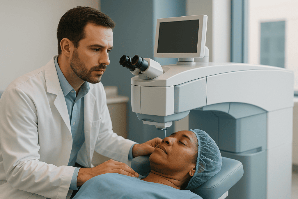
For astigmatism specifically, the laser removes tissue in an oval rather than a circular pattern, taking more from the steep axis. Modern systems track the eye during treatment and register the axis of astigmatism, which is why the pre-operative axis measurement has to be accurate to within a degree or two.
Mayo Clinic describes the experience: numbing drops, a lid holder to stop blinking, a suction ring that produces a feeling of pressure and a brief dimming of vision during flap creation, then the laser itself, which patients hear as clicking and sometimes smell as a faint singed odor. Mayo Clinic puts the whole procedure at about 30 minutes or less for both eyes, with the laser portion lasting under a minute per eye. The technology is impressive, but it is only as good as the map it is given.
Why a contact lens break before LASIK is the first step, not an afterthought
A soft contact lens drapes over the cornea like a wet cloth over a bowl; a rigid lens sits on it like a lid. Both alter the cornea’s shape and its oxygen supply, and both change how the cornea bends light. The medical term is corneal warpage: a temporary, lens-induced distortion of the corneal surface that can mimic or mask astigmatism. Warpage can also produce a false-positive pattern that resembles keratoconus, a progressive thinning disorder described in the next sections, and a surgeon who cannot tell the two apart has to postpone or decline treatment.
Mayo Clinic’s pre-operative guidance is direct: contact lenses can distort the shape of the cornea, so you will need to stop wearing them completely and rely on glasses for at least a few weeks before both the evaluation and the surgery, with the exact period set by your surgeon according to lens type and how long you have worn lenses.
The consequences of skipping the break are practical. The consultation scans may show irregular astigmatism that is not really yours. The prescription measured for the laser may be off by a fraction of a diopter or a few degrees of axis, which for astigmatism can be the difference between crisp and slightly smeared. And the surgeon may lose confidence in the stability of your readings, which pushes the timeline back further than the break itself would have.
The break also gives the corneal surface a chance to recover from years of lens wear, which matters for the dry-eye risk discussed later. Think of it less as a delay and more as the first part of the treatment: you are resetting the eye to baseline so that everything measured afterward is trustworthy.
How long the contact lens break usually lasts, by lens type
There is no single number that applies to everyone, and any article that gives you one without a caveat is oversimplifying. The principle is consistent: the more a lens reshapes the cornea, the longer the cornea takes to return to its own form. Soft daily or monthly lenses change the surface least. Toric soft lenses, which are weighted to sit in a fixed orientation for astigmatism, sit slightly differently on each blink. Rigid gas-permeable lenses and orthokeratology lenses (worn overnight deliberately to flatten the cornea) change shape most, and recovery from them is slowest and least predictable.

| Lens type | Effect on corneal shape | Typical approach to the break |
|---|---|---|
| Soft spherical or toric | Mild, reversible flattening and surface change | The shorter end of the range Mayo Clinic describes as at least a few weeks |
| Extended-wear soft | Mild to moderate, plus oxygen-related surface stress | Often longer than daily soft, at the surgeon’s discretion |
| Rigid gas-permeable | Marked molding of the corneal curve | Substantially longer; often extended if repeat scans still shift |
| Orthokeratology | Deliberate, sustained reshaping | The longest breaks; stability must be proven on serial scans |
What ends the break is not the calendar but the measurements. Surgeons commonly take corneal maps at the first visit, then repeat them after further lens-free weeks; if the two sets match within the instrument’s tolerance, the cornea is considered settled. If they still differ, the break is extended and the scans are repeated again. This is why a person who has worn rigid lenses for decades may be asked to wait longer than a friend who wore soft lenses for two years.
Plan for the break the way you would plan for a minor injury: sort out a current pair of glasses, expect a period of feeling less sharp, and avoid scheduling important visual tasks, such as a long night drive, for the first days of the switch.
Corneal topography before laser eye surgery: what the machines are measuring
Corneal topography is a photograph-based map of the front surface of the cornea, produced by projecting rings of light onto the eye and analyzing how they reflect. It shows steep areas in warm colors and flat areas in cool colors, and it locates the axis of astigmatism to the degree. Corneal tomography goes a step further: it images a cross-section of the cornea, mapping both the front and back surfaces and producing a thickness map across the whole dome. Pachymetry is the measurement of corneal thickness at specific points, usually the thinnest one, given in micrometers (thousandths of a millimeter).
Alongside these, the team measures refraction, both with and without pupil-dilating drops so that the eye’s focusing muscle cannot hide part of the prescription; pupil size in dim light; tear film quality; and the overall health of the retina and optic nerve. Mayo Clinic lists detailed corneal measurement as a core part of the evaluation because the laser plan is generated from it and because the residual cornea left after tissue removal has to remain thick enough to stay structurally sound.
Why so many instruments for one dome of tissue? Each catches something the others miss. Topography reads the front surface superbly but cannot see the back surface, where the earliest signs of keratoconus often appear. Tomography sees both surfaces but is less precise about the tear film. The refraction tells the surgeon what you actually see, which does not always match what the cornea alone predicts, because the lens inside the eye contributes its own astigmatism.
You do not need to interpret the color maps yourself. What you are entitled to is a clear explanation of whether your corneas are regular in shape, whether the front and back surfaces agree, and whether thickness leaves a comfortable margin for the treatment proposed.
Why the measurements are repeated, and what "stable" really means
Repeat corneal measurements do two separate jobs. The first is to confirm that the contact lens break has done its work: if maps taken at visit one and visit two are essentially identical, the cornea has stopped changing shape and the readings reflect the eye’s true form. If they differ, the break continues. The second job is more serious. Keratoconus, and a related group of conditions called corneal ectasia, involve progressive thinning and bulging of the cornea. Cleveland Clinic describes keratoconus as a condition that typically begins in the teens or twenties and worsens over years, and notes that it is diagnosed and monitored with corneal topography.
A single scan can look mildly irregular for innocent reasons: a dry patch, a blink, residual lens warpage. Two scans weeks apart that show the same steepening in the same place, or a thinnest point that has moved or shrunk, tell a different story. Laser surgery removes tissue and weakens the cornea by design; performing it on a cornea already prone to thinning risks accelerating that process, a complication called post-refractive ectasia. This is the main reason surgeons repeat scans even when the first set looks acceptable.
Refraction stability is checked the same way. Mayo Clinic notes that surgeons generally want a prescription that has held steady, typically for at least a year, before operating, since a still-changing prescription would be corrected to a value that is about to drift. Bring old glasses prescriptions or optician records to the consultation; they are evidence.
“Stable” therefore has a precise meaning: two or more sets of readings, taken lens-free and separated by time, that agree within the instrument’s known margin of error. It is not a feeling, and it cannot be rushed by wanting the surgery sooner.
Preparing for astigmatism laser surgery: what happens at the consultation
Arrive lens-free for the agreed period, with your glasses and any records of past prescriptions. The visit is long, often longer than the surgery itself, because most of it is measurement. Expect vision testing, the scans described above, an eye pressure check, a tear film assessment, and dilating drops that blur near vision and increase light sensitivity for several hours. Arrange a lift home or plan for public transport, and bring sunglasses.
The medical history matters as much as the machines. Be candid about dry eye symptoms, autoimmune conditions, diabetes and how well it is controlled, previous eye injuries or surgery, medicines that affect healing or tear production, pregnancy or breastfeeding, and any family history of keratoconus. Mayo Clinic and the NHS both flag several of these as reasons to delay or avoid surgery, and none of them is a judgment on you; they are variables that change the risk arithmetic.
Preparing for astigmatism laser surgery also means preparing your expectations. A good consultation covers which technique is being proposed and why, how much astigmatism the plan is designed to correct, how much residual prescription might remain, the likelihood of needing reading glasses later regardless of surgery, and the specific risks for your corneal thickness and pupil size. You should leave with a written summary and the name of the person to contact with questions.
If the surgeon says the readings are not yet stable and asks you to return after more lens-free weeks, take that as the system working. The most useful mindset is the one Maya eventually adopted: the goal is not to get to the laser quickly; it is to arrive with measurements nobody has to second-guess.
Who laser surgery for astigmatism is usually for, and who is asked to wait
Mainstream guidance from Mayo Clinic, the NHS and MedlinePlus converges on a similar profile. Suitable candidates are adults, generally at least 18 and often older, whose prescription has been stable for around a year, whose corneas are regular in shape and adequately thick, whose eyes are otherwise healthy, and who understand that the aim is reduced dependence on glasses rather than guaranteed freedom from them. Moderate astigmatism combined with nearsightedness or farsightedness is routinely treated; very high astigmatism, or astigmatism that is irregular rather than symmetrical, may push the surgeon toward a different procedure or toward advising against surgery.
People commonly asked to wait rather than declined outright include those whose prescription changed within the past year, those still within a contact lens break whose repeat scans have not yet matched, anyone who is pregnant or breastfeeding (hormonal shifts alter corneal shape and refraction, and Mayo Clinic lists both as reasons to postpone), and people with active eye surface inflammation or significant untreated dry eye. Waiting in these situations is not a soft refusal; it is the honest answer that the measurements needed to plan safely do not yet exist.
Age brings its own nuance. From the mid-forties onward, the eye’s natural lens stiffens and near focus fades, a normal change called presbyopia. Laser surgery does not prevent it. Someone in that age range may be offered a plan that leaves one eye slightly nearsighted, or may be steered toward lens-based surgery instead. Later still, early cataract makes corneal laser treatment less logical, since the cloudy lens will eventually need replacing and that operation can correct astigmatism at the same time.
The decision always rests with the treating team, who weigh all of these together rather than any single one.
What disqualifies you from laser eye surgery? The reasons surgeons decline
Some findings are firm reasons not to operate on the cornea with a laser, and it helps to know them in advance so a decline does not feel arbitrary. Keratoconus or other corneal ectasia is the clearest. Because laser correction thins the cornea further, operating on a cornea that is already weak invites progressive bulging; this is precisely what repeat topography is designed to catch. Corneas that are too thin to leave a safe residual bed after the planned tissue removal fall into the same category, even when their shape is regular.
Uncontrolled autoimmune or connective tissue disease can impair healing of the corneal surface, and Mayo Clinic lists these among the conditions that may make surgery inadvisable. Poorly controlled diabetes carries a similar healing concern. Severe dry eye that has not responded to management is another frequent reason to decline, because laser treatment disrupts corneal nerves and commonly worsens dryness for months. Active eye infection, significant cataract, uncontrolled glaucoma, and certain retinal conditions also weigh against proceeding. Some surgeons are cautious about very large pupils in dim light because of the risk of halos and glare.
Unrealistic expectations count too. A person who expects perfect vision at every distance for life, or who would regard any need for glasses afterward as a failure, is likely to be disappointed by even a technically excellent result, and many surgeons will say so.
Being declined for one technique is not necessarily being declined for all vision correction. Surface treatments, implantable lenses placed inside the eye, or lens replacement surgery may be discussed as alternatives, each with its own suitability rules. The NHS overview describes lens surgery as an option when laser surgery is not suitable. Whether any of them fits your eyes is a judgment only your treating team can make after examination.
How painful is astigmatism surgery? An honest account of sensation
People searching this question deserve specifics rather than reassurance. During the procedure itself, numbing drops mean the cornea does not register sharp pain. What Mayo Clinic and Johns Hopkins describe, and what patients consistently report, is pressure: the suction ring used in flap creation presses on the eye firmly enough that vision dims or blacks out for several seconds, which is startling rather than painful. The laser is silent to the eye and audible to the ear as rapid clicking. Some people notice a faint smell. Anxiety is the main discomfort in the room, and teams are used to talking patients through it.
The hours afterward are where the techniques diverge. After LASIK, Mayo Clinic describes burning, itching, grittiness and watering for several hours, often eased by keeping the eyes closed and resting; discomfort typically settles within the first day. After PRK or LASEK, the surface layer has to regrow, so the eyes are usually more sore and light-sensitive for several days, and a soft bandage contact lens is often placed to protect the healing surface. Pain relief is managed by the surgical team with drops and, where needed, oral medicines they will specify; do not add your own.
Pain that is severe, worsening after the first day, or accompanied by a sudden drop in vision is not part of normal recovery and is covered in the red-flag section below.
The honest summary: the operation is uncomfortable rather than painful, the first evening after LASIK is irritating, and the first few days after PRK can be genuinely sore. Knowing which technique you are having tells you which of those to plan for.
How long to rest after eye laser treatment: the first days and weeks
Recovery timelines are technique-dependent, and every figure here is a typical range, not a promise for your eyes. Mayo Clinic advises that after LASIK, vision is usually blurry or hazy on the day and improves over the following days, with a follow-up visit typically within the first day or two so the surgeon can check the flap and look for infection. Many people return to desk work within a day or two, but the eyes tire quickly on screens, and frequent breaks help. The NHS notes that most people are able to resume normal activities within a few days of laser surgery.
After PRK or LASEK, the surface layer takes several days to regrow, and vision is often blurry and fluctuating for the first week, sometimes longer, so time off work is usually measured in days rather than one afternoon.
Protective habits matter more than bed rest. Mayo Clinic’s after-care points include: do not rub the eyes, especially in the first weeks while a LASIK flap is knitting down; wear the shields provided for sleep; keep water, soap, sweat and cosmetics out of the eyes; avoid swimming, hot tubs and dusty or smoky environments in the early weeks; and postpone contact sports until your surgeon clears you. Driving resumes only when vision meets the legal standard and your team agrees.
Dry eye is the most common lingering symptom. Mayo Clinic notes it can persist for several months while corneal nerves recover, and lubricating drops are routinely used. Vision typically continues to fine-tune over two to three months, which is why a decision about any enhancement treatment is deferred until the measurements have settled again, repeating, in miniature, the stability logic that governed the surgery itself.
What people often get wrong about preparing for astigmatism laser surgery
“Astigmatism can’t be treated with a laser.” This was true of early systems decades ago and is now a persistent myth. Mayo Clinic lists astigmatism, alongside nearsightedness and farsightedness, as a refractive error that laser surgery is designed to correct. Suitability depends on the amount, regularity and stability of the astigmatism, not on its mere presence.
“A few days without lenses is enough.” Corneal warpage from lenses, especially rigid ones, resolves over weeks, not days. Mayo Clinic’s guidance is at least a few weeks, extended at the surgeon’s discretion. Cutting the break short risks planning the laser on a shape that is still changing.
“If the first scan looked fine, repeating it is a formality.” A single map cannot show progression. Repeat topography is how early keratoconus, the most important reason to avoid corneal laser surgery, is distinguished from a one-off irregular reading.
“The surgery fixes your eyes for life.” Laser surgery reshapes the cornea; it does nothing to stop the natural stiffening of the lens that brings reading glasses in midlife, and it does not prevent cataract. Some people also experience small regression of their prescription over years.
“Dry eye is rare and brief.” Mayo Clinic describes dry eye as common after LASIK, often lasting for months. Anyone with pre-existing dryness should raise it before surgery, not after.
“The numbers in adverts describe my odds.” Headline percentages come from selected populations and specific prescriptions. Your realistic outcome range depends on your corneal measurements and prescription, and only your surgeon, looking at your scans, can describe it honestly.
Is laser eye surgery worth it if you have astigmatism? Weighing alternatives
“Worth it” is a personal calculation, and the honest answer is that it depends on what you value, what your measurements allow, and how you feel about permanence. The evidence base described by Mayo Clinic, MedlinePlus and Johns Hopkins is consistent on the broad picture: laser surgery reduces dependence on glasses or contact lenses for most suitable candidates, carries a small risk of complications including dry eye, glare and halos, undercorrection or overcorrection, flap problems and, rarely, ectasia or infection, and does not remove the eventual need for reading glasses.
Set that against the alternatives. Glasses correct astigmatism with zero surgical risk and can be updated as the eye changes. Toric contact lenses correct it without surgery but carry infection risk with imperfect hygiene and can be less stable in vision than spherical lenses because they rotate slightly. Implantable collamer lenses, placed inside the eye in front of the natural lens, correct higher prescriptions and are removable, but they are intraocular surgery with their own risks. Lens replacement with a toric intraocular lens, the same operation used for cataract, corrects astigmatism at the lens rather than the cornea and is generally reserved for older adults or those already developing cataract.
The people who tend to describe surgery as worthwhile share some features: stable, moderate prescriptions; healthy tear films; realistic goals; and a clear preference for not managing lenses daily. The people who tend to regret it often had marginal corneas, significant pre-existing dryness, or expectations that no procedure could meet.
None of this is a recommendation. It is the frame within which you and your treating team can place your own scans, your own prescription and your own priorities.
Questions to ask your care team before you agree to a date
A prepared patient changes the tone of a consultation. Take these questions in writing, and expect answers you can understand.
- How long a contact lens break do you want for my lens type, and will you repeat the corneal scans after it before finalizing the plan?
- Were my two sets of corneal maps consistent with each other? Is there anything on the front or back surface, or in the thickness map, that gives you pause?
- How thick are my corneas, and how much tissue will remain after the planned treatment?
- Which technique are you proposing for my astigmatism, and why that one rather than the alternatives?
- How much of my astigmatism is corneal and how much comes from the lens inside my eye? Does that change the plan?
- What is a realistic range of outcomes for someone with my prescription and measurements, and how likely is it that I would still need glasses for some tasks?
- How do you assess dry eye, and what did my tear film tests show?
- If my prescription drifts afterward, what are the options, and how long do you wait before considering them?
- What symptoms after surgery should prompt me to call you the same day, and how do I reach someone out of hours?
- Who will perform the procedure, who will see me at follow-up, and how many follow-up visits are planned?
Notice that none of these asks the surgeon to sell you anything. They ask for evidence about your own eyes, and a team confident in its process will welcome them. If an answer is vague, or if you are being hurried toward a date before the repeat scans are done, that in itself is information worth weighing.
When to call your doctor: red-flag signs before and after surgery
Most of the pre-operative period is uneventful, but a few situations deserve a prompt call rather than a wait for the next appointment. During the contact lens break, a sudden change in vision, new eye pain, marked redness or discharge, or a feeling that something is in the eye that does not clear with blinking should be reported, since surface infection or inflammation would delay surgery and needs assessment in its own right. If you develop a new medical diagnosis, start a new medicine, or become pregnant between the consultation and the surgery date, tell the team; each can change suitability.
After surgery, the early days should bring gradual improvement. Contact the surgical team the same day, or the emergency service they have given you, if you experience any of the following:
- Severe or worsening eye pain, particularly pain that increases after the first day rather than easing
- A sudden drop in vision, or vision that was improving and then deteriorates
- Increasing redness, thick discharge, or swelling of the lids
- New sensitivity to light that is markedly worse than the day before
- Any injury to the eye, a hard rub, or the sense that the LASIK flap has moved, sometimes described as a sudden crinkled or doubled image
- Flashes of light, a curtain or shadow across the vision, or a sudden shower of floaters
Mayo Clinic advises that the early follow-up visit exists specifically to catch infection and flap problems, so do not skip it, even if the eye feels fine. Do not treat these symptoms with leftover drops or someone else’s medicine, and do not wait to see if they settle overnight. Infection and flap displacement are uncommon, but both are far more manageable when treated early. Your surgical team, not an internet search, is the right first call.
Frequently asked questions
How long is the contact lens break before LASIK for astigmatism?
Mayo Clinic advises stopping lenses entirely for at least a few weeks before both the evaluation and the surgery, with your surgeon setting the exact length based on lens type and years of wear. Soft lenses generally need the shortest break; rigid gas-permeable and orthokeratology lenses need considerably longer. The break ends when repeat corneal scans match, not when a set number of days has passed.
Why is corneal topography before laser eye surgery repeated more than once?
A single corneal map cannot show whether the cornea is still changing. Repeating topography after further lens-free weeks confirms that contact lens warpage has resolved and that the readings are stable enough to program the laser. It is also the standard way to detect early keratoconus, a progressive thinning condition that Cleveland Clinic notes is diagnosed by topography and that rules out corneal laser surgery.
What disqualifies you from laser eye surgery?
Firm reasons include keratoconus or other corneal thinning, corneas too thin to leave a safe residual thickness, uncontrolled autoimmune disease or diabetes that impairs healing, severe dry eye that has not responded to management, active eye infection, significant cataract and unstable prescriptions. Mayo Clinic also lists pregnancy and breastfeeding as reasons to postpone. Being unsuitable for one technique does not always rule out other forms of vision correction.
How painful is astigmatism surgery?
The procedure itself is uncomfortable rather than painful because numbing drops are used; the main sensation is firm pressure from the suction ring, which briefly dims vision. After LASIK, Mayo Clinic describes burning, itching and grittiness for several hours that usually settles within a day. After PRK or LASEK, soreness and light sensitivity commonly last several days while the surface layer regrows. Severe or worsening pain is not normal and should be reported.
How many days should I rest after eye laser treatment?
It depends on the technique. After LASIK, Mayo Clinic notes that vision improves over a few days and many people return to desk work within a day or two, with a follow-up visit in the first day or two. After PRK or LASEK, blurry and fluctuating vision often lasts about a week, so time off is usually longer. Your surgeon’s advice for your eyes takes precedence over any general figure.
Is laser eye surgery worth it if you have astigmatism?
For suitable candidates with stable, moderate astigmatism and healthy tear films, laser surgery reduces dependence on glasses or contact lenses, but it carries risks including dry eye, glare, halos and, rarely, corneal weakening, and it does not prevent the need for reading glasses in midlife. Whether that trade is worthwhile is personal, and it should be weighed against glasses, toric contact lenses and lens-based surgery with your treating team.
Can I wear my glasses during the contact lens break?
Yes, glasses are exactly what surgeons want you to use during the break, because they do not touch the cornea and so do not alter its shape. Make sure the prescription is current enough for safe driving and work. If your only glasses are very old, an optician can update them; bring the old and new prescriptions to your consultation, since a record of stability over time is useful evidence for the surgeon.
Does astigmatism come back after laser surgery?
The cornea’s new shape is generally lasting, but small changes can occur as the eye heals, and some people experience mild regression of their prescription over years. Astigmatism can also arise from the natural lens inside the eye, which the laser does not treat and which changes with age and cataract. Surgeons wait until measurements have settled, typically over two to three months according to Mayo Clinic, before considering any enhancement.
Why does my surgeon want my prescription stable for a year first?
The laser corrects the prescription measured at the time of planning. If the eye is still changing, that correction will be right on the day and wrong within months. Mayo Clinic notes that surgeons generally look for a prescription that has held steady for at least a year. Bringing old optician records to the consultation helps the team judge stability without waiting longer than necessary.
What if my repeat scans still show irregular astigmatism?
The surgeon will usually extend the lens-free period and repeat the maps again, since residual lens warpage can take longer to resolve in long-term or rigid lens wearers. If irregularity persists on serial scans with no lens exposure, keratoconus or another corneal condition is considered, and corneal laser surgery is typically not offered. Other options, such as monitoring, specialty lenses or lens-based surgery, would then be discussed with your treating team.
References
This article is for general information only and is not a substitute for professional medical advice. Please consult a qualified doctor about your individual situation.

More from the Blog

Cloudy Vision Months After Cataract Surgery: Why It Happens and How It Is Treated
Cloudy vision that returns months after cataract surgery is most often posterior capsule opacification, a haze that forms on the thin membrane holding the…
Is There an Age Limit for LASIK? Prescription Stability, Presbyopia and Older Eyes
There is no fixed upper age limit for LASIK. In the United States the lasers are approved for adults 18 and older, and surgeons…

Presbyopia Treatment Options Ranked by Invasiveness: Readers, Contacts, Drops, Laser, Lenses
Presbyopia treatment options, from least to most invasive, are over-the-counter or prescription reading glasses, progressive or bifocal lenses, multifocal or monovision contact lenses, prescription…

Why Do Most Eye Surgeries Share the Same Rules? Drops, No Rubbing, No Swimming, No Lifting
Most eye surgeries share the same recovery rules because they protect the same three vulnerabilities: a fresh wound in or on the eye, a…
Can Amblyopia Be Treated in Teenagers and Adults? What Eye Doctors Consider
Amblyopia can often be improved in teenagers and, to a smaller and less predictable degree, in adults. The visual brain keeps some flexibility beyond…

What Multifocal Lens Implants Really Deliver: Halos, Reading Light and Adaptation
Multifocal lens implants split incoming light into two or more focal points so many people read and see distance with less need for glasses…