

Draining a Bursa (Aspiration): How the Procedure Is Done and What the Fluid Tells Your Doctor
Key Takeaways
- A bursa is a lubricating sac between bone and soft tissue, and the body has more than 150 of them, according to Cleveland Clinic.
- Clinicians drain a bursa for two reasons that often overlap: to test the fluid for infection or crystals, and to relieve pressure from a tense swelling.
- Fluid appearance is a first clue only; early septic bursitis can look clear and a gout flare can look like pus, so laboratory analysis decides.
- The laboratory typically runs a white cell count, a rapid Gram stain, a slower culture that identifies the bacteria, and a crystal examination for gout.
- Refilling within days is common because the irritated lining that produced the fluid is unchanged; compression and removing the original pressure reduce recurrence.
- Fever, spreading redness or red streaks up the limb after aspiration are red flags that call for same-day or emergency assessment.
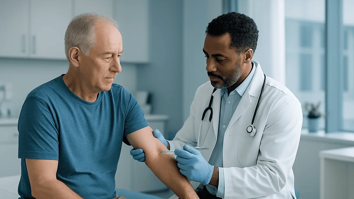
Bursa aspiration is a short office procedure in which a clinician cleans the skin over a swollen bursa, numbs the area if needed, and draws the excess fluid out through a fine needle into a syringe. The fluid's color, thickness and laboratory results help distinguish simple irritation from gout crystals, bleeding or infection, which guides what treatment your care team recommends next.
The swelling arrived quietly. A week of long shifts spent propped on one elbow, and now the tip of it looks like someone slipped a small water balloon under the skin. It doesn’t hurt much. It just wobbles when pressed, and it will not fit under a shirt cuff. At the appointment, the phrase comes up almost casually: “We could take that fluid off with a needle.”
That is bursa aspiration, and the word tends to land harder than the procedure itself. People picture a large syringe and a long afternoon. What actually happens is closer to a blood draw in reverse, usually finished before the coffee in the waiting room has cooled.
The more interesting question is what happens to the fluid afterward. A syringe of pale yellow liquid and a syringe of cloudy, thick material tell two very different stories, and the laboratory can read details the eye cannot. This is what to expect, what the results can and cannot show, and where the decisions belong.
What is a bursa, and why does it fill with fluid?
A bursa is a thin, slippery sac lined with tissue that produces a small amount of lubricating fluid. Think of a tiny, nearly empty water balloon tucked between a bone and the skin, tendon or muscle sliding across it. Its whole purpose is to reduce friction. Cleveland Clinic counts more than 150 of them in the body, clustered around the shoulders, elbows, hips, knees and heels, exactly where structures rub.
Most of the time a bursa holds so little fluid that you would never know it existed. Bursitis is what happens when that lining becomes irritated and starts producing far more fluid than it can absorb. The sac stretches, the skin over it bulges, and a joint that felt fine last month becomes stiff or tender.
The triggers are familiar. Repetitive pressure is the classic one: kneeling on hard floors, leaning on an elbow at a desk, throwing a ball for hours. A direct blow can inflame a bursa in a single afternoon. Inflammatory conditions such as gout or rheumatoid arthritis can flare inside a bursa the same way they flare inside a joint. Less commonly, bacteria find their way in through a scrape or crack in the skin and the fluid becomes infected, a situation clinicians call septic bursitis.
Mayo Clinic notes that the shoulder, elbow and hip are the most frequent sites, though the knee, heel and base of the big toe are also common. The bursae just under the skin, at the point of the elbow and the front of the kneecap, are the ones most often drained, simply because they are easy to reach and their swelling is hard to ignore.
What is bursa aspiration, and why would a doctor suggest it?
Aspiration means drawing fluid out with a needle and syringe. Bursa aspiration, then, is the removal of excess fluid from an inflamed bursa. Clinicians reach for it for two reasons that often overlap, and it helps to know which one applies to you.

The first reason is diagnostic. A swollen bursa that is warm, red or painful raises the possibility of infection, and no examination from the outside can settle that question reliably. Mayo Clinic lists analysis of aspirated fluid among the tests used to pinpoint the cause of bursitis. Sending the fluid to the laboratory answers the questions that matter most: Are there bacteria? Are there crystals that point to gout? Is the white cell count in a range that suggests inflammation rather than simple mechanical irritation?
The second reason is relief. A tense, overfilled bursa can be uncomfortable simply because of its size. Emptying it takes the pressure off, and for some people that is enough to make an elbow bend freely again. When the fluid is not thought to be infected, some clinicians will inject a corticosteroid, an anti-inflammatory medicine, into the sac in the same sitting; whether that is appropriate is a judgment for the person holding the syringe.
What aspiration is not is a routine first step. NHS guidance describes most bursitis settling with rest, protection of the area and time. The needle comes into the conversation when swelling is large, when infection needs to be ruled in or out, or when a bursa keeps refilling despite the basics. Your treating team weighs those factors against the modest risks, which are covered further on.
How bursa aspiration is actually done, step by step
The procedure almost always happens in a clinic room or emergency department rather than an operating theater. You will be asked to sit or lie so the swollen area is stable and easy to reach: elbow bent on a table, or leg extended for a kneecap.
The clinician first examines and sometimes marks the fullest part of the swelling. If the bursa is deep or the borders are unclear, a handheld ultrasound probe may be used to see the fluid pocket and steer the needle. Ultrasound is simply sound waves bounced off tissue to make a live picture; it is painless and involves no radiation.
The skin is cleaned with an antiseptic solution, usually in widening circles, and left to dry. This step is unglamorous but it is the main protection against carrying skin bacteria into the sac. Some clinicians then numb the skin with a spray or a small injection of local anesthetic, a medicine that blocks nerve signals in one small area for a short while. Others go straight in, reasoning that one needle is kinder than two when the bursa sits directly under thin skin.
A needle attached to a syringe is passed into the sac. Fluid is drawn back steadily; the clinician may gently press around the swelling to coax the last of it toward the needle. The whole collection typically takes a minute or two. The needle is withdrawn, pressure is applied, and a firm dressing or elastic wrap goes on to discourage the space from refilling straight away.
The syringe contents are labeled and sent for testing. You will usually be shown the fluid, because its appearance is part of the conversation about what comes next.
Does bursa aspiration hurt? What the needle actually feels like
Honest answer: a little, briefly, and usually less than people fear. The bursae that get drained most often lie just beneath the skin, so the needle does not have to travel through thick muscle. Most people describe a sharp sting at the surface followed by a pushing or pulling sensation as the fluid moves. If the sac is very tense, the moment the pressure releases can feel like relief rather than pain.
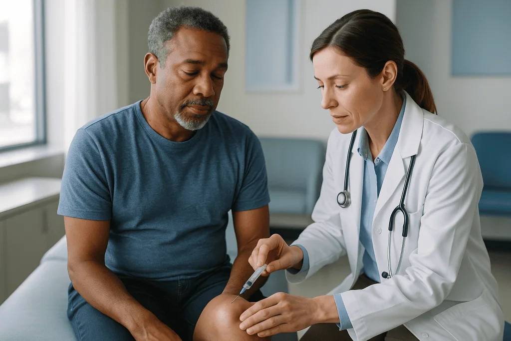
Three things shape the experience. First, how inflamed the skin is. An angry, red, infected bursa is tender before anyone touches it, and the antiseptic wipe alone can smart. Second, whether local anesthetic is used. Numbing adds one small needle prick and a minute of waiting, in exchange for a duller sensation during the main draw. Third, depth. A hip bursa sits under layers of muscle and is generally approached with imaging guidance and anesthetic; a kneecap bursa is barely below the surface.
Tell the clinician if you have fainted with needles before or if you tend to feel lightheaded during blood tests. Lying down rather than sitting is an easy adjustment and prevents the rare dramatic slump.
Afterward, the site can ache in the way a bruise does. That soreness is expected and short-lived. What is not expected is pain that escalates over the following day, spreading redness, or heat, all of which are described in the red-flag section. If you take a blood-thinning medicine, mention it before the procedure; it does not automatically rule aspiration out, but the clinician may adjust technique or pressure time, and any change to your prescription is their call, never something to do on your own.
Who is usually offered aspiration, and who is usually asked to wait
The needle is offered when it will change what happens next. A few situations tend to tip the decision toward draining.
Suspected infection is the clearest. Warmth, spreading redness, fever, or a break in the skin over the swelling all raise the stakes, because septic bursitis is treated differently from the irritated kind and the laboratory is the only way to be sure. Cleveland Clinic notes that infection of the bursa is a particular concern when the skin over it has been cut or scraped.
A very large, tight swelling that limits movement is another. So is bursitis in someone with known gout or an inflammatory arthritis, where confirming crystals or cell counts can redirect the whole treatment plan. Swelling that has failed to settle after the standard weeks of rest, padding and pressure relief that NHS guidance describes also earns a closer look.
Others are usually asked to wait, at least initially. Mild, soft swelling with no warmth or redness after a clear episode of overuse often shrinks on its own. A child with a bumped knee is generally watched rather than needled unless infection is a worry. Anyone with an active skin infection spreading across the area, rather than contained within the bursa, may need that treated first so the needle does not pass through infected tissue. Bursae lying close to an artificial joint are approached cautiously and often by the surgical team that placed the implant.
None of these are absolute rules. They are the factors your treating team balances, and the final decision belongs to them and to you, made together with the specifics of your history in front of you.
Reading the fluid: what color and clarity suggest
The first analysis happens in the room, by eye. Experienced clinicians hold the syringe up to the light because appearance offers a rough sorting before any laboratory result returns. The table below summarizes the common patterns, with an important caveat underneath.
| What the fluid looks like | What it commonly suggests | What the lab usually checks next |
|---|---|---|
| Clear to pale yellow, thin, easy to draw | Mechanical irritation from pressure or overuse; low inflammation | Cell count; sometimes culture as a precaution |
| Cloudy yellow, thicker | Inflammatory process, such as gout or rheumatoid flare, or early infection | Cell count, crystal examination, Gram stain, culture |
| Thick, opaque, creamy or greenish | Infection more likely | Gram stain and culture prioritized; cell count |
| Red or brown, bloody | Bleeding into the bursa after injury, or a small vessel nicked by the needle | Assessment of whether blood is old or fresh; often repeat sampling if unclear |
Here is the caveat, and it matters more than the table: appearance alone cannot confirm or exclude infection. Early septic bursitis can produce fluid that looks deceptively clear, and a fierce gout flare can produce fluid that looks alarmingly like pus. Mayo Clinic frames fluid analysis as a laboratory test for exactly this reason. Clinicians use the visual impression to decide how urgently to act while the formal results are pending, not as a final verdict.
Fluid that is bloody after a fall is often just that: bleeding into the sac. Fluid that is bloody in someone on a blood-thinning medicine, with no injury, prompts a more careful look. In every case, the syringe in the room is a first chapter, not the whole book.
Bursa fluid analysis in the lab: cell counts, crystals and cultures
Once the syringe reaches the laboratory, the fluid is usually divided into several tubes because each test needs different handling. Knowing what each one asks makes the eventual phone call easier to follow.
The cell count measures white blood cells, the immune cells that gather wherever the body is fighting something. Higher counts lean toward infection or intense inflammation; lower counts lean toward mechanical irritation. The laboratory also notes which type of white cell dominates, since a heavy majority of the type called neutrophils raises concern for bacteria. Exact thresholds are interpreted by your clinician alongside the rest of the picture, and bursal fluid behaves differently from joint fluid, so joint-based rules of thumb do not transfer neatly.
The Gram stain is a rapid test in which a drop of fluid is dyed and examined under a microscope for bacteria. It can return within hours and, when positive, is a strong signal. When negative, it does not rule infection out; bacteria may simply be too sparse to spot on one slide.
Culture places fluid on nutrient plates and waits for any bacteria to multiply into visible colonies. This is the definitive test for infection and the one that identifies which organism is present, which in turn shapes antibiotic choice. Because bacteria need time to grow, culture results arrive after the stain, and your team will tell you when to expect them.
Crystal analysis uses a polarizing microscope to look for needle-shaped urate crystals, the signature of gout, or the rhomboid crystals of the related condition sometimes called pseudogout. Finding crystals can redirect treatment toward controlling those conditions rather than simply protecting the bursa. Some laboratories also measure glucose or protein levels, which add supporting detail without deciding the case on their own.
Septic bursitis treatment: when the fluid shows infection
Septic bursitis means bacteria are multiplying inside the sac. Cleveland Clinic and Johns Hopkins both describe it as most often following a cut, scrape or insect bite over the elbow or knee, where only a thin layer of skin separates the bursa from the outside world. The most frequent culprit is Staphylococcus aureus, the same organism that lives harmlessly on many people’s skin until it finds an opening.
When the Gram stain or culture confirms infection, or when suspicion is high enough to act before results return, treatment shifts. Antibiotics become the foundation. Which medicine, how it is given and for how long are decisions for the prescribing clinician, guided by the organism identified, the severity of the infection and your health history. What patients can reasonably expect is that the plan may start broadly and then be narrowed once the culture names the bacteria.
Aspiration itself often becomes part of the treatment as well as the diagnosis. Removing infected fluid reduces the bacterial load and the pressure. Some people need the bursa drained more than once as it refills during the first days of treatment. Where fluid keeps returning thick and cloudy, or the infection is not responding, a surgeon may recommend a small operation to open and wash out the bursa, or to remove it entirely, a procedure called bursectomy.
Warning signs that infection is spreading beyond the bursa include fever, chills, redness tracking up the limb, or feeling generally unwell. Those belong in the emergency category rather than the wait-and-see one. Uncomplicated septic bursitis, caught early, is usually managed as an outpatient; people with weakened immune systems, diabetes or severe symptoms may be admitted for closer monitoring. Your team will explain which path fits your situation.
Risks of draining a bursa, stated plainly
Aspiration is a minor procedure, but minor is not the same as risk-free, and a fair explanation covers what can go wrong.
Introducing infection is the risk clinicians think about most. Any needle that crosses the skin can carry bacteria inward, which is why the antiseptic preparation is done so deliberately and why a sterile bursa is not drained casually. The risk is low with good technique, but it is not zero, and it is one reason a soft, painless swelling is often left alone.
Bleeding into the bursa or a bruise around the site is possible, particularly for people taking anticoagulant medicines or with bleeding disorders. Pressure after the needle comes out addresses most of this. Pain at the site for a short period is expected rather than a complication.
Recurrence is common enough to deserve its own mention. The bursa lining that overproduced fluid in the first place is still there after the syringe is full, so the sac may refill, sometimes within days. This is not a failed procedure; it is the nature of an irritated bursa, and it is why compression and removing the original pressure matter as much as the draw itself.
Less common problems include a persistent drainage tract, where fluid keeps leaking through the needle hole, most often at the elbow. If a corticosteroid is injected, Mayo Clinic notes possible skin thinning, lightening of skin color at the site, or a temporary flare of pain. Rarely, a needle can contact nearby tendon or nerve, which is a reason deeper bursae are approached with imaging guidance.
Put beside the risk of leaving a possibly infected bursa untested, most clinicians consider these manageable. Your team will walk through them before asking for your consent.
Alternatives to aspiration: rest, protection, medicines and surgery
Aspiration sits in the middle of a range of options, and understanding the rest of the range helps you ask the right questions.
At the gentle end is what NHS guidance describes as the first line for most bursitis: rest the area, avoid the activity or posture that provoked it, apply ice wrapped in a cloth for short periods, and cushion the spot. Elbow pads for desk workers and kneeling pads for people who work on floors are unglamorous but often decisive, because they remove the cause rather than the consequence. A soft compression wrap can discourage swelling from building.
Medicines come next. Non-steroidal anti-inflammatory drugs, a class that reduces inflammation by blocking certain enzymes, are commonly used for pain and swelling; whether they suit you depends on your kidneys, stomach, heart history and other prescriptions, all of which your clinician weighs. A corticosteroid injected directly into the bursa delivers a concentrated anti-inflammatory effect to the sac itself and is sometimes combined with aspiration when infection has been excluded.
Physical therapy has a role when a bursa is inflamed because of the way a nearby joint moves. A therapist can identify a tight muscle or weak stabilizer that keeps loading the same spot and work on correcting it, which addresses recurrence rather than the current swelling.
At the far end is surgery. Bursectomy, removing the bursa, is reserved for cases that keep recurring despite everything else or for infections that do not clear. Mayo Clinic lists it among the options for bursitis that does not improve with conservative measures. Bodies generally do well without a single bursa; the surrounding tissue adapts.
None of these excludes the others. Many people move through several steps, and the sequence is set by your treating team.
The days and weeks after a bursa is drained
The first hours are mostly about the dressing. Keep it clean and dry and leave the compression in place for as long as you were told. If the wrap feels tight enough to cause numbness, tingling or color change in the fingers or toes, loosen it and let the clinic know; that is a fit problem, not a sign of trouble in the bursa.
Soreness at the site is normal and fades. Ice wrapped in cloth, applied in short spells, helps both the ache and any residual swelling. Try not to test the area by pressing on it to see whether the fluid has returned; every time an elbow is leaned on or a knee is knelt on, the lining is invited to produce more.
Laboratory results arrive in stages. The cell count and Gram stain usually come first, often the same day; culture needs longer because bacteria grow at their own pace. Your team should tell you how and when you will hear, and what would change if the culture turns positive. Write the number down.
Over the following week or two, the shape of recovery depends on the cause. Bursitis from pressure or overuse tends to settle over a few weeks once the pressure is removed, according to NHS and Mayo Clinic guidance, though refilling in the early days is common and does not mean the procedure failed. Bursitis driven by gout or arthritis follows the course of that condition. Septic bursitis follows the antibiotic plan, and you should expect a follow-up check to confirm the redness and warmth are retreating.
A return to sport or manual work is a conversation with your clinician rather than a fixed date. The usual advice is to rebuild gradually and to keep the padding that protects the bursa when the activity resumes.
What people often get wrong about draining a bursa
“If it comes back, the doctor did it wrong.” Refilling reflects a lining that is still irritated, not a missed target. The needle removes fluid; it does not reset the tissue that made it. Compression, padding and addressing the cause are what reduce recurrence.
“Clear fluid means there is definitely no infection.” Early infection can look innocent. That is why clinicians send even reassuring-looking fluid to the laboratory when the story includes a skin break, fever or warmth.
“Pus means surgery.” Most septic bursitis is managed with antibiotics chosen by the prescriber, sometimes with repeat aspiration. Surgery is held for infections that do not respond or keep returning.
“I can drain it at home with a clean needle.” Please do not. Home attempts bypass proper antiseptic preparation, risk injecting skin bacteria into a sterile space, and can turn simple bursitis into the infected kind. There is also no way to send the fluid for analysis, which is half the point.
“A steroid injection is always given at the same time.” Steroids are generally avoided when infection has not been ruled out, because they dampen the immune response inside the sac. Whether one is appropriate is decided case by case.
“Bursitis is a joint problem.” The bursa sits outside the joint. Bursal fluid is not joint fluid, and the two are interpreted differently. This distinction is why a swollen elbow tip with a fully mobile joint reassures clinicians in a way a swollen joint would not.
“Once drained, I can go straight back to what I was doing.” The thing you were doing is usually what filled the bursa. Returning to it unchanged is the most reliable route back to the clinic.
Questions to ask your care team before and after bursa aspiration
A short list, carried into the appointment, turns a rushed consultation into a useful one. These are the questions clinicians tend to welcome.
- Are you draining this mainly to test the fluid, mainly to relieve pressure, or both? The answer tells you how much weight the laboratory results will carry.
- Do you suspect infection, and if so, will treatment start before the culture returns or after?
- Will you use ultrasound to guide the needle, and will you numb the skin first?
- Are you planning to inject anything into the bursa at the same time? If so, what is it and why is it appropriate in my case?
- I take these medicines, including any blood thinners or supplements. Does anything need to change, and who will tell me if it does?
- How will I receive the results, how long should I expect to wait for the culture, and what would a positive result change?
- How long should I keep the compression on, and can I shower with the dressing?
- What would make you want to see me again urgently, and what is the quickest way to reach the team?
- What is the most likely reason this bursa filled, and what should I change to stop it refilling?
- When can I go back to work, sport or driving, and what protection should I use when I do?
- If it comes back, what is the next step: repeat aspiration, injection, therapy or a surgical opinion?
Take notes or bring someone who can. Results delivered by phone are easier to interpret when you already know what each test was for, and the section on laboratory analysis above can serve as a reference while you listen. If any instruction you receive conflicts with something you read here, the instruction from your treating team wins.
When to call your doctor after bursa aspiration
Most people leave the clinic with a dressing, a follow-up plan and an aching but otherwise unremarkable elbow or knee. A small number develop signs that need prompt attention, and knowing them in advance removes the guesswork.
Contact your care team the same day if the redness around the site is spreading rather than shrinking, if the skin feels hot to the touch, if the pain is escalating rather than easing after the first day, or if fluid or pus keeps leaking from the needle hole. Swelling that returns rapidly and feels tense and hot is a different situation from swelling that drifts back slowly and stays soft.
Seek urgent care, through an emergency department or emergency number, for any of the following: a fever or shaking chills; red streaks tracking up the arm or leg from the site; feeling generally unwell, confused or faint; a limb that becomes numb, pale or cold below a dressing that cannot be loosened; or rapidly increasing pain out of proportion to what you were told to expect. These can signal infection spreading beyond the bursa or a circulation problem, and both are treated far more easily when caught early.
For people with diabetes, a weakened immune system, or a joint replacement near the drained bursa, the threshold for calling should be lower still, because infection in these settings can move faster and present more quietly. Mayo Clinic and Cleveland Clinic both flag fever with a swollen, red bursa as a reason to seek medical attention without delay.
When in doubt, call. A quick conversation that turns out to be unnecessary costs a few minutes; a delayed one can cost considerably more. The team that drained the bursa knows what they saw in the syringe and is best placed to judge what your symptoms mean.
Frequently asked questions
How long does bursa aspiration take?
The needle part usually lasts only a minute or two. Preparation, including positioning, cleaning the skin, any local anesthetic and, if used, ultrasound to locate the fluid, adds several more minutes. Most appointments are finished within a short clinic visit, and you can generally leave once a dressing is applied and you have been told how and when to expect laboratory results.
Will the fluid come back after the bursa is drained?
It can, and often does in the first days, because aspiration removes fluid without changing the inflamed lining that produced it. This is not a sign the procedure failed. Compression after the draw, padding the area, and stopping the pressure or repetitive movement that caused the swelling are what reduce recurrence. Persistent refilling is a reason to discuss next steps with your treating team.
Is elbow bursa drainage different from draining a knee or hip bursa?
The principle is identical, but the elbow and kneecap bursae sit just beneath the skin, so they are often drained without imaging and with minimal anesthetic. Hip bursae lie under thick muscle, so clinicians typically use ultrasound guidance and local anesthetic. The elbow is also the site where a persistent leaking tract after the needle is most often reported, which is why firm dressing and avoiding leaning matter there.
What does cloudy bursa fluid mean?
Cloudy fluid suggests an inflammatory process, which could be infection, a gout or pseudogout flare, or a rheumatoid flare. Appearance alone cannot tell these apart, so the laboratory checks white cell counts, looks for bacteria with a stain and culture, and examines the fluid for crystals. Your clinician will often decide how urgently to act based on the look of the fluid while awaiting the formal results.
Can a steroid be injected at the same time as bursa aspiration?
Sometimes, when infection has been excluded or is considered very unlikely. A corticosteroid is an anti-inflammatory medicine, and placing it inside the sac aims to calm the lining that overproduces fluid. Clinicians usually avoid it when infection is possible, because steroids dampen the immune response. Whether it suits your situation, and any side effects such as skin thinning or lightening, are for your treating clinician to weigh with you.
Do I need to stop blood thinners before having a bursa drained?
Do not stop or change any prescribed medicine on your own. Tell the clinician performing the procedure about every anticoagulant, antiplatelet medicine and supplement you take. Superficial bursae are often drained without any change to these medicines, with extra pressure applied afterward, but the decision depends on your bleeding risk and the site involved and rests with your prescribing clinician and the person doing the procedure.
Is bursa aspiration the same as joint aspiration?
No. A bursa sits outside the joint, and its fluid is interpreted differently from joint fluid. A swollen elbow tip with a fully mobile, painless elbow joint points to bursitis, whereas swelling inside the joint limits movement and raises different concerns. Laboratory thresholds used for joint fluid do not transfer directly to bursal fluid, which is one reason your clinician interprets results in context rather than by a single number.
How soon will I get the results of bursa fluid analysis?
Results arrive in stages. The cell count and Gram stain, which look for bacteria under the microscope, are often available the same day. Culture, which grows any bacteria to identify them, takes longer because organisms need time to multiply. Crystal analysis is usually quick. Your team should tell you exactly how you will be contacted and what a positive culture would change about your treatment.
Can I drain a bursa at home?
No, and attempting it is one of the more reliable ways to turn simple bursitis into an infected one. Home attempts lack proper antiseptic preparation and sterile equipment, risk introducing skin bacteria into a sterile space, and give no way to send fluid for analysis. If a swollen bursa is bothering you, the safe route is an appointment where the fluid can be removed and tested properly.
Does every case of bursitis need to be drained?
No. NHS guidance describes most bursitis settling with rest, protection of the area, ice and time. Aspiration is offered when infection needs to be ruled in or out, when a large tense swelling limits movement, when gout or inflammatory arthritis is suspected, or when swelling persists despite conservative care. A soft, painless swelling after clear overuse is often simply watched, a decision your treating team makes with you.
References
This article is for general information only and is not a substitute for professional medical advice. Please consult a qualified doctor about your individual situation.

More from the Blog
The Brostrom Procedure: Fixing Chronic Ankle Instability, Step by Step
The Brostrom procedure is an outpatient operation that tightens and reattaches the stretched outer ankle ligaments, usually the anterior talofibular and calcaneofibular ligaments, to…
When an ACL Graft Fails: Re-Rupture Causes and How Revision ACL Reconstruction Differs
ACL graft failure means a reconstructed ligament has torn, stretched or loosened enough that the knee is unstable again. Causes fall into three groups:…

How Quickly Does Tenosynovitis Settle? Healing Phases From Splint to Full Use
Tenosynovitis usually settles over weeks rather than days. With early rest and a splint, mild overuse cases often ease within a few weeks, and…

Shoulder Impingement vs Rotator Cuff Tear: How Doctors Tell the Two Apart and Why It Matters
Shoulder impingement is irritation of the rotator cuff tendons and the bursa as they pass beneath the bony roof of the shoulder blade; a…
Anesthesia for Arthroscopy: General, Regional or Spinal and Why Same-Day Discharge Is Usual
Arthroscopy is usually done under general anesthesia (fully asleep), spinal anesthesia (numb from the waist down, often with sedation), or a regional nerve block…

Arthroscopic Rotator Cuff Repair Step by Step: Anchors, Sutures and Reattaching the Tendon
The arthroscopic rotator cuff repair procedure reattaches a torn shoulder tendon to bone through several small incisions using a camera and thin instruments. The…