Bursitis Treatment
Bursitis treatment relieves inflammation of fluid-filled bursae around joints, commonly in the shoulder, hip, elbow or knee, using medication, injections, physical therapy and activity modification.

Quick answer
Bursitis is inflammation of a bursa, a small fluid-filled sac that cushions tendons, muscles and skin where they move over bone. It causes localised pain, swelling and stiffness, most often at the shoulder, hip, elbow or knee. Most cases settle with activity modification, anti-inflammatory medication and physiotherapy; aspiration, image-guided injections, antibiotics for infection or, rarely, surgery are used when conservative care is not enough.
What Is Bursitis?
Bursitis is inflammation of a bursa, a small fluid-filled sac that lets tendons, muscles, skin and bone glide over one another with less friction. When a bursa is irritated, it produces extra fluid, swells and becomes painful, and the movements it normally cushions, reaching overhead, climbing stairs, kneeling, lying on one side, begin to hurt. Bursitis most often affects the shoulder, hip, elbow and knee, although it can develop anywhere repeated pressure or friction loads soft tissue against bone. Most episodes of bursitis improve without surgery. The real work lies in confirming that the bursa genuinely is the source of pain, calming the inflammation, and correcting whatever provoked it, so that the problem does not simply return.
Bursitis usually starts quietly. A tender shoulder after a weekend of painting a ceiling. An aching outer hip that wakes you when you roll onto that side. A soft lump on the tip of the elbow after weeks of leaning on a desk. Knee pain that makes stairs and squatting awkward. The discomfort is frustrating precisely because it interferes with ordinary movement that once felt automatic, and because joint pain has many possible causes: tendon injury, arthritis, nerve irritation, crystal disease such as gout, infection, or pain referred from the spine. A careful diagnosis matters, since the most effective treatment for bursitis depends on identifying the exact source of symptoms and understanding why the bursa became inflamed in the first place.
Treatment for bursitis is not a single procedure applied to every patient. It is a structured approach that may combine rest from aggravating activities, anti-inflammatory medication, protective padding, physiotherapy and, when appropriate, aspiration of excess fluid or an image-guided injection. Antibiotics are used if the bursa is infected, and surgery is reserved for a small minority of persistent or complicated cases. In plain terms, treatment aims to do three things. It reduces pain and swelling so the joint can move comfortably. It addresses the cause of irritation, whether that is repetitive pressure, abnormal movement patterns, tendon overload or occupational strain. And it works to prevent recurrence by strengthening the muscles around the joint, improving flexibility and teaching you how to return to activity safely.
Where Are Bursa Sacs Located?
A bursa sits wherever the body needs a low-friction interface, and there are dozens of them, from the shoulders down to the feet. Some lie just beneath the skin: the olecranon bursa over the tip of the elbow and the prepatellar bursa in front of the kneecap are the two best known, which is why these sites are the most likely to swell visibly and the most exposed to direct injury and infection. Others lie deeper, between tendon and bone. The subacromial bursa cushions the rotator cuff beneath the bony roof of the shoulder. The trochanteric bursa covers the bony prominence on the outer side of the hip. The pes anserine bursa sits on the inner side of the knee just below the joint line, the iliopsoas bursa at the front of the hip, the retrocalcaneal bursa behind the heel, and the ischial bursa under the sitting bones. The location of the inflamed bursa shapes both the symptoms and the treatment. A superficial bursa often responds to pressure relief and padding, while an inflamed deep bursa usually says something about how the tendons and muscles moving over it are working.
What Causes Bursitis?
Bursitis is caused by anything that irritates the lining of a bursa, and the triggers fall into a few recognisable groups. Often more than one factor is at work in the same joint.
- Repetitive pressure or friction: prolonged kneeling in flooring, tiling, plumbing or gardening work; habitually leaning on the elbows at a desk; long periods of sitting on hard surfaces.
- Overuse and training errors: a sudden increase in running distance, a new overhead sport or gym routine, or a rapid return to activity after a long break, all of which overload tendons and the bursae beneath them.
- Direct injury: a fall onto the elbow, knee or hip, or a blow to the joint, which can inflame a bursa or cause bleeding into it.
- Mechanical and tendon problems: rotator cuff irritation in the shoulder, gluteal tendinopathy at the hip, muscle imbalance, altered gait, leg length differences and posture habits that increase friction over a bursa with every movement.
- Inflammatory and crystal disease: rheumatoid arthritis, gout and pseudogout can all inflame bursae directly, sometimes at several sites over time.
- Infection: bacteria entering through a graze, cut or cracked skin over a superficial bursa, particularly at the elbow or kneecap. The risk is higher in people with diabetes, immune suppression or skin conditions.
- Age and occupation: bursae and the tendons around them tolerate friction less well with age, and jobs or hobbies with repetitive positions concentrate load on the same tissue day after day.
Understanding the cause is not academic. A kneeling tradesman with prepatellar bursitis, a runner with outer hip pain and an office worker with an elbow lump need genuinely different plans, even though all three have bursitis.
Symptoms of Bursitis
The typical symptoms of bursitis are localised pain around a joint, tenderness when the affected spot is pressed, swelling, warmth, stiffness and pain with specific movements rather than with every movement. That specificity is a useful clue. Bursitis tends to hurt when the inflamed bursa is compressed or when the tendon gliding over it is loaded, and to ease when that particular pressure is removed. Pain that is constant, worsening at rest or spreading beyond the joint deserves closer attention, because it points towards other diagnoses.
The pattern varies by site. Shoulder bursitis commonly causes pain when lifting the arm to the side, reaching behind the back or sleeping on the affected shoulder. Hip bursitis produces pain over the outer hip that flares when walking, climbing stairs or lying on that side at night. Elbow bursitis often appears first as a soft lump over the tip of the elbow, sometimes a painless swelling in the early stage, that becomes sore with pressure. Knee bursitis causes swelling and discomfort with kneeling, squatting or bending, with the exact spot depending on which bursa is involved.
What Are the Symptoms of Severe Bursitis?
Severe bursitis announces itself with more than mechanical pain. Fever, chills, spreading redness over the joint, marked warmth, rapidly worsening swelling, drainage from the skin, inability to move the joint at all, or intense pain following an injury are the features clinicians take most seriously. These signs point away from simple inflammation and towards septic bursitis, fracture or tendon rupture. Septic bursitis most often affects the superficial bursae of the elbow and kneecap, because bacteria can enter through small breaks in the overlying skin, and it is treated as a clinical priority: an infected bursa can worsen quickly and the infection can spread into surrounding tissue. Severity also has a chronic face. A bursa that has been inflamed for months may become thickened, permanently swollen and painful at night, and at that stage inflammation alone is no longer the whole problem: the tissue itself has changed, and rehabilitation takes correspondingly longer.
The Joints Bursitis Affects Most Often
Each site has its own anatomy, its own typical triggers and its own look-alike conditions. Knowing which bursa is inflamed, and why, is the foundation of every sensible treatment decision.
What Is Bursitis of the Hip?
Hip bursitis usually means inflammation of the trochanteric bursa, the sac that covers the greater trochanter, the bony prominence you can feel on the outer side of your hip. The classic picture is aching or sharp pain on the outside of the hip that flares when walking, climbing stairs, standing up from a low chair or lying on the affected side in bed. Modern clinicians often use the broader term greater trochanteric pain syndrome, because in many patients the gluteal tendons that attach nearby are irritated as well as, or instead of, the bursa itself. That distinction matters for treatment: tendon problems respond to progressive strengthening, not rest alone. Bursitis around the hip can also involve the iliopsoas bursa at the front of the joint, which causes deep groin discomfort with hip flexion, or the ischial bursa under the sitting bone, aggravated by prolonged sitting on hard surfaces. The most important diagnostic task is separating true outer-hip bursitis from arthritis of the hip joint itself, which typically causes groin pain and stiffness, and from pain referred from the lumbar spine, which may travel down the outer thigh in a similar distribution.
Can Hip Bursitis Be Healed Quickly?
There is no reliable shortcut, and it helps to know that from the start. The fastest dependable route with hip bursitis is to stop compressing the irritated tissue, avoid lying on the affected side, place a pillow between the knees at night, avoid sitting with crossed legs or standing with the weight hung on one hip, while inflammation is calmed under a doctor’s guidance and, once pain allows, a progressive gluteal strengthening programme begins. An image-guided injection may be considered when pain is blocking rehabilitation, but it accelerates the plan rather than replacing it. Established cases usually improve over weeks rather than days, and rushing back to the same walking volume, training load or sleeping position tends to reignite symptoms just as they were settling.
Shoulder Bursitis
Shoulder bursitis is inflammation of the subacromial or subdeltoid bursa, the sac that cushions the rotator cuff tendons as they pass beneath the bony roof of the shoulder. It rarely occurs in isolation. In most patients it accompanies rotator cuff irritation or shoulder impingement, in which the tendons and bursa are pinched during overhead movement. Typical complaints are pain when lifting the arm through the middle of its arc, difficulty reaching behind the back, and night pain when lying on the affected side. Repetitive overhead work or sport is the usual driver; calcific deposits within the cuff tendons can also irritate the bursa, sometimes intensely. Shoulder bursitis can additionally follow injury, including the altered mechanics that sometimes persist after a shoulder dislocation. Because the bursa is caught up in a wider mechanical story, effective treatment addresses shoulder blade control, cuff strength and movement patterns, not only the inflamed sac. Injecting or resting the bursa while ignoring the mechanics behind the irritation is the most common reason shoulder symptoms return.
Knee Bursitis
Knee bursitis can involve several different bursae, and the location tells much of the story. Prepatellar bursitis, swelling directly in front of the kneecap, historically called housemaid’s knee, is the signature condition of kneeling trades and is also the knee bursa most vulnerable to infection through the skin. Infrapatellar bursitis sits just below the kneecap around the patellar tendon and is more common in jumping sports. Pes anserine bursitis causes tenderness on the inner side of the knee below the joint line and frequently accompanies knee osteoarthritis, tight hamstrings and altered gait. That overlap matters: when arthritis of the joint itself is the dominant problem, treating the bursa alone brings limited relief, and the conversation shifts to arthritis care, which in advanced cases can extend to knee arthroplasty, including robotic-assisted knee replacement. Bursitis on its own, however, never requires joint replacement: the two conditions simply coexist often enough that a careful examination is needed to work out which one is producing the pain.
Elbow Bursitis
Elbow bursitis, medically olecranon bursitis, produces a visible, often golf-ball-like swelling over the tip of the elbow. It frequently begins as a painless swelling and only becomes sore with pressure or further irritation. The usual causes are habitual leaning on the elbow, a direct blow or fall, gout, rheumatoid arthritis or infection. Because the bursa lies immediately under the skin, it is one of the most commonly infected bursae in the body; redness, warmth, increasing pain and fever change the diagnosis from a mechanical nuisance to septic bursitis, which is managed very differently. For non-infected cases, the mainstays are pressure avoidance, elbow padding and time, with aspiration considered when the swelling is large, uncomfortable or diagnostically unclear.
Heel and Foot Bursitis
Bursitis in the foot and heel most often involves the retrocalcaneal bursa, which sits between the Achilles tendon and the heel bone, or the superficial bursa between the tendon and the skin. Stiff or rigid-backed footwear, a sudden increase in running or hill training, Achilles tendon problems and inflammatory conditions are the usual contributors. Pain is felt at the back of the heel, worse at the start of movement and with direct shoe pressure. A bony prominence at the upper edge of the heel bone, sometimes called a Haglund deformity, can add to the pressure on the retrocalcaneal bursa, and where it is present, shoes with a soft or open back and a small heel lift often relieve symptoms noticeably. Clinicians are also deliberately cautious with corticosteroid injections in this region because of the closeness of the Achilles tendon. Footwear modification, load management and calf rehabilitation carry most of the treatment.
Inflammatory, Crystal-Related and Septic Bursitis
Not all bursitis is mechanical. Gout and pseudogout can deposit crystals inside a bursa and trigger abrupt, intensely painful swelling, most memorably at the elbow. Rheumatoid arthritis and related conditions can inflame bursae as part of a wider disease process, sometimes at several sites over the years. In these patients, treating the local bursa without addressing the underlying disease invites repeated episodes, so care is coordinated with rheumatology where appropriate. Septic bursitis, bacterial infection within the sac, stands apart from everything else on this page: it requires antibiotics, sometimes drainage of the infected fluid, and close monitoring, and a corticosteroid injection is avoided whenever infection is suspected.
How Bursitis Is Diagnosed
Diagnosis begins with a detailed history and physical examination, and in many patients that is enough. The physician asks when symptoms started, which activities worsen or relieve them, whether there was an injury or a change in routine, and whether conditions such as diabetes, gout, rheumatoid arthritis, kidney disease or immune suppression are present. The examination maps the exact point of tenderness, tests range of motion and strength, looks for swelling, warmth and skin changes, and checks for signs that the tendon or the joint itself, rather than the bursa, is the true source of pain. For hip and knee complaints, watching how you walk can be as informative as any scan.
Imaging is chosen to answer a specific question, not ordered by default. X-rays show bone: arthritis, bone spurs, calcific deposits and injury. Ultrasound shows the soft tissue in real time, fluid within the bursa, tendon changes, inflammation, and has the practical advantage of guiding a needle precisely if aspiration or injection is planned. Magnetic resonance imaging is reserved for persistent, complex or atypical cases, where a tendon tear, a deep soft-tissue problem or an alternative diagnosis needs to be confirmed or excluded.
Laboratory testing enters the picture when infection or crystal disease is possible. Fluid drawn from the bursa can be analysed for white blood cells, bacteria and crystals, which distinguishes septic bursitis from gout and from simple inflammation, three conditions that can look similar from the outside and demand entirely different treatment. Blood tests may support the assessment when systemic inflammation is suspected.
The differential diagnosis is where experience earns its keep. Outer hip pain may originate in the hip joint or the lumbar spine rather than the trochanteric bursa. Shoulder bursitis overlaps with rotator cuff tears and frozen shoulder. Knee swelling can reflect a meniscus injury, ligament strain or arthritis rather than a bursa. Getting this step right prevents months of treating the wrong structure.
How to Treat Bursitis
Treatment is layered, starting with the simplest effective measures and escalating only when the response is insufficient or the diagnosis demands it. The sequence below reflects how most non-infected bursitis is actually managed.
How Do You Make Bursitis Go Away?
You make bursitis go away by removing the irritation, calming the inflammation and then rebuilding the strength and mechanics around the joint, in that order. Skipping the first or last step is why bursitis so often comes back. In practice, the sequence looks like this:
- 1. Identify and stop the trigger. Kneeling, leaning, overhead repetition, a training spike, a sleeping position, something started the irritation, and it must pause while healing happens.
- 2. Protect the bursa. Padding for superficial bursae, position changes for deep ones, and ice or heat depending on the stage of symptoms.
- 3. Calm the inflammation. Anti-inflammatory medication chosen by your doctor around your medical history, or an image-guided injection in selected persistent cases.
- 4. Rule out infection and crystal disease where the picture suggests them, because those change everything about the plan.
- 5. Rehabilitate the mechanics. Strengthen and retrain the muscles and movement patterns that overload the bursa.
- 6. Return to activity in stages, increasing load gradually rather than resuming the old routine on the first pain-free day.
Activity Modification and Protection
Reducing irritation does not mean complete rest, and in most cases prolonged rest is counterproductive. Gentle movement is usually encouraged while the specific aggravating load is removed. A patient with knee bursitis avoids kneeling and uses protective padding when kneeling is unavoidable. A patient with elbow bursitis stops leaning on the elbow and pads the desk edge. A patient with shoulder bursitis temporarily limits overhead lifting while keeping the shoulder moving through comfortable ranges. Small ergonomic changes, workstation height, chair cushioning, footwear, sleep position, often matter more than they appear to. Ice tends to help recent swelling and acute flares; heat may ease the muscle tightness that gathers around a chronic problem. Compression can be useful over some superficial bursae, applied carefully and never in a way that increases pain or restricts circulation.
Medication
Medication for bursitis usually means non-steroidal anti-inflammatory drugs, paracetamol (acetaminophen) or topical anti-inflammatory preparations, selected by the treating doctor around the patient’s medical history. Kidney disease, stomach ulcers, blood-thinning treatment, heart disease and several other conditions change what is safe, which is why the choice belongs in a consultation rather than a pharmacy aisle. If infection is suspected or confirmed, antibiotics are prescribed according to the likely organism and the severity of the picture. Pain relief is calibrated deliberately: the aim is comfort that allows movement and rehabilitation, not medication that masks a condition needing more urgent attention.
Physiotherapy and Movement Rehabilitation
Physiotherapy, physical therapy, in North American usage, is central to bursitis treatment whenever symptoms trace back to mechanics, muscle imbalance, tendon overload or posture, which is most of the time in the shoulder, hip and knee. It is not merely a sheet of exercises; it is a guided process of teaching the joint to move with less friction over the bursa.
For shoulder bursitis, the programme typically rebuilds shoulder blade control, rotator cuff strength and flexibility while avoiding the positions that pinch the bursa. For hip bursitis, the emphasis falls on gluteal strengthening, pelvic stability, core control and a graded return to walking or running. For knee bursitis, therapists address quadriceps and hamstring flexibility, hip strength, kneeling technique and activity progression. For elbow bursitis, education about pressure habits often achieves more than any exercise. Therapists combine manual techniques, stretching, strengthening and functional retraining, adjusting the load week by week according to the pain response. A well-designed programme is also the best recurrence prevention available, because it changes the mechanical environment around the bursa rather than only quieting the inflammation inside it.
Aspiration and Injection Treatments
If a bursa is significantly swollen, the physician may recommend aspiration: drawing off the fluid with a sterile needle after cleaning the skin, sometimes with local anaesthetic. Aspiration relieves pressure and, just as importantly, produces fluid that can be analysed for white blood cells, bacteria and crystals when infection or gout is a possibility. For a tense, uncomfortable olecranon or prepatellar bursa, it can be both treatment and test in a single step.
Corticosteroid injection is considered when inflammation persists despite conservative care, when pain is blocking rehabilitation, or when rapid symptom control is medically appropriate. Ultrasound guidance is often used to place the medication precisely into or around the inflamed bursa, and it is particularly valuable for deep bursae such as the trochanteric or iliopsoas, where landmark-based injection is less reliable. The decision is individualised. Repeated steroid injections are not suitable for every patient and carry risks, including local tissue irritation, temporary blood sugar elevation in people with diabetes, skin changes at the injection site and, rarely, infection. An injection buys a window of comfort in which rehabilitation can progress; it is not a substitute for correcting the cause of irritation, and treating it as one is a common route to disappointment.
Treatment for Septic Bursitis
Septic bursitis is treated with antibiotics matched to the likely organism and the severity of the infection, alongside drainage of the infected fluid when needed, sometimes more than once. Steroid injection is avoided whenever infection is suspected, which is one reason fluid analysis precedes injection in uncertain cases. Patients with diabetes, immune suppression or broken skin over the bursa are monitored more closely, and in stubborn or complicated infections, surgical drainage or removal of the bursa may become necessary.
When Is Surgery Considered?
Surgery is uncommon in bursitis and sits at the end of the pathway, not the beginning. It is considered when symptoms remain persistent, recurrent and genuinely disabling despite properly conducted non-surgical care, or when an infected bursa fails to resolve with antibiotics and drainage. The operation depends on the site and the cause. The inflamed bursa itself may be removed, a bursectomy, most often at the elbow or in front of the kneecap for chronic recurrent swelling. In the shoulder, surgery may address the associated impingement or tendon pathology when imaging clearly shows it, since the bursa there is rarely the whole problem. The decision is made after reviewing imaging, the record of previous treatment, medical risks and what the patient actually needs the joint to do. For the large majority of patients, the pathway remains non-surgical: accurate diagnosis, targeted inflammation control and rehabilitation.
How Long Does It Take for Bursitis to Heal?
It depends on the joint, the cause and how long the bursa has been irritated. A mild, recent episode may improve within days to a few weeks once the trigger is removed and inflammation is treated. Persistent bursitis, particularly when tendon irritation or movement problems are involved, typically needs several weeks of structured rehabilitation. After an injection, some patients notice improvement within a few days, while others improve more gradually as the inflammation settles and therapy progresses. Infected bursitis follows its own timeline, set by the severity of the infection, the need for drainage and the response to antibiotics.
| Time Period | What Patients Can Expect |
|---|---|
| Day 1 | Evaluation focuses on diagnosis, pain control and identifying the activities to modify. If aspiration or injection is performed, the area may feel sore for a short time afterwards. |
| First week | Swelling and pain often begin to ease with medication, ice, protection and reduced irritation. Gentle movement is usually encouraged while the aggravating activity stays off the menu. |
| First month | Physiotherapy builds flexibility, strength and corrected movement patterns. Many patients gradually return to normal daily activities, guided by how symptoms respond. |
| Six to twelve weeks | Persistent cases typically improve with consistent rehabilitation and staged activity progression. Sport, heavy work and repeated kneeling, lifting or overhead activity return more gradually. |
| Longer term | Prevention takes over: maintaining strength, using protective equipment where needed and adjusting repetitive activities to keep flare-ups from returning. |
Recovery is judged by function rather than pain alone. The target is walking, sleeping, lifting, working or training without repeatedly provoking the bursa, and activity is increased step by step towards that target. Returning too quickly to the same triggering movements is the single most common cause of recurrence, even when the pain initially seemed to have gone.
Benefits of Structured Bursitis Treatment
The benefits depend on the cause and severity of the condition, but a properly structured plan aims at pain relief, safer movement and lasting prevention rather than temporary quiet.
| Benefit | What It Means for You |
|---|---|
| Reduced pain and inflammation | Treatment calms the irritated bursa, making daily movements such as walking, reaching, bending and sleeping on the affected side more comfortable. |
| More accurate diagnosis | A structured evaluation distinguishes bursitis from tendon injury, arthritis, infection and referred pain, so treatment targets the true source of symptoms. |
| Improved mobility | Medication, therapy and guided activity changes restore range of motion and reduce stiffness around the affected joint. |
| Lower risk of recurrence | Rehabilitation and ergonomic advice address the movement patterns, pressure points and training errors that caused the problem in the first place. |
| Safer return to activity | A staged plan lets you resume work, exercise and sport without overloading the bursa too soon. |
| Appropriate care for infection | If septic bursitis is present, prompt antibiotics and drainage where needed reduce the risk of the infection spreading or complicating. |
Why Acting Early Matters
Many people wait, because bursitis can come and go, and mild cases genuinely may settle with sensible self-care. But persistent or recurrent symptoms deserve evaluation, particularly when pain has started to change how you move. A painful joint invites compensation. A sore shoulder stiffens and weakens as you stop using it. Hip bursitis alters gait and quietly overloads the back, the knee and the opposite hip. Knee pain shrinks activity levels and deconditions the muscles that were supposed to protect the joint. The longer these compensations run, the more the eventual rehabilitation has to undo.
Early assessment matters for a second reason: not all swelling around a joint is simple bursitis. Infection, gout, inflammatory arthritis, tendon rupture, fracture and nerve-related pain can all mimic it convincingly. A delayed diagnosis can allow an infection to worsen or a tendon injury to progress while the wrong problem is being rested and iced. Superficial bursitis at the elbow or kneecap with increasing redness, warmth or fever is the scenario where the difference between simple and septic matters most.
Treating bursitis early also means catching it before movement patterns become entrenched, when a short course of protection, medication and targeted exercise is usually all that is required. It is also the stage at which practical guidance, ergonomics, training adjustments, footwear, sleep position, has the greatest preventive effect, because the habits that provoked the bursa have not yet had months to harden into routine.
What Influences a Good Outcome
A good result in bursitis treatment means more than a quiet fortnight. It means understanding why the bursa became irritated and choosing treatment that fits your medical profile, your joint mechanics and your life. Several factors consistently shape how well that goes.
Accurate diagnosis is the strongest of them. If hip pain is actually coming from the spine, or shoulder pain is primarily a rotator cuff tear, treating only the bursa delivers limited and temporary relief. Careful examination and appropriately chosen imaging keep the plan pointed at the right structure.
The presence of infection changes both the urgency and the toolkit. Septic bursitis needs antibiotics and sometimes repeated drainage; steroid injection is off the table while infection is suspected; and patients with diabetes, immune suppression or open skin need closer follow-up.
Duration of symptoms sets realistic expectations. Recent inflammation may respond within days to protection and medication. Chronic bursitis often comes with thickened tissue, tendon involvement, weakness and entrenched movement habits, and needs a correspondingly longer rehabilitation arc.
Adherence to activity modification is where outcomes are quietly won or lost. If the same pressure or repetition that caused the inflammation continues, symptoms return regardless of how good the injection was. The effective adjustments are usually small: elbow padding, a changed workstation height, a modified running plan, a knee cushion, a different sleeping position.
Physical conditioning around the joint determines how much friction the bursa faces every day. For hip bursitis, gluteal strength and pelvic control carry the load. For shoulder bursitis, scapular mechanics and rotator cuff endurance decide long-term comfort. For knee bursitis, lower-limb alignment, flexibility and kneeling habits are the levers that matter.
Underlying medical conditions such as gout, rheumatoid arthritis, thyroid disease, kidney disease and diabetes influence inflammation, healing and which medications are safe. Treating the bursa while ignoring these conditions is a recipe for repeat episodes, which is why complicated cases benefit from input across rheumatology, endocrinology or infectious disease when needed.
Judicious use of injections protects the long game. Injections help selected patients, especially when pain is blocking rehabilitation, but timing, technique, medication choice and frequency all deserve deliberate thought rather than routine repetition.
Your own goals shape the plan more than any protocol. A recreational walker with hip bursitis, a manual worker with a swollen elbow and an overhead athlete with a sore shoulder need different strategies, different timelines and different definitions of success. Treatment works best when it is built around what the joint actually has to do.
Preventing Recurrence
Bursitis prevention is unglamorous and effective. It means permanently changing whatever loaded the bursa: knee pads for kneeling work, elbow padding or a rearranged desk, graduated training increases instead of sudden spikes, supportive footwear for heel bursitis, a pillow between the knees for a sensitive hip, and regular strength work for the muscles that shield the joint. Because bursitis is fundamentally a friction problem, anything that reduces friction, better mechanics, stronger stabilising muscles, softer interfaces, smarter load progression, reduces the odds of another episode.
It is worth saying something honest about the quick-fix stories that circulate online, in which one stretch, supplement or gadget made someone’s bursitis vanish. Those accounts usually describe mild, recent inflammation that would have settled with almost any sensible care, or a case that was never bursitis to begin with. What reliably reduces recurrence is duller: identifying the trigger, respecting the healing timeline, rebuilding strength and returning to load in stages. Patients who do those four things tend to stop thinking about their bursa; patients who chase shortcuts tend to meet it again.
Bursitis Care at Acibadem
At Acibadem, bursitis is assessed with attention to the whole clinical picture rather than the bursa in isolation. Orthopaedics, physical medicine and rehabilitation, radiology, infectious disease, rheumatology and pain management can each be drawn into a case when it requires them: a multidisciplinary approach that earns its value when bursitis is recurrent, complicated, tangled with another medical condition or difficult to distinguish from the conditions that mimic it.
Diagnostic pathways are built around specific questions: imaging where it changes the decision, ultrasound-guided procedures where precision matters, laboratory analysis of bursal fluid where infection or crystal disease is on the table, and rehabilitation planning coordinated with the diagnosis rather than bolted on afterwards. The purpose of the technology is practical, to confirm what is actually wrong, guide treatment accurately and avoid unnecessary interventions.
Because bursitis so often reflects a wider mechanical issue, physicians evaluate the joint and soft tissues around the inflamed bursa: the gluteal tendons behind an aching hip, the rotator cuff behind a sore shoulder, the arthritis or kneeling habits behind a swollen knee. Treatment plans are individualised and explained in a form that supports continuity, what to expect in the first days after an aspiration or injection, how exercises should progress, and which changes would warrant reassessment. Sometimes the answer is a targeted injection and a therapy programme; sometimes it is treatment of a different underlying condition; rarely, it is surgery.
Living Well After Bursitis
Bursitis is common, and most people who have it once return to everything they did before. But persistent joint pain should not be waved away as inevitable. With an accurate diagnosis and a plan matched to the cause, the majority of patients can reduce pain, restore comfortable movement and understand their own joint well enough to keep the problem from returning. The right treatment may be as simple as padding, medication and a strengthening programme, or it may involve imaging, aspiration, injection, antibiotics or specialist input, what matters is that it fits the actual cause.
A well-managed episode ends with more than a quiet bursa. It ends with a joint that moves better than it did before the trouble started: stronger supporting muscles, smarter habits around pressure and load, and a clear sense of which early signs mean it is time to ease off. That, rather than any single treatment, is what keeps bursitis in the past tense.
Preparation
- A specialist reviews symptoms, medical history and the affected joint, and may request imaging or blood tests to rule out infection or other conditions. Patients should report current medications, allergies and any recent injury or fever. Avoiding activities that worsen pain may be advised before the visit.
Aftercare
- After treatment, rest, ice, prescribed medication and gradual return to activity help reduce inflammation. Physical therapy may be recommended to improve flexibility, strength and joint mechanics. Patients should seek medical advice if pain worsens, swelling increases or fever develops.
Frequently Asked Questions
What affects the cost of bursitis treatment?
The final cost depends on the joint involved, whether imaging is needed, the suspected cause, the type of treatment recommended, the need for physiotherapy, and whether aspiration, injection, laboratory testing, or follow-up care is required.
How can I get a personalised quote for bursitis treatment in Turkey?
You can request a free consultation by sharing your symptoms, medical history, previous imaging, test results, and details of any treatments already tried. A specialist can then advise which services may be needed and the international patient team can prepare a personalised estimate.
Is imaging always required before treatment?
Not always. Some cases can be assessed clinically, while others may need ultrasound, X-ray, MRI review, or additional tests to confirm the diagnosis, exclude other joint problems, or guide an injection safely.
Are injections and physiotherapy usually included in the same package?
Package content varies by hospital and by medical need. Some plans may include consultation, imaging review, injection if appropriate, medication guidance, and physiotherapy recommendations, while other services may be listed separately.
Can bursitis be treated during a short medical trip?
Many non-surgical bursitis treatments can be planned within a coordinated outpatient visit, depending on diagnosis and test requirements. Suspected infection, complex joint disease, or surgical needs may require a different schedule.
Is this information medical or financial advice?
No. This is general educational information. Treatment choice and cost can only be confirmed after specialist assessment, so a free consultation is recommended for a personalised care plan and quote.
Medically reviewed by the Acıbadem International Medical Board September 1, 2026
See our medical review board →
Update history
- PublishedJune 8, 2026
- Medical review approvedSeptember 1, 2026
- Last content updateSeptember 8, 2026
Trusted care for international patients
From the Acibadem Blog

Does Bursitis Come Back? Why Flares Recur and What Long-Term Results Usually Look Like
Yes, bursitis can come back. Most flares settle within a few weeks with rest, ice and reduced pressure on the area, but recurrence is…
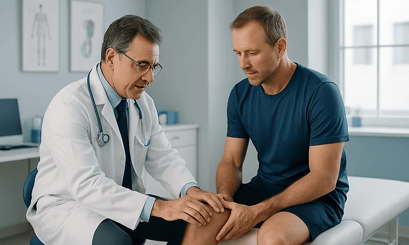
Bursitis in Runners and Cyclists: Knee and Hip Hot Spots and How Training Is Adjusted
Bursitis in runners and cyclists most often affects the inner knee (pes anserine bursa), the front of the knee, and the outer or front…

Steroid Injections for Bursitis: When They Are Offered, How They Work and What Follows
A bursitis steroid injection places a corticosteroid, often mixed with a local anesthetic, into an inflamed bursa to calm inflammation and ease pain. It…

Draining a Bursa (Aspiration): How the Procedure Is Done and What the Fluid Tells Your Doctor
Bursa aspiration is a short office procedure in which a clinician cleans the skin over a swollen bursa, numbs the area if needed, and…