

Follow-Up After Thyroid Cancer: What Blood Tests and Ultrasound Track Over Time
Key Takeaways
- Thyroglobulin is made only by thyroid cells, so after total thyroidectomy its trend across several tests, measured in the same laboratory, is the main blood marker for differentiated thyroid cancer.
- Anti-thyroglobulin antibodies can make thyroglobulin read falsely low, so an undetectable result alongside positive antibodies is not the all-clear and neck ultrasound carries more of the surveillance load.
- Neck ultrasound judges lymph nodes by shape and internal features rather than size, and very small suspicious nodes are often rescanned rather than biopsied straight away.
- TSH appears on cancer follow-up reports because thyroid cells respond to it; the target is matched to risk and is usually relaxed once the response is judged excellent.
- Medullary thyroid cancer is tracked with calcitonin and CEA rather than thyroglobulin, and radioactive iodine has no role in treating or scanning it.
- Most recurrences are found on scheduled tests in people who feel well, which is why surveillance continues for years even after an excellent response.
After treatment for differentiated thyroid cancer, follow-up usually combines a blood test for thyroglobulin, a protein made only by thyroid tissue, with a check for anti-thyroglobulin antibodies and thyroid hormone levels, plus periodic neck ultrasound. A sustained rise in thyroglobulin or a suspicious lymph node prompts closer evaluation. Test frequency depends on the original risk category and how results evolve, decided by the treating team.
The envelope from the lab sits on the kitchen counter for most of an afternoon before Maria opens it. Eight months after surgery for papillary thyroid cancer, she is well, back at work, and still unsure what the row of abbreviations is supposed to tell her. Tg, TgAb, TSH. Someone said the first one was the cancer test. Nobody explained the second.
That gap is common. Thyroid cancer follow up blood tests are among the most misread results in cancer care, not because they are complicated but because the logic behind them is rarely spelled out. Add a neck ultrasound every so often, a hormone tablet whose target keeps shifting, and a lead specialist who is not an oncologist, and it is easy to feel monitored without feeling informed.
This explainer walks through what each test measures, why the numbers are read as a pattern rather than a verdict, who needs close watching and who can breathe out, and which changes should send you to the phone.
What thyroid cancer follow up blood tests actually measure
Three numbers do most of the work on a post-treatment lab report, and only one of them is a cancer marker. Thyroglobulin is a protein made exclusively by thyroid cells, whether those cells are normal or cancerous. Once the whole gland has been removed by total thyroidectomy (surgery that takes out the entire thyroid), very little tissue is left to make the protein, so a low or undetectable level is the reassuring result and a rising one asks a question that needs answering (MedlinePlus).
The second number, anti-thyroglobulin antibodies, is not a cancer test at all. These are immune proteins that some people produce against thyroglobulin, and their presence can hide the true thyroglobulin level. Laboratories run both together for that reason, so the result can be read honestly.
The third pair, TSH (thyroid-stimulating hormone, the pituitary signal that tells thyroid tissue to grow and work) and free T4 (the main thyroid hormone), checks that hormone replacement is doing its job. After the gland is gone, levothyroxine, a synthetic version of the body’s own thyroid hormone, takes over. TSH also matters for the cancer itself, because any remaining thyroid cells, malignant or not, respond to it.
Some teams add calcium and parathyroid hormone in the early weeks, since the four tiny parathyroid glands sit behind the thyroid and can be bruised during surgery, causing temporary or occasionally lasting low calcium (NHS).
None of these tests diagnoses cancer on its own. Thyroid cancer follow up blood tests are trend instruments: a single value means less than the direction it takes over several visits, ideally measured in the same laboratory each time.
How thyroglobulin levels after thyroidectomy work as a marker
Think of thyroglobulin as smoke from a fireplace. If the chimney has been dismantled and no smoke appears, the house is quiet. If a wisp keeps rising, someone goes looking for the ember. The analogy has limits, and those limits are where interpretation gets interesting.
A small amount of normal thyroid tissue almost always survives surgery, tucked near the voice-box nerves or the parathyroid glands where a surgeon deliberately stays cautious. That remnant makes a little thyroglobulin. If radioactive iodine, a treatment in which thyroid cells absorb a radioactive form of iodine and are destroyed from within, was given afterward, the remnant usually fades and the level falls further (NHS). Someone treated with surgery alone may run a stable low value for life with no disease present. The word that matters is stable.
Laboratory sensitivity has changed the picture. Older assays needed thyroglobulin stimulated into view, either by pausing hormone replacement for a period so the body’s own TSH climbed, or by giving a synthetic form of TSH by injection, because a high TSH pushes any thyroid cell to release more protein. Modern high-sensitivity assays can often detect tiny amounts while a person stays on their usual tablets, so many low-risk patients are now followed with unstimulated values alone. Whether stimulation is still worthwhile depends on the original risk and the assay the laboratory uses, and that call belongs to the treating team.
One practical caution: switching laboratories mid-course can create a false rise, because assays are not perfectly interchangeable. Clinicians prefer the same lab, and the same method, at every visit (MedlinePlus).
Why anti-thyroglobulin antibodies can muddy the numbers
A positive antibody result frustrates patients more than almost any other finding, mostly because nobody warned them it might appear. Anti-thyroglobulin antibodies form when the immune system treats thyroglobulin as foreign. They are common in people with autoimmune thyroid conditions such as Hashimoto’s thyroiditis, and papillary thyroid cancer often sits alongside that background inflammation.
The problem is mechanical. Most thyroglobulin assays work by sandwiching the protein between detection molecules. Antibodies already bound to thyroglobulin block those sites, so the reading comes back falsely low, sometimes undetectable, even when thyroglobulin is present. An undetectable thyroglobulin beside positive antibodies therefore cannot be read as the all-clear that the same result would mean in an antibody-negative person (MedlinePlus).
Clinicians handle this in three ways. The antibody level itself becomes a surrogate marker: after complete removal of thyroid tissue, antibodies tend to decline over months to years because their target has gone, so a level that keeps falling is broadly reassuring, while one that plateaus or climbs is a prompt to look harder. Neck ultrasound carries more of the surveillance load, since imaging does not care about antibodies. And some laboratories offer an alternative measurement method that is less affected by interference, which the team may request in selected cases.
What antibodies do not mean is that the cancer is back. They are an immune signature, not a tumor product, and plenty of people carry them for years with no evidence of disease. The honest summary is that antibodies make one tool less reliable, so the team leans on the others. If your report shows them, ask how your team intends to track you, and expect ultrasound to feature prominently in the answer.
What a neck ultrasound after thyroidectomy is looking for
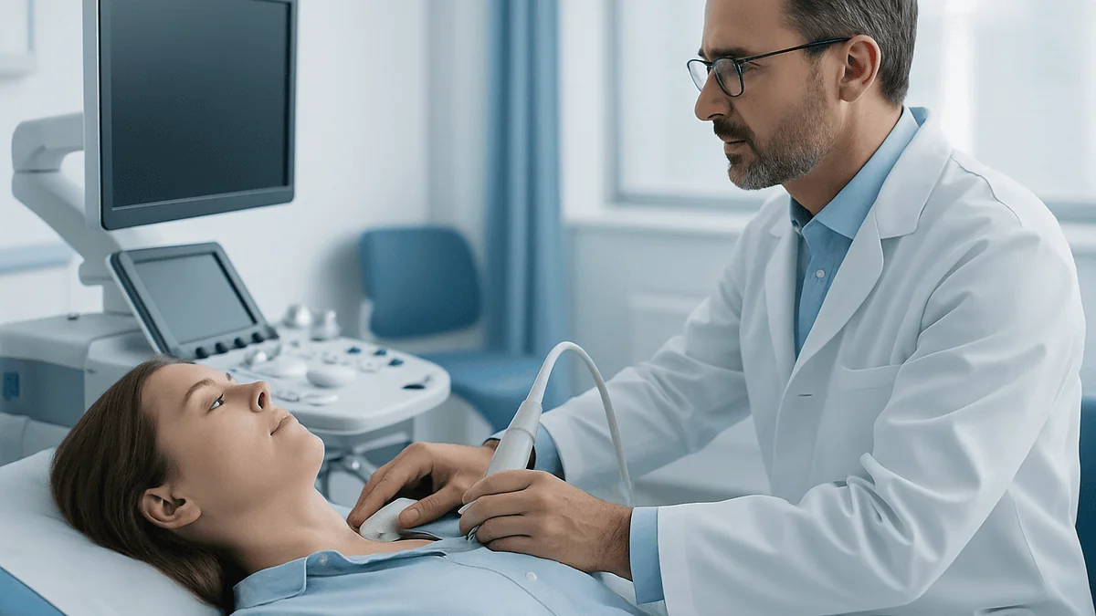
Ultrasound is the quiet workhorse of thyroid cancer follow-up. A handheld probe glides over gel on the front of the neck during a short outpatient appointment; there is no radiation, no injection and no preparation. What the sonographer is mapping is the thyroid bed, the empty space where the gland used to be, and the chains of lymph nodes running down both sides of the neck and just above the collarbones.
Lymph nodes are small filtering stations of the immune system, and the neck has dozens of them. Almost everyone has visible nodes on a neck scan, so size alone is a poor guide. Radiologists look instead at shape and internal structure. A healthy node is oval, with a bright central stripe of fat called the hilum. Features that raise concern include a rounded shape, loss of that fatty center, tiny bright specks suggesting calcification, cystic areas, or an unusual blood-flow pattern on Doppler, the ultrasound mode that shows moving blood (Mayo Clinic).
Finding a suspicious node is not the same as confirming recurrence. Guidelines generally favor watching very small suspicious nodes with a repeat scan rather than acting immediately, because many stay unchanged for years and surgery in a previously operated neck carries its own risks. When a node is larger or growing, the next step is usually a fine-needle aspiration, a thin-needle sample of cells, sometimes with thyroglobulin measured in the needle washout to strengthen the answer.
Scan and blood test complement each other. Thyroglobulin can climb from disease anywhere in the body; ultrasound can only see the neck, but the neck is where differentiated thyroid cancer most often returns (Cleveland Clinic). For antibody-positive patients, this scan is frequently the primary surveillance tool.
Who gets close monitoring, and who is asked simply to wait and watch
Intensity of follow-up is not decided by how frightened someone feels. It is set by two questions: how risky did the cancer look at the start, and how has the body responded since?
The first is called initial risk stratification. Pathologists and surgeons weigh tumor size, whether it grew through the thyroid capsule into surrounding tissue, how many lymph nodes were involved and how large they were, whether the cells showed aggressive features under the microscope, and whether cancer was found beyond the neck. The result sorts patients into low, intermediate or high initial risk, which guides whether radioactive iodine is offered and how tightly TSH is controlled (NCI).
The second is dynamic risk stratification, a reassessment usually made after the first year or two of results. In plain terms the team asks: with thyroglobulin, antibodies and ultrasound in hand, does this person now show an excellent response, an indeterminate response, a biochemical incomplete response (rising thyroglobulin with nothing visible on imaging), or a structural incomplete response (disease that can be seen)? Someone who started at intermediate risk but shows an excellent response is often stepped down to less frequent checks. Someone who started low risk but whose thyroglobulin drifts upward is watched more closely.
People asked to wait rather than test more are usually those with an excellent response: undetectable thyroglobulin, negative antibodies and a clean neck scan. For them, extra scans tend to turn up harmless incidental findings that cause worry and further tests without changing outcomes. Those who had only half the gland removed, a hemithyroidectomy, sit in a different lane: thyroglobulin is less informative because normal tissue remains, so ultrasound and clinical review carry more weight (Mayo Clinic). Where you land is a conversation, not a formula.
Thyroid cancer follow up blood tests and scans at a glance
The pattern below reflects how guideline-based teams commonly structure surveillance for differentiated thyroid cancer. Your schedule may look different, and that is usually deliberate.
| Test | What it measures | Typical use | What a change may mean |
|---|---|---|---|
| Thyroglobulin | Protein made only by thyroid cells | Each visit after total thyroidectomy | A sustained rise prompts imaging; a stable low value is generally reassuring |
| Anti-thyroglobulin antibodies | Immune proteins that interfere with the thyroglobulin assay | Always run alongside thyroglobulin | Falling levels are reassuring; rising levels prompt closer imaging |
| TSH and free T4 | Adequacy of hormone replacement and degree of TSH control | Each visit, and after any change to the tablet | Out-of-range values are adjusted by the prescriber |
| Calcium, parathyroid hormone | Parathyroid function after surgery | Early weeks; longer if low | Low calcium may need supplements under supervision |
| Neck ultrasound | Thyroid bed and lymph node structure | Within the first year, then according to risk | A suspicious node may be watched or sampled |
| Radioactive iodine scan | Iodine-avid thyroid tissue anywhere in the body | Selected cases after treatment or with rising markers | Uptake outside the neck points to distant disease |
| CT, MRI or PET | Structural or metabolic imaging | When markers rise but ultrasound is clear | Locates disease that iodine scans cannot show |
| Calcitonin and CEA | Markers for medullary thyroid cancer | Replace thyroglobulin in medullary cancer | Doubling time guides imaging |
Two things the table cannot show. Tests are read together: a slightly raised thyroglobulin beside a clean ultrasound and falling antibodies tells a different story from the same number beside a new node. And frequency shrinks over time for most people; the first year or two is the busiest, and visits often spread to yearly once the response is judged excellent (Mayo Clinic). A quiet schedule is a sign the numbers earned it.
What the first weeks, the first year and the years after usually look like
The calendar of surveillance has a shape, even if the exact dates belong to your team.
In the first days and weeks after thyroidectomy, blood tests concentrate on calcium. Tingling around the lips or fingertips is the classic sign of a temporary dip, which most people recover from as the parathyroid glands settle (NHS). Hormone replacement usually begins straight away, and the first TSH check follows several weeks later because the pituitary responds slowly to changes in thyroid hormone. Thyroglobulin drawn very early is hard to interpret, since protein released during surgery takes time to clear; a baseline is usually taken once things have settled.
If radioactive iodine is planned, it typically follows surgery within a few months, sometimes with a scan a few days afterward that doubles as the first look for tissue outside the neck (NHS). Not everyone needs it; low-risk cancers are increasingly treated with surgery alone.
The first year brings the busiest rhythm: thyroglobulin and antibodies with each hormone check, and the first post-treatment neck ultrasound, which many teams schedule within 6–12 months (Mayo Clinic). Around the one- to two-year mark, the team formally reassesses response using the dynamic categories described earlier.
After that, most people with an excellent response move to annual visits, and some guidance allows ultrasound to be dropped if thyroglobulin stays undetectable and antibodies remain negative. Follow-up nonetheless continues for many years, because differentiated thyroid cancer can return late (NCI). Expect the visits to become shorter and calmer rather than to disappear. Anyone with an indeterminate or incomplete response stays on a tighter cycle until the picture clarifies.
Why TSH suppression after thyroid cancer is part of surveillance
Levothyroxine after total thyroidectomy does two jobs, and only one of them is replacing a missing hormone. The other is tactical. Thyroid cells, cancerous ones included, carry receptors for TSH; when TSH is high, those cells are urged to grow and to produce thyroglobulin. Keeping TSH at the low end of, or just below, the usual range removes that growth signal. This is what clinicians mean by TSH suppression, and it is why the TSH result sits on a cancer follow-up report rather than being filed as routine hormone housekeeping (NCI).
The degree of suppression is matched to risk. Higher-risk or incompletely treated disease tends to earn a lower target, while people with an excellent response are usually eased toward a normal range over time, because holding TSH low for years is not free. Too little TSH means slightly too much thyroid hormone, which can thin bone, quicken the heart and nudge the rhythm toward atrial fibrillation, particularly in older adults (Cleveland Clinic). Balancing those risks against the cancer risk is the prescriber’s call, revisited as results come in.
Practically, this is why TSH is rechecked a few weeks after any change to the tablet, why consistency matters (same time of day, apart from calcium or iron supplements, which impair absorption), and why palpitations, heat intolerance or unexpected weight change are worth mentioning even if they seem unrelated to cancer. Never adjust the tablet yourself in response to a number; the target that suits your risk category is set by the team, and a value outside range is information for them, not a fault.
Pregnancy, or planning one, changes the targets, so tell the team early.
Medullary and anaplastic thyroid cancer: different tests, different rhythm
Everything above assumes differentiated thyroid cancer, the papillary and follicular types that arise from hormone-producing follicular cells. Cleveland Clinic notes papillary alone accounts for around 8 in 10 thyroid cancers, so most readers are in that group. Two rarer types play by different rules.
Medullary thyroid cancer grows from C cells, which make calcitonin, a hormone involved in calcium regulation, rather than thyroglobulin. Because C cells do not take up iodine, radioactive iodine treatment and scans have no role, and thyroglobulin is irrelevant. The blood markers are calcitonin and CEA (carcinoembryonic antigen, a protein several cancers release). The useful figure here is not the absolute level but the doubling time, meaning how quickly a rising level doubles, which helps the team decide how urgently to image and where. A slowly drifting marker may simply be watched; a fast doubling prompts CT or other scans of the neck, chest and liver, where medullary cancer tends to travel (Johns Hopkins). Some medullary cancers are inherited through a gene change, so close relatives may be offered genetic testing.
Anaplastic thyroid cancer is rare and fast-growing, and its follow-up is built around clinical examination and cross-sectional imaging at short intervals rather than blood markers, coordinated by a medical oncology team (NCI).
The lesson for anyone reading a report: know which type you had. Asking why your thyroglobulin is not being checked makes perfect sense for a papillary patient and no sense for a medullary one. Your pathology report states the type near the top, and your team can translate anything unfamiliar.
Do I need to see an oncologist for thyroid cancer follow-up?
In many countries the specialist who leads long-term follow-up for differentiated thyroid cancer is an endocrinologist, a physician who manages hormone conditions, rather than a medical oncologist. That surprises people used to thinking every cancer needs a chemotherapy doctor. The reason is that surveillance is mostly hormone management plus marker interpretation, and standard chemotherapy plays little part in differentiated thyroid cancer.
Behind the endocrinologist sits a multidisciplinary team, the group of specialists who review complex cases together: the surgeon who operated, a nuclear medicine physician who handles radioactive iodine, a radiologist who reads the ultrasounds, and a pathologist. The NHS describes this team model as standard for thyroid cancer decisions (NHS). A medical oncologist typically joins when disease no longer responds to iodine and targeted drugs, medicines that block growth signals inside cancer cells, are being considered, or for medullary and anaplastic types, where they often lead from the outset.
So, do you need an oncologist? For most people with papillary or follicular cancer showing a good response, not routinely, and being followed by an endocrinologist is not a downgrade. What matters more than the specialist’s title is that one clinician clearly owns your surveillance plan, that results are reviewed against your history rather than in isolation, and that there is a route back to the full team if the picture changes.
Some centers run combined clinics where blood is drawn, the ultrasound performed and the consultation held in a single visit. Others separate these steps by days or weeks. Neither model is superior in itself; ask which yours uses, so you know when to expect results and who to call about them.
How often does thyroid cancer come back or spread?
The question hides two different questions, and the honest answer separates them.
Spread at diagnosis is common for papillary cancer in one specific sense: lymph nodes in the neck are frequently involved when the cancer is first found, and this alone does not move most patients into a high-risk category (NCI). Spread to distant sites such as lungs or bones at diagnosis is far less common and is mainly seen with larger, more aggressive or follicular-type tumors. These are patterns rather than percentages; the figures vary widely by age, tumor type and how thoroughly nodes are examined, so no single number applies to an individual.
Recurrence after treatment follows the same geography. When differentiated thyroid cancer returns, it most often does so in neck lymph nodes, which is precisely why ultrasound is part of routine follow-up, and why a thyroglobulin rise with a clear neck scan sends the team looking further afield (Cleveland Clinic). Most recurrences appear within the first several years, but late recurrence is well documented, which underpins guidance for long-term surveillance (NCI).
What does recurrence feel like? Frequently, like nothing at all. Most recurrences are found on a blood test or scan in people who feel entirely well. When symptoms do occur they tend to be local: a new lump in the neck, a change in the voice, difficulty swallowing, or a persistent cough. Bone pain or breathlessness that does not settle would raise the question of distant disease. None of these is specific to cancer, and an anxious hand on the neck is a poor detector; the structured tests exist because they catch change earlier than symptoms do.
What people often get wrong about thyroid cancer surveillance
Some misunderstandings show up in almost every clinic.
‘Detectable thyroglobulin means the cancer is back.’ Not necessarily. A small stable value is common after surgery without radioactive iodine, because normal remnant tissue makes the protein. Direction over time, not presence, is the signal.
‘Undetectable thyroglobulin means I am clear.’ Usually reassuring, but only if antibodies are negative. With positive antibodies, an undetectable result may simply be blocked (MedlinePlus).
‘More scans are safer.’ Ultrasound is harmless, yet over-scanning low-risk patients tends to find tiny nodes that are almost always benign, leading to biopsies and anxiety without improving outcomes. Guideline groups have deliberately moved toward less imaging for excellent responders.
‘Radioactive iodine is always needed.’ Increasingly, low-risk cancers are treated with surgery alone, and the decision is individualized (NHS).
‘A normal TSH means everything is fine.’ TSH reflects hormone replacement, not cancer status, and in some risk categories a normal TSH may sit above the intended target.
‘If I feel well, I can skip follow-up.’ Most recurrences are found in people who feel well; feeling fine is not a test.
‘Normal thyroid blood tests rule out thyroid cancer.’ Thyroid cancer rarely disturbs hormone levels at diagnosis, which is why it is found by ultrasound and needle biopsy, not by a hormone panel (Mayo Clinic).
‘Supplements or diets keep thyroglobulin down.’ No evidence supports any supplement lowering recurrence risk, and some, such as large amounts of iodine or biotin, can interfere with tests or treatment. Tell the team about everything you take.
Each of these drives behavior: skipped visits, unnecessary scans, or a home-made adjustment to a tablet. The evidence supports steady, proportionate follow-up rather than vigilance at either extreme.
Questions to ask your care team
Bringing a short list to the first follow-up visit changes the tone of the appointment from receiving news to sharing a plan. These are the questions clinicians most often wish patients asked.
- What type of thyroid cancer did I have, and what initial risk category did the team assign, based on which features?
- Which markers will you follow for me: thyroglobulin and antibodies, or calcitonin and CEA?
- Do I have anti-thyroglobulin antibodies, and if so, how does that change the way you monitor me?
- What is my thyroglobulin baseline, and what kind of change would prompt further tests?
- Will my thyroglobulin be measured while I stay on my tablets, or will I need a stimulated test?
- What TSH range are you aiming for, and when might that be relaxed?
- When is my first neck ultrasound, and how often will it repeat if results stay reassuring?
- Which laboratory do you want my blood drawn at, so results stay comparable?
- When will you formally reassess my response, and what would an excellent response look like for me?
- Who owns my follow-up plan, and how do I reach that person between visits?
- Does my cancer type carry any inherited component that matters for my family?
- Should anything change if I am planning a pregnancy?
Write the answers down, or ask whether they can be recorded in a letter you keep. Follow-up for thyroid cancer often spans many years and several clinicians; a plan you understand is the one most likely to be kept, and a plan you can quote is the one most likely to be honored when a new doctor picks up your file.
When to call your doctor
Follow-up is designed so that most change is caught on a schedule, quietly. A few situations should not wait for the next appointment.
Contact your team promptly if you notice a new or enlarging lump in the neck or above the collarbone, a hoarse or changed voice that persists beyond a couple of weeks, new or worsening difficulty swallowing, a persistent cough or coughing up blood, or shortness of breath that is new for you. Bone pain that does not settle, particularly at night, or an unexplained fracture also warrants a call.
Symptoms that suggest a hormone problem deserve attention too. A racing or irregular heartbeat, tremor, marked heat intolerance or unexplained weight loss can mean hormone replacement is running high; profound tiredness, feeling cold, constipation or unexpected weight gain can mean it is running low. Either is a reason to have TSH checked sooner, not to change the tablet yourself.
In the weeks after surgery, tingling around the mouth or in the fingers, muscle cramps, or twitching may signal low calcium and should be reported the same day (NHS).
Seek emergency care immediately for difficulty breathing, noisy breathing, a rapidly swelling neck, chest pain, fainting, or severe muscle spasms with numbness spreading up the limbs.
For anything less urgent, a message to the clinic is always acceptable. Teams would much rather hear about a change that turns out to be nothing than learn about it months later. Deciding what a symptom means, and what to do next, sits with the clinicians who know your history; your part is to make sure they hear about it in time.
Frequently asked questions
What labs would indicate thyroid cancer?
No blood test diagnoses thyroid cancer. Thyroid hormone levels are usually normal at diagnosis, and the cancer is found by ultrasound followed by a fine-needle biopsy of the nodule. Thyroglobulin only becomes a useful marker after the thyroid has been removed. The exception is medullary thyroid cancer, where a raised calcitonin level can point toward the diagnosis before surgery, according to Mayo Clinic and Cleveland Clinic.
What are papillary thyroid cancer recurrence symptoms?
Most recurrences cause no symptoms and are picked up on thyroglobulin testing or neck ultrasound in people who feel well. When symptoms occur they are usually local: a new neck lump, a persistent change in voice, difficulty swallowing or a lingering cough. Bone pain or breathlessness that does not settle can suggest distant disease. None of these is specific to cancer, so report them rather than trying to interpret them.
How often does thyroid cancer spread?
Papillary thyroid cancer frequently involves nearby neck lymph nodes at diagnosis, and this alone does not usually place someone in a high-risk group. Spread to distant organs such as lungs or bones at diagnosis is much less common and tends to occur with larger or more aggressive tumors. Exact figures vary widely by age, type and how thoroughly nodes are examined, so no single percentage applies to an individual (NCI).
Do I need to see an oncologist for thyroid cancer?
Not always. Long-term follow-up for papillary and follicular thyroid cancer is often led by an endocrinologist, supported by a multidisciplinary team that includes the surgeon, a nuclear medicine physician and a radiologist. A medical oncologist usually becomes involved when disease no longer responds to radioactive iodine and targeted drugs are considered, or for medullary and anaplastic types. What matters most is that one clinician clearly owns your plan.
Why is my thyroglobulin still detectable after my whole thyroid was removed?
A small amount of normal thyroid tissue almost always remains after surgery, often deliberately left to protect the voice-box nerves and parathyroid glands, and that remnant produces a little thyroglobulin. Without radioactive iodine, a low stable value can persist for life without any disease present. Clinicians focus on the trend across several tests in the same laboratory rather than on whether the value is zero.
What do positive thyroglobulin antibodies after thyroidectomy mean?
They mean your immune system makes proteins against thyroglobulin, which can block the laboratory assay and make thyroglobulin read falsely low. They do not mean the cancer has returned. Teams respond by tracking the antibody level itself, which tends to fall over months to years once thyroid tissue is gone, and by relying more on neck ultrasound. A rising antibody trend is a prompt for closer imaging (MedlinePlus).
How long does follow-up for thyroid cancer continue?
Longer than most people expect. The first year or two is the busiest, with blood tests at each visit and a first neck ultrasound commonly within 6–12 months of treatment (Mayo Clinic). After an excellent response, visits usually become yearly and simpler, but some form of follow-up generally continues for many years because differentiated thyroid cancer can return late (NCI). Your team sets the schedule based on your risk category.
Can I stop having neck ultrasounds if my thyroglobulin is undetectable?
Possibly, if antibodies are negative and the ultrasound has been clear, since some guidance allows imaging to be reduced or paused for people with an excellent response. Routine scanning of low-risk patients often finds tiny benign nodes that lead to unnecessary biopsies. The decision depends on your initial risk, the sensitivity of the assay your laboratory uses and your team’s judgment, so ask rather than assume.
How long does TSH suppression after thyroid cancer last?
It depends on risk and response. People with higher-risk or incompletely treated disease usually keep a lower TSH target for longer, while those with an excellent response are typically eased toward a normal range over time, because prolonged suppression can affect bone density and heart rhythm (Cleveland Clinic). The target is reviewed at each reassessment and set by the prescribing clinician; never adjust the tablet yourself.
Is follow-up different if I only had half my thyroid removed?
Yes. After a hemithyroidectomy, the remaining lobe keeps producing thyroglobulin, so that blood test is far less informative as a cancer marker. Follow-up leans on neck ultrasound of the remaining lobe and lymph nodes, along with clinical review and thyroid hormone checks, since some people eventually need hormone replacement. Radioactive iodine is not used unless the rest of the gland is removed (Mayo Clinic).
References
This article is for general information only and is not a substitute for professional medical advice. Please consult a qualified doctor about your individual situation.

More from the Blog

Neck Dissection Preparation: Imaging, Nutrition and How Prior Radiotherapy Changes the Plan
Preparing for neck dissection usually involves imaging to map which lymph node groups need removal, an anesthetic assessment, and attention to nutrition, smoking and…

Is a Second Opinion Worth It for Lymphocytic Leukemia? Questions a Hematologist Expects
A second opinion is often worthwhile for chronic lymphocytic leukemia (CLL), a slow-growing blood cancer in which experienced hematologists can reasonably disagree about when…
Who Is a Candidate for Mohs Surgery? Skin Cancer Types, Locations and What Surgeons Consider
Candidates for Mohs surgery are usually people with basal cell or squamous cell skin cancer that sits in a high-risk or cosmetically sensitive area…
Does Surgery Come First or Chemotherapy? How Surgical Oncology Teams Sequence Cancer Treatment
Neither order is automatically better. Surgery first or chemotherapy first depends on the cancer type, its stage, how fast it tends to spread, and…

Can You Work and Travel During Immunotherapy? Planning Life Around Infusion Cycles
Many people can work during immunotherapy, often with reduced hours or flexible days around infusion cycles, because most checkpoint-inhibitor infusions take under a few…

How Remission Is Confirmed After Lymphoma Treatment and Then Monitored Over Time
Remission after lymphoma treatment is usually confirmed with an end-of-treatment PET-CT scan read against a standard scale, plus a physical examination and blood tests,…