

Planning Melanoma Skin Cancer Treatment by Stage: Surgery, Drug Therapy or Both?
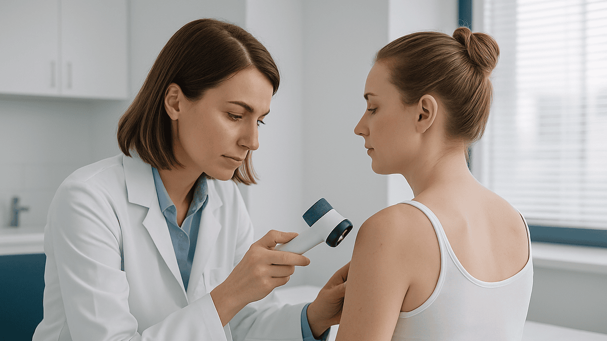
Key Takeaways
- Breslow thickness, measured in millimeters under the microscope, is the single strongest factor deciding a skin-confined melanoma's stage and its surgical margin.
- Stage 0, stage 1 and stage 2A melanomas are standardly treated with surgery alone; drug therapy has not been shown to help once these tumors are fully removed.
- Guideline excision margins range from about half a centimeter for melanoma in situ to about 2 centimeters for thicker tumors, and wider cuts do not improve outcomes.
- Sentinel lymph node biopsy is a staging test, not a treatment; a positive result moves the stage to 3 and opens the discussion of adjuvant drug therapy.
- Adjuvant immunotherapy or targeted therapy after stage 3 surgery is typically given for about a year, with targeted therapy reserved for tumors carrying a BRAF mutation.
- There is no fixed timeline for melanoma to progress from stage 1 to stage 4; growth rate varies by subtype from weeks to years, so change in a lesion matters more than elapsed time.
Melanoma treatment is planned by stage. Stage 0, 1 and most stage 2 melanomas are usually treated with surgery alone, removing the tumor with a margin of healthy skin. Stage 3, where cancer has reached nearby lymph nodes, often combines surgery with drug therapy such as immunotherapy or targeted therapy. Stage 4 relies mainly on drug therapy, with surgery in selected cases. Your oncology team decides based on your pathology report.
The pathology report arrives before the appointment does. Someone sits at a kitchen table reading words like “Breslow thickness” and “ulceration absent” next to a stage written in Roman numerals, and one question crowds out all the others: is this a surgery problem or a medicine problem? Or both?
That question is exactly the right one, because melanoma treatment by stage is not a slogan. It is the way oncologists actually work. A melanoma no thicker than a credit card is handled very differently from one that has already traveled to a lymph node, and the difference is driven by measurements a pathologist makes under a microscope, not by how alarming the mole looked in the mirror.
This explainer walks through what those measurements mean, what a surgeon removes and why, where immunotherapy and targeted drugs enter the picture, and which widely repeated beliefs about melanoma stages the evidence does not support. It is written to help you follow the conversation with your care team, who make the final call.
What does melanoma treatment by stage actually mean?
Staging is a shared language. When a dermatologist, surgeon and medical oncologist look at the same melanoma, the stage tells all three how far the disease has spread and, by extension, which tools are worth using. The Mayo Clinic describes the system in plain terms: stage 0 sits in the top layer of skin only, stages 1 and 2 are confined to the skin at increasing thickness, stage 3 has reached nearby lymph nodes or the skin between the tumor and those nodes, and stage 4 has spread to distant organs.
Treatment planning follows the same ladder. The earlier the stage, the more the plan leans on surgery alone, because removing the tumor removes the problem. The later the stage, the more the plan leans on medicines that travel through the bloodstream to reach cancer cells surgery cannot see.
Two things about this system surprise many patients. First, the stage is not final on the day of diagnosis. A biopsy gives a provisional stage; the wide excision and, where indicated, a lymph node procedure may move it up or down. Second, the stage is not the only input. Age, general health, the tumor’s location, genetic features of the cancer cells and a patient’s own priorities all feed into the plan. Two people with the same stage can reasonably receive different recommendations.
Melanoma treatment by stage, then, is best understood as a starting framework rather than a rulebook. It narrows the options so that the conversation with your team can focus on the choices that genuinely apply to you.
Melanoma stages explained: what depth, ulceration and lymph nodes tell the team
Three findings do most of the work in melanoma staging. The first is Breslow thickness, the depth of the tumor measured in millimeters from the top of the skin to its deepest cancer cell. Cleveland Clinic and the Mayo Clinic both note that thickness is the single strongest predictor of how a skin-confined melanoma is likely to behave, which is why staging uses cut points such as 1 millimeter, 2 millimeters and 4 millimeters to separate stage 1 from stage 2.
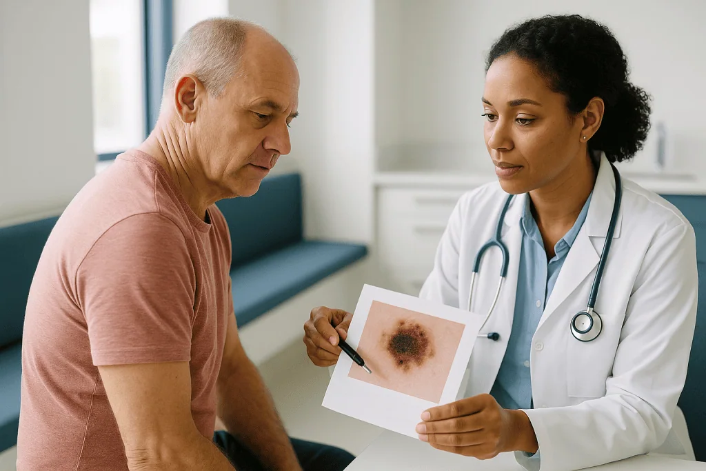
The second is ulceration, meaning the skin over the tumor has broken down under the microscope. An ulcerated melanoma is staged one notch higher than a non-ulcerated one of the same depth, because it tends to behave more aggressively.
The third is lymph node involvement. Lymph nodes are small filters along the drainage channels of the body; melanoma cells that leave the skin usually pass through the nearest group first. Cancer in those nodes defines stage 3. Spread beyond them, to organs such as the lungs, liver or brain, defines stage 4.
You may also see letters after the stage number, such as 1A or 3C. These substages refine the picture using the same variables and can influence, for example, whether a lymph node biopsy or drug therapy is offered. If your report uses terms you do not recognize, ask your team to walk through it line by line. The report is written for other clinicians, and there is no expectation that you decode it alone.
How melanoma surgery works: what actually happens in a wide local excision
Most melanomas are diagnosed by an initial biopsy that removes the visible lesion. The definitive operation that follows is called a wide local excision: the surgeon removes the scar from the biopsy plus a rim of normal-looking skin around it and the tissue beneath, down to the layer of fat over the muscle. The NHS describes this as the main treatment for melanoma that has not spread.
Why remove healthy skin? Melanoma cells can extend microscopically beyond the edge of what is visible. The margin is a safety zone, and its width is set by the tumor’s thickness rather than by the surgeon’s preference. The excised tissue goes back to the pathology laboratory, where the edges are checked to confirm they are free of cancer.
For smaller excisions the wound is closed with stitches in a straight line. Larger ones, or those on the face, hands or lower leg where skin is tight, may need a skin flap (nearby skin rotated to cover the gap) or a skin graft (skin borrowed from another site). Cleveland Clinic notes that most wide excisions are performed under local anesthetic as day procedures, although general anesthetic is used when a lymph node procedure is done at the same time.
If staging suggests the nearest lymph nodes should be checked, a sentinel lymph node biopsy is often added. A small amount of tracer is injected around the tumor site; it drains to the first node or nodes, which are then removed and examined. This tells the team whether stage 3 disease is present without removing the entire node group.
How deep do they cut for stage 1 melanoma?
People searching for how deep surgeons cut are usually asking two separate things: how wide, and how deep. The answers differ.
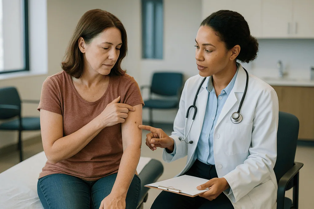
Width is governed by guideline margins that scale with Breslow thickness. Melanoma in situ (stage 0) takes the narrowest margin, roughly half a centimeter to a centimeter of surrounding skin. Stage 1 melanomas up to about 1 millimeter thick generally take a margin of around 1 centimeter, and thicker skin-confined tumors take up to about 2 centimeters, according to guidance summarized by the Mayo Clinic and the NHS. Research over several decades found that wider margins than these did not add benefit, which is why modern excisions are considerably less extensive than those performed a generation ago.
Depth is more consistent. The surgeon removes the full thickness of skin and the underlying fat down to the fascia, the thin sheet of connective tissue over the muscle. Going deeper is not necessary for skin-confined disease.
What this means in practice: a stage 1A melanoma measuring 5 millimeters across might leave a scar 3 to 4 centimeters long once the elliptical excision is closed, because a circle of skin cannot be sewn shut without lengthening the wound into an oval. That surprises many patients who expected a tiny mark.
The margin recommendation for your tumor will be recorded in your surgical plan. Ask what width has been chosen and why; the answer should trace directly back to the thickness on your pathology report.
Stage 0 and stage 1 melanoma treatment: why surgery alone is usually the whole plan
For melanoma in situ and stage 1 disease, the treatment conversation is usually short, and that is good news. The tumor has not spread beyond the skin, and removing it completely with an appropriate margin is the standard of care described by MedlinePlus, the Mayo Clinic and the NHS. Drug therapy is not routinely offered, because there is no evidence it improves outcomes when the cancer is already fully removed.
The one refinement at this stage concerns the lymph nodes. Sentinel lymph node biopsy is not generally recommended for very thin melanomas, since the chance of finding cancer in a node is low and the procedure carries its own small risks. Cleveland Clinic notes it is typically discussed for stage 1B tumors and above, particularly those approaching 1 millimeter with ulceration or a high rate of dividing cells. Your team may present this as an option rather than a requirement, and reasonable people choose differently.
What follows surgery is surveillance rather than treatment. Skin checks with a clinician at intervals set by your team, plus your own monthly self-examination, are the main tools. Anyone who has had one melanoma has a higher chance of developing another, so this vigilance is lifelong.
It bears saying plainly: a stage 1 diagnosis is frightening, and the word melanoma carries weight. But the plan for most people at this stage is a single operation and a commitment to follow-up, not months of medical therapy.
Stage 2 melanoma: sentinel lymph node biopsy and the question of adjuvant therapy
Stage 2 means the melanoma is thicker, ulcerated, or both, but no cancer has been found in the lymph nodes. Surgery remains the foundation, with wider margins than stage 1. The added consideration is that thicker tumors have a higher chance of having seeded cells that scans and examination cannot detect.
Sentinel lymph node biopsy is routinely offered at this stage, according to Mayo Clinic guidance. If the sentinel node is clear, the stage stays at 2. If cancer is found, the stage moves to 3, and the treatment discussion changes. This is why the procedure is best understood as a staging tool rather than a treatment in itself.
The newer question at stage 2 is adjuvant therapy, meaning medicine given after surgery to reduce the chance of the cancer returning. Immunotherapy has been studied in higher-risk stage 2B and 2C disease, and some guidelines now list it as an option to discuss. The trade-off is real: a proportion of people treated would never have relapsed anyway, and immunotherapy can cause lasting side effects, including permanent thyroid or other hormone gland problems. Whether the balance favors treatment depends on the specific substage and on how a person weighs risk, and this is a decision that sits squarely with the oncologist and patient together.
Follow-up for stage 2 usually includes more frequent examinations than stage 1 and may include imaging, depending on local practice and substage.
Stage 3 melanoma treatment options: surgery, drug therapy, or both?
Stage 3 is where the title question truly applies, because the answer is often both. Melanoma has reached the regional lymph nodes or the skin between the primary tumor and those nodes, but not distant organs. The NHS and the Mayo Clinic describe the standard sequence as surgery to remove the primary tumor and the involved nodes, followed by adjuvant drug therapy.
Two drug approaches are used. Immunotherapy, specifically immune checkpoint inhibitors, blocks a braking signal that cancer uses to switch off immune cells, allowing the immune system to attack melanoma. Targeted therapy, for the roughly half of melanomas carrying a mutation in a gene called BRAF, blocks the abnormal protein driving the cancer’s growth. In guideline-based practice both are typically given for about a year after surgery, as summarized by the Mayo Clinic, with the choice guided by the tumor’s genetic test result, other medical conditions and side-effect profiles.
Surgery itself has become more conservative. Complete removal of an entire lymph node basin, once routine when a sentinel node was positive, is now often replaced by close monitoring with ultrasound, because trials found it did not improve survival while carrying a real risk of lymphedema, a persistent swelling of the limb.
Some stage 3 patients are offered neoadjuvant therapy, drug treatment before surgery, within or outside clinical trials. Early evidence is encouraging, but this remains an evolving area rather than a settled standard in every setting, and your team can tell you whether it applies to your situation.
Immunotherapy for melanoma stage 4: how it works and what it does not do
Stage 4 melanoma has spread to distant sites, and the treatment center of gravity shifts to medicines that reach the whole body. Surgery still has a role, for example removing a single accessible metastasis or a tumor causing symptoms, but it is rarely the main strategy.
Immunotherapy has changed this stage more than any other. Checkpoint inhibitors do not attack cancer directly; they remove the molecular signals that stop T cells, the immune system’s specialist killers, from recognizing melanoma. Because the effect relies on the immune system rather than the drug alone, responses can take weeks to months to appear and can persist after treatment stops. The Mayo Clinic and Cleveland Clinic describe this as a treatment that can control disease for extended periods in some people, while acknowledging that it does not work for everyone and that no clinician can predict in advance who will benefit.
Side effects reflect the mechanism. When the brakes come off the immune system, it may attack healthy tissue, causing inflammation of the bowel, liver, lungs, skin or hormone glands. These effects can appear at any point during treatment and occasionally afterward, which is why patients are asked to report new symptoms promptly.
For BRAF-mutant melanoma, targeted therapy offers an alternative or a sequence partner. It often works faster than immunotherapy, which matters when disease is causing symptoms, but cancers commonly develop resistance over time. Radiation therapy, particularly focused techniques for brain metastases, is another tool. Clinical trials are a mainstream option at this stage, not a last resort.
Who is usually offered drug therapy, and who is usually asked to wait
The line between watching and treating is drawn by evidence about who benefits enough to justify the risks. Broadly, drug therapy after surgery is offered when the chance of the cancer returning is high enough that the expected reduction outweighs the expected harm.
People usually offered drug therapy include those with stage 3 disease of any substage, those with stage 4 disease, and increasingly those with higher-risk stage 2B or 2C tumors, according to current guideline summaries reflected by the Mayo Clinic. Within these groups, targeted therapy is considered only when a BRAF mutation is confirmed on genetic testing of the tumor.
People usually asked to wait, meaning to follow a surveillance schedule without medication, include those with stage 0, stage 1 and stage 2A disease. For them, the melanoma has been fully removed and studies have not shown that adding medicine improves outcomes. Waiting here is not neglect; it is the evidence-based standard.
A third group deserves mention: people for whom drug therapy might be considered but who have conditions that make it hazardous. Active autoimmune disease, prior organ transplant, or significant heart, lung or liver disease can change the balance of immunotherapy, because the same immune activation that fights cancer can flare these conditions. Frailty and personal priorities matter too. Someone who wants to avoid a year of treatment visits after a stage 3A diagnosis is making a legitimate choice, provided it is informed.
The point is that eligibility is a conversation, not a verdict. If you are unsure which group you fall into, ask your team to say so explicitly and to explain what would change the answer.
Melanoma treatment by stage at a glance
The table below summarizes typical guideline-based approaches. It is a map of the usual territory, not a prescription; your team may depart from it for sound reasons specific to you.
| Stage | What it means | Usual main treatment | Drug therapy usually considered? |
|---|---|---|---|
| 0 (in situ) | Cancer cells in the top skin layer only | Surgical excision with a narrow margin | No |
| 1 | Skin-confined, up to 2 mm thick | Wide local excision; sentinel node biopsy discussed for 1B | No |
| 2 | Skin-confined, thicker or ulcerated, nodes clear | Wide local excision plus sentinel node biopsy | Discussed for higher-risk 2B and 2C |
| 3 | Spread to regional lymph nodes or nearby skin | Surgery to primary site and involved nodes | Yes, usually about a year of immunotherapy or targeted therapy after surgery |
| 4 | Spread to distant organs | Immunotherapy and/or targeted therapy; surgery or radiation for selected sites | Yes, as the main treatment |
Sources for the stage definitions and typical approaches: Mayo Clinic, NHS and Cleveland Clinic patient guidance listed in the references. A few notes on reading the table. The stage that matters is the final one after surgery and any node procedure, which may differ from the stage first mentioned at diagnosis. “Discussed” in the drug therapy column means precisely that: an option weighed with you, not an automatic recommendation. And the distinction between stages 3 and 4 is the most consequential in the whole system, because it marks the shift from treatment given to prevent recurrence to treatment given to control disease that is already present.
What the following days and weeks usually look like
The rhythm of melanoma care is set largely by the pathology laboratory, and waiting is a legitimate frustration. After a biopsy, results typically take one to two weeks, according to the NHS. After wide excision, margin and sentinel node results follow on a similar timescale. Cancer pathways in many health systems aim to move from diagnosis to treatment within a matter of weeks rather than days, and melanoma rarely requires emergency surgery.
Recovery from wide local excision is usually brief. Cleveland Clinic describes most people returning to normal activity within a few days, with stitches removed after about one to two weeks depending on the site. Wounds on the lower leg heal more slowly; those closed with grafts need longer protection. Numbness around the scar is common and often improves over months. A sentinel node biopsy adds a small second wound, usually in the groin, armpit or neck, and a modest chance of a fluid collection called a seroma that resolves on its own or with aspiration.
If adjuvant drug therapy is planned, it typically begins within about three months of surgery, once wounds have healed and baseline blood tests, including thyroid function, are done. Immunotherapy is given as an intravenous infusion at intervals of several weeks; targeted therapy is taken by mouth daily. Both involve regular clinic visits and blood monitoring for around a year.
Follow-up after treatment continues for at least five years and often longer. Your schedule will be tailored to your stage, and the NHS notes that the intensity of surveillance decreases over time as the risk of recurrence falls.
What people often get wrong about melanoma stages and treatment
“Melanoma moves from stage 1 to stage 4 on a predictable schedule.” It does not. Growth rate varies enormously between tumors. Nodular melanomas can thicken measurably over weeks to months, while superficial spreading melanomas may stay thin for years before invading downward, as the Mayo Clinic describes. There is no reliable conversion from time elapsed to stage reached, and no one can tell you how long your melanoma took to develop. Attention to change in a lesion matters more than any calendar.
“Waiting a few weeks for surgery lets it spread.” Early melanoma should be removed promptly, but there is no evidence that the normal interval between biopsy and excision in a functioning cancer pathway changes outcomes. The biopsy has already removed the visible tumor; the excision addresses microscopic remnants. Anxiety about waiting is understandable, but it is not a medical emergency.
“Stage 1 means it is gone forever.” Stage 1 melanoma carries a low risk of recurrence, which is why surgery alone is standard. Low is not zero, and anyone treated for melanoma remains at higher risk of a second primary. Follow-up exists for a reason.
“Immunotherapy is chemotherapy.” The two work in opposite ways. Chemotherapy poisons rapidly dividing cells; immunotherapy activates the immune system. Their side effects, timelines and monitoring are different, and chemotherapy now plays a minor role in melanoma.
“Wider surgery is safer surgery.” Trials established that margins beyond guideline widths add scarring without benefit. A surgeon who follows the recommended margin is not being conservative; they are following the evidence.
Questions to ask your care team
A melanoma consultation covers a great deal in a short time, and most people think of their sharpest questions in the car afterward. Bringing a written list helps, as does bringing a second person to listen. These questions draw on what patients commonly report wishing they had asked.
- What is my Breslow thickness, is there ulceration, and what stage does that give me before any further surgery?
- What excision margin are you recommending, and which guideline does it follow?
- Is a sentinel lymph node biopsy recommended, optional or not indicated for me, and what would the result change?
- Has my tumor been tested for a BRAF mutation? If not, when would that happen?
- If drug therapy is being discussed, what is the estimated reduction in recurrence risk for someone with my stage, and what are the most common and most serious side effects?
- Is there a clinical trial I could be considered for?
- What will my follow-up schedule look like, and who do I contact between appointments?
- What symptoms after surgery or during treatment should prompt me to call the same day?
- How will this affect my ability to work, drive, exercise or travel in the coming weeks?
- Is it reasonable for me to take time to decide, and is a second opinion something you would support?
None of these questions is an imposition. Teams that treat melanoma expect them, and a question that feels awkward to ask is often the one whose answer matters most. If any answer is unclear, ask for it in writing or ask who else on the team can explain it.
When to call your doctor
Melanoma care includes long stretches of watching and waiting, and knowing which changes need same-day attention takes some of the anxiety out of that time. The signs below are drawn from NHS and Mayo Clinic patient guidance; when in doubt, call.
After surgery, contact your team promptly for spreading redness, increasing pain, warmth or pus at the wound; a fever; bleeding that does not stop with firm pressure for ten minutes; or a wound edge that has opened. A rapidly enlarging, tense swelling under a node biopsy site also warrants a call.
During immunotherapy or targeted therapy, the Mayo Clinic advises reporting new diarrhea, especially with blood or more than a few episodes a day; severe fatigue, dizziness or fainting; yellowing of the skin or eyes; a new cough or breathlessness; a widespread rash or blistering; persistent headache with vision change; or confusion. These can signal immune-related inflammation that is far easier to manage when caught early. Never pause or adjust a prescribed medicine on your own; call and let the prescribing clinician decide.
At any point after treatment, report a new or changing mole, a lump under the skin near the scar or in the neck, armpit or groin, unexplained weight loss, persistent bone pain, or new neurological symptoms such as weakness or seizures.
Seek emergency care for chest pain, severe breathlessness, a sudden severe headache, a seizure, or signs of a serious allergic reaction during an infusion.
Your team would rather hear from you about something minor than miss something serious. That is the arrangement, and it is worth using.
Frequently asked questions
How long does it take for melanoma to go from stage 1 to stage 4?
There is no reliable timeline, because melanoma growth varies enormously between tumors. Nodular melanomas can thicken over weeks to months, while superficial spreading melanomas may remain thin for years before invading deeper, according to Mayo Clinic guidance. Stage also depends on whether cells reach lymph nodes or distant organs, which does not follow a schedule. Rather than counting time, clinicians focus on whether a lesion is changing in size, shape, color or texture.
How deep do they cut for melanoma?
Surgeons remove the full thickness of skin and the fat beneath it, down to the connective tissue layer over the muscle. The width of healthy skin removed around the tumor, called the margin, depends on Breslow thickness: roughly half a centimeter to a centimeter for melanoma in situ, around 1 centimeter for thin stage 1 tumors, and up to about 2 centimeters for thicker melanomas, per guidance summarized by the Mayo Clinic and NHS. Wider margins have not been shown to help.
What stage of melanoma is most treatable?
Stage 0 and stage 1 melanomas are the most treatable, because the cancer is confined to the skin and complete surgical removal is the standard and usually the only treatment needed. The NHS and MedlinePlus note that outcomes are best when melanoma is detected before it thickens or reaches lymph nodes. This is why regular skin checks and prompt evaluation of a changing mole matter more than any later intervention.
How quickly should stage 1 melanoma be removed?
Stage 1 melanoma should be excised promptly, usually within a few weeks of the biopsy result, which is the pace most organized cancer pathways aim for. It is not an emergency, however. The biopsy has already removed the visible tumor, and there is no evidence that the routine interval before wide excision changes outcomes. If your surgery date feels distant, ask your team about the timeline; they can tell you whether it falls within expected practice.
What are the stage 3 melanoma treatment options?
Stage 3 melanoma is usually treated with surgery to remove the primary tumor and involved lymph nodes, followed by about a year of adjuvant drug therapy, either immunotherapy or, for BRAF-mutant tumors, targeted therapy, as the Mayo Clinic and NHS describe. Some centers offer drug therapy before surgery within trials. Full removal of a lymph node basin after a positive sentinel node is now often replaced by ultrasound surveillance. Your oncology team tailors the combination to your substage and health.
Can melanoma stages be explained simply?
Yes. Stage 0 means cancer cells sit only in the top skin layer. Stages 1 and 2 mean the melanoma is confined to the skin, with stage 2 being thicker or ulcerated. Stage 3 means it has reached nearby lymph nodes or the skin between the tumor and those nodes. Stage 4 means it has spread to distant organs. The Mayo Clinic notes that thickness, ulceration and node status drive these assignments, and substage letters refine them.
Does immunotherapy for melanoma stage 4 work for everyone?
No. Immune checkpoint inhibitors help the immune system recognize and attack melanoma, and the Mayo Clinic and Cleveland Clinic describe them as able to control stage 4 disease for extended periods in some people. Responses vary, can take weeks to months to appear, and cannot be predicted in advance for an individual. Side effects arise from immune activation against healthy tissue. Targeted therapy, radiation, surgery for selected sites and clinical trials are other options your team may discuss.
Is a sentinel lymph node biopsy always necessary?
No. It is generally not recommended for very thin stage 1A melanomas, where the chance of finding cancer in a node is low, and it is usually offered from stage 1B upward and routinely for stage 2, according to Cleveland Clinic and Mayo Clinic guidance. The procedure stages the disease rather than treating it. Your team will weigh the information it provides against its small risks, including infection, seroma and, rarely, lymphedema, and present it as a shared decision.
What happens if the surgical margins are not clear?
If the pathologist finds melanoma cells at or near the edge of the excised tissue, a further operation is usually recommended to remove more skin until the margins are clear. This is not unusual, particularly for melanoma in situ on sun-damaged skin where the tumor’s edges are indistinct. A second excision does not by itself change the stage or the overall plan; it completes the surgical part of treatment your team had already intended.
Will I need scans after melanoma surgery?
It depends on stage. For stage 0 and 1 melanoma, routine imaging is generally not recommended because the chance of hidden spread is low and scans can produce false alarms. For stage 2B and above, and for all stage 3 and 4 disease, imaging such as CT or PET-CT is commonly used at diagnosis and at intervals during follow-up, as the NHS describes. Your team will explain which schedule applies to you and why.
References
This article is for general information only and is not a substitute for professional medical advice. Please consult a qualified doctor about your individual situation.

More from the Blog

Targeted Therapy for Chronic Myeloid Leukemia: How Daily Treatment and Monitoring Work
Targeted therapy for chronic myeloid leukemia uses daily oral tyrosine kinase inhibitors (TKIs) that block BCR-ABL1, the abnormal enzyme driving the disease. Treatment is…
Why a Dental Assessment Comes Before Head Cancer Radiotherapy and What It Involves
A dental assessment before head and neck radiotherapy is a specialist check of the teeth, gums and jaw carried out before treatment starts. Radiation…

What Happens After TURBT? How Pathology Results and Follow-Up Cystoscopy Guide the Next Steps
After TURBT, the removed tissue goes to a pathologist, whose report on stage, grade and the presence of bladder muscle in the sample decides…
Bone Marrow Transplant in Older Adults: Reduced-Intensity Conditioning and What Age Changes
Older adults can and do receive bone marrow transplants; fitness, organ function and the specific blood cancer matter more than a birthday. Reduced-intensity conditioning…

Who May Be a Candidate for HIPEC? Peritoneal Spread, Imaging and the Surgical Evaluation
People considered for HIPEC usually have cancer confined to the lining of the abdomen, a primary tumor type where the approach is supported by…
Your First Chemotherapy Infusion Day: Check-In, Blood Work, Pre-Medication and the Infusion
On a first chemotherapy infusion day, most people check in, have weight and vital signs recorded, give a blood sample, and wait for results…