

Recovery After Melanoma Surgery: Stitches, Skin Grafts and Getting Back to Activity
Key Takeaways
- The scar from a wide local excision is typically about three times longer than the margin is wide, because an elliptical wound is needed to close in a straight line.
- Surface stitches are usually removed within one to three weeks depending on location, but healing skin regains its strength gradually over weeks, so stretching restrictions outlast the stitches.
- A skin graft creates a second wound at the donor site, which often hurts more than the graft itself in the first week and re-covers within roughly two to three weeks for a split-thickness graft.
- The NHS "2 week rule" is a referral standard for how quickly a suspected melanoma should be seen by a specialist, not a biological countdown to spread.
- Pain that increases after day three, spreading redness, or discharge distinguishes a wound problem from the normal pink edge of a healing incision.
- Sun protection over a new scar for at least a year matters twice: it prevents permanent uneven pigmentation and reduces the risk of a second melanoma.
Recovery after melanoma surgery usually takes about one to three weeks for a wound closed with stitches, and several weeks longer when a skin graft or sentinel lymph node biopsy is involved. Most people return to desk work within days and to full exercise once the wound has healed. Timing depends on the site, depth of excision, closure method and the treating team's advice.
The bandage comes off in the bathroom mirror on day two, and the first reaction is nearly always the same: the cut is longer than expected. A mole the size of a pencil eraser has become a line of stitches the length of a house key. That gap between what was removed and what was taken away with it is where most questions about healing begin.
People ask about melanoma surgery recovery time before they ask about almost anything else, partly because the operation is often done under local anesthetic in an afternoon and feels, at first, like a small thing. The wound tells a different story over the following weeks, especially when a skin graft has been used or lymph nodes were sampled at the same time.
This article walks through what actually happens to the skin, why the surgeon cuts wider and deeper than the visible spot, how stitches, flaps and grafts heal differently, and when it is reasonable to lift, run, swim or fly again. Every timeline here is a typical range, not a promise, and the final call always sits with the team caring for you.
What decides melanoma surgery recovery time?
Four things shape how long healing takes, and only one of them is under your control.
The first is the size of the excision. A melanoma removed with a 1 cm margin on the upper back leaves a wound that can be closed in a straight line and heals like any tidy surgical cut. The same tumor on a shin, where skin is tight and blood supply is comparatively poor, may need a graft and weeks of careful protection. The NHS notes that wider margins are used for thicker melanomas, so depth on the pathology report indirectly predicts the size of the scar.
The second is the closure method. Stitches alone are generally removed within one to three weeks depending on location, according to MedlinePlus guidance on surgical wounds. A flap, where nearby skin is moved to cover the gap, adds swelling and a longer settling period. A skin graft adds a second wound at the donor site.
The third is whether a sentinel lymph node biopsy was done. Sampling a node in the armpit or groin means a second incision, some stiffness and, occasionally, fluid collection under the skin.
The fourth is general health. Smoking, uncontrolled diabetes, obesity and certain medicines slow wound healing through well-described mechanisms: nicotine narrows small blood vessels, high glucose impairs immune cells, and steroids blunt the inflammatory response that builds new tissue.
Only that last group is modifiable, and even then only partly. The honest answer to “how long will I take to heal” is therefore a range: about two weeks for a small stitched wound, four to eight weeks or longer where grafts or node surgery are involved, with the scar itself continuing to change for up to a year, based on Mayo Clinic and MedlinePlus descriptions of post-surgical healing.
How does wide local excision actually work?
Wide local excision is the removal of the melanoma together with a rim of visibly normal skin around it, cut down to the layer of fat beneath. It is the standard operation for melanoma that has not spread, according to the NHS and Mayo Clinic.
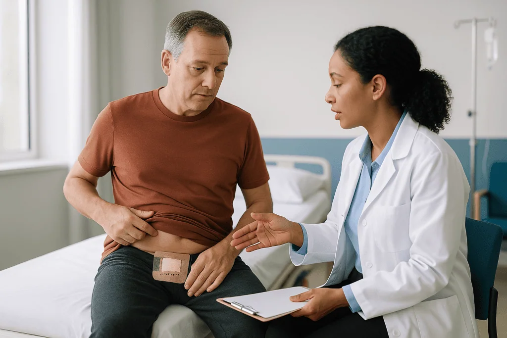
The logic is simple. Melanoma cells can extend microscopically beyond the edge you can see. Taking a measured margin of healthy-looking skin removes those stray cells and gives the pathologist a clear border to examine.
On the day, the surgeon draws the planned excision with a skin marker, usually as an ellipse rather than a circle, because an ellipse can be sewn into a straight line without puckering at the ends. That is why the scar is roughly three times longer than the margin is wide.
Local anesthetic is injected around the site. This is the part most people describe as the sharpest moment of the whole procedure; the stinging fades within a minute or two as the nerve endings are numbed. The tumor and margin are lifted out as one piece, oriented with a small stitch so the lab knows which edge is which, and sent for examination.
Bleeding points are sealed with a heat probe. The wound is then closed in layers: dissolving stitches below the surface take the tension, and finer stitches, staples or skin glue close the top.
Most excisions on the trunk or limbs take under an hour and are day procedures. Excisions on the face, hands or feet, or those combined with a lymph node biopsy, may be done under general anesthetic in an operating room, as the NHS describes. The pathology report typically returns within one to three weeks and confirms whether the margins are clear.
How deep do they cut to remove melanoma?
The surgeon cuts down through the full thickness of the skin to the subcutaneous fat, and sometimes to the fibrous layer over the muscle. That depth is fixed by anatomy. The width of the margin is what changes with the tumor.
Guidelines used across the NHS and North American practice link margin width to Breslow thickness, which is the pathologist’s measurement of how far the melanoma has grown down from the skin surface, recorded in millimetres. The NHS describes margins of roughly 1 cm for early stage 1 melanoma and around 2 cm for thicker or stage 2 tumors. Very thin, non-invasive melanoma (melanoma in situ) is usually treated with a narrower margin.
Why not simply take more? Larger margins mean larger wounds, more grafts and slower recovery, and studies summarized in the guideline literature have not shown that going wider than these recommended ranges improves outcomes for early melanoma. The margins represent a balance struck on evidence rather than caution alone.
People are often surprised to learn that the initial biopsy that diagnosed the melanoma was not the treatment. That first procedure removes enough tissue to establish the diagnosis and measure depth. Wide local excision is a second, planned operation, usually scheduled once the pathology is known, so the surgeon can choose the right margin.
On a practical level, depth matters for two reasons. Reaching the fat layer means the wound needs layered closure and cannot simply be taped. It also means tension on the stitches is real, which is why lifting, stretching and heavy activity are restricted early on. A wound on the shoulder blade pulled open by an overhead reach is one of the more common reasons for a delayed recovery.
Who has surgery straight away, and who is usually asked to wait?
Almost everyone with a newly diagnosed melanoma is offered wide local excision, because it is the treatment with the clearest evidence for early disease. The NHS describes surgery as the main treatment for stage 1 and 2 melanoma. A short delay between biopsy and excision is normal and allows the pathology report to guide the margin.
Some situations do change the timing or the plan.
- People taking blood thinners such as anticoagulants or antiplatelet medicines may be asked to continue, pause or adjust them. This decision belongs to the prescribing clinician, because stopping a blood thinner carries its own clot risk, while continuing raises the chance of bleeding under the wound.
- Melanoma on the face, ear, fingertip or genital skin often needs a plastic or specialist surgeon and may be scheduled separately so a flap or graft can be planned.
- Where imaging or examination suggests spread to lymph nodes or beyond, the team may arrange staging scans and a multidisciplinary discussion first. Surgery may still go ahead, but sometimes alongside or after drug treatment.
- People who are pregnant can usually still have local excision, since local anesthetic is considered acceptable, though sentinel node procedures using radioactive tracer are discussed case by case.
- Active skin infection at the site, poorly controlled diabetes or recent heart events may lead to a short postponement to reduce complications.
The waiting group, in other words, is not waiting because the melanoma is being ignored. They are waiting because the team is arranging the safest version of the operation. If the delay feels long, ask what is being organized and whether there is anything you can do to prepare, such as smoking cessation support, which improves healing in every wound-healing study the evidence base contains.
Stitches, flaps or a skin graft: how the wound gets closed
The closure method determines the shape of your recovery more than almost any other factor, so it is worth understanding the three options.
Primary closure means the two edges of the ellipse are stitched directly together. It works where skin is loose enough to stretch, which is most of the trunk, upper arms and thighs.
A local flap moves a tongue of neighboring skin, still attached to its blood supply, to fill the defect. Flaps are common on the face and scalp because the color and thickness match, but they create a longer, sometimes zigzag scar and swell noticeably in the first two weeks.
A skin graft lifts a thin sheet of skin from elsewhere and lays it over the wound, where it must grow a new blood supply. MedlinePlus describes grafts as either split-thickness, taking the top layers only, or full-thickness. Grafts are typical on the lower leg, where skin cannot stretch.
| Closure | Typical sites | Stitch removal or dressing change | Main recovery issue |
|---|---|---|---|
| Primary closure | Back, chest, arms, thighs | About 1 to 3 weeks (MedlinePlus) | Tension on the line; avoid stretching |
| Local flap | Face, scalp, nose, ear | About 1 to 2 weeks on the face | Swelling and bruising; scar length |
| Skin graft | Shin, foot, hand, scalp | First dressing left about 5 to 7 days | Two wounds; immobilization; graft take |
Dissolving stitches under the surface usually disappear over several weeks and can occasionally poke through as a small white thread, which is harmless and simply trimmed at clinic. Skin glue peels off on its own within a week or two. Staples, used mainly on the scalp, are removed in clinic with a small tool that takes seconds per staple.
Skin graft after melanoma removal: what recovery involves
A graft turns one wound into two, and each heals on its own schedule.
The grafted site is the delicate one. For the first days the sheet of skin survives by absorbing fluid from the tissue below, then tiny blood vessels grow into it. MedlinePlus notes that this process begins within about 36 hours and that the graft is usually protected under a firm dressing, often with a foam bolster, for the first week. Movement, knocks or fluid collecting underneath can lift the graft before those vessels connect, which is why a graft on the lower leg comes with strict instructions to keep the leg elevated and walking to a minimum.
The donor site, most often the thigh or behind the ear, heals like a graze. A split-thickness donor area typically re-covers itself within about two to three weeks; a full-thickness donor site is stitched closed and heals as a straight-line scar. Many people report the donor site as the more uncomfortable of the two in the first week, because the raw surface has exposed nerve endings.
A grafted area will look alarming at first dressing change: purple, mottled, sometimes with blistered edges. Color evens out over months. The graft may remain slightly paler, shinier or flatter than the surrounding skin permanently, and sensation returns slowly and incompletely.
Practical points that come up repeatedly:
- Moisturize the healed graft daily once the team allows; grafted skin lacks normal oil glands and dries easily.
- Protect it from sun for at least a year, as healing skin burns quickly and pigments unevenly.
- Expect the graft to feel tight or numb; gentle massage after full healing, if approved, helps soften it.
Partial graft loss happens and is managed with dressings rather than repeat surgery in most cases. Ask your team what percentage of the graft has “taken” at each visit so you understand the trajectory.
Is melanoma removal painful?
Less than most people fear, and in a different way than they expect.
During the operation, the only sharp sensation is the anesthetic injection, which stings for a moment and then numbs the area for one to several hours depending on the agent used. Pressure and tugging are felt; cutting is not. Under general anesthetic, nothing is felt at all.
The first evening, as the numbness wears off, the wound aches and throbs. Mayo Clinic and the NHS describe this as manageable with simple pain relievers for most people, and the team will advise which class suits you, since some over-the-counter anti-inflammatory medicines can increase bleeding under a fresh wound. Do not add or swap medicines without checking.
Pain generally peaks in the first 48 hours and falls steadily. By day three or four most stitched wounds are more itchy and tight than painful. A graft donor site tends to burn for longer, often a week, because it is an open surface. A sentinel node biopsy in the armpit or groin produces a deep ache and stiffness that improves as you gently move the joint.
Some sensations are normal and easily mistaken for problems. Shooting or electric twinges around the scar reflect small nerves regrowing and can continue for months. Numbness beyond the scar edge is common and often permanent to some degree. Firmness under the line is scar tissue maturing, not a lump returning.
Pain that increases after day three, rather than decreasing, is the signal that matters. Together with spreading redness, heat or discharge, it points to infection or a collection of blood or fluid under the wound, and it should be assessed rather than waited out.
Melanoma surgery recovery time week by week
Timelines vary with the closure method, so treat this as a map rather than a schedule, and defer to the instructions you were given.
Days 1 to 3. Keep the dressing dry and undisturbed unless told otherwise. Ache, mild swelling and bruising are expected. Elevate a limb wound above heart level when sitting. Sleep on the opposite side.
Days 4 to 7. Many teams allow a brief shower with the dressing removed or changed, patting the wound dry. The line may look red at the edges and slightly raised; this is inflammation building new tissue, not infection, unless it spreads. Graft dressings are usually left untouched until the first clinic check around this point, per MedlinePlus.
Weeks 1 to 2. Surface stitches on the face come out early in this window; trunk and limb stitches toward the end of it or into week three, according to MedlinePlus guidance. Skin glue peels. Pathology results typically arrive, and the team confirms whether margins are clear or a further excision is needed.
Weeks 2 to 4. The wound is closed but weak. Healing skin regains strength gradually over weeks, so this is the period when overstretching pulls scars wide. Light activity resumes; heavy lifting and contact sport usually do not.
Weeks 4 to 8. Most stitched wounds are fully sealed and firm. Grafts are typically stable, though still fragile to knocks. Swimming, running and gym work are commonly cleared around here, depending on the site.
Months 3 to 12. The scar reddens, then thickens, then softens and fades. Numbness slowly recedes. Regular skin checks begin, which the NHS describes as continuing for several years depending on stage.
People with a graft or a node biopsy should stretch each phase by roughly one to two weeks; those with a small facial excision often move faster.
Looking after the wound at home, and what your scar will do
Good wound care is mostly restraint. The skin knows how to heal; the job is to avoid interrupting it.
Keep the dressing on for as long as instructed, usually 24 to 48 hours for a stitched wound and about a week for a graft. When cleaning is allowed, use lukewarm water, no soap directly on the line, and pat rather than rub. MedlinePlus advises against soaking baths, hot tubs and swimming until the surface has fully closed, because prolonged wetness softens the edges and lets bacteria in.
Apply a thin layer of plain petroleum-based ointment if your team recommends it; a moist wound heals faster and scars less than a dry crusted one. Antibiotic ointments are not routinely needed and cause allergic reactions in a meaningful minority of people.
Leave scabs alone. Picking exposes tissue that has not finished rebuilding and widens the eventual scar.
Once the wound is fully closed, the scar takes over as the project. The pattern is predictable: pink and flat in month one, redder and firmer through months two to four as collagen is laid down, then gradually paler and softer over the rest of the year. Silicone gel or sheeting, started after the wound has sealed, has moderate evidence for flattening scars and is often suggested by surgical teams; massage with a bland moisturizer, a few minutes daily, helps soften firmness.
Sun protection is not optional. Healing skin produces pigment unpredictably, and ultraviolet exposure on a young scar can leave it permanently darker or blotchy. Cover the area or use a broad-spectrum sunscreen for at least the first year, which also aligns with the broader advice every melanoma patient receives about ongoing skin protection from the CDC and NHS.
Raised, itchy, thickened scars, called hypertrophic or keloid scars, are more common on the chest, shoulders and in people with darker skin tones. They can be treated; mention them early.
Exercise after melanoma excision: work, driving and getting back to activity
The wound does not know whether you are at a desk or at the gym. It knows tension, impact and moisture. Plan activity around those three.
Desk-based work is usually possible within one to three days for a stitched wound, once the initial ache settles. Physical jobs involving lifting, reaching or kneeling generally wait until stitches are out and the team confirms the wound has sealed, often two to three weeks, and longer after a graft.
Driving depends on whether you can perform an emergency stop and turn the wheel without hesitation. After a back, shoulder or arm excision, that may be a few days; after a groin node biopsy or a leg graft, considerably longer. Insurers may also ask that you are not under restriction from your surgeon.
Walking is encouraged from day one for almost everyone. It reduces the risk of blood clots in the legs, which the American Heart Association identifies as a hazard whenever people are less mobile than usual after surgery. The exception is a lower-leg graft, where walking is deliberately limited to protect the graft; in that case, ankle circles and calf squeezes while seated keep blood moving.
Stretching and resistance exercise need the most caution. A wound crosses skin that moves with the joint below it, and repeated pulling in the first month widens scars and can open them. As a rough guide drawn from surgical wound-healing timelines on MedlinePlus, gentle range-of-motion work can restart once stitches are out, and progressive loading from about four weeks for stitched wounds and six to eight weeks for grafts, always with the team’s agreement.
Swimming waits for a fully sealed surface with no scab, since pools and open water carry bacteria. Contact sport waits longest, because a direct blow to a graft can shear it off weeks after it looks healed.
Flying is generally acceptable once you are mobile and the wound is dressed; for longer flights within the first weeks, move regularly and stay hydrated to reduce clot risk.
Sentinel lymph node biopsy: how it changes recovery
A sentinel lymph node biopsy identifies and removes the first lymph node, or small group of nodes, that drains fluid from the melanoma site, so the pathologist can check whether cancer cells have reached it. The NHS and Mayo Clinic describe it as an option offered when the melanoma is thicker or has other higher-risk features, and it is usually performed at the same operation as the wide local excision.
Before surgery, a small amount of radioactive tracer and sometimes blue dye are injected around the melanoma. These travel along lymph channels to the sentinel node, which the surgeon locates with a handheld probe through a separate small incision, typically in the armpit, groin or neck.
Recovery is different in kind from the skin wound. Expect:
- A deep ache and stiffness in the nearby joint for one to two weeks; gentle movement helps, and physiotherapy is sometimes suggested.
- Bruising and, if dye was used, a blue-green tinge to the skin and urine for a day or two, which is harmless.
- A soft, fluid-filled swelling under the incision called a seroma. It is common, usually reabsorbs over weeks, and may be drained with a needle in clinic if uncomfortable.
- Numbness on the inner arm or upper thigh from small sensory nerves near the node, which may be permanent.
Lymphedema, a chronic swelling of the limb from disrupted lymph drainage, is a recognized but uncommon complication after removing a single sentinel node; it is more likely if further nodes are later removed. Report any persistent limb swelling, heaviness or tightness in rings and sleeves.
Results from the node typically return with the excision pathology. A negative sentinel node is reassuring; a positive one leads to a multidisciplinary discussion about closer surveillance, imaging or drug treatment. Which path follows is a decision for the team and you together, not something recovery instructions can predict.
What people often get wrong about melanoma surgery recovery
“The biopsy removed it, so the second operation is optional.” The biopsy established the diagnosis. Wide local excision removes the margin where microscopic cells may remain. The NHS describes it as the standard treatment precisely because the biopsy alone is not considered sufficient.
“The 2 week rule means I have two weeks before it spreads.” The phrase comes from the NHS urgent referral pathway, under which someone with a suspected melanoma should be seen by a specialist within two weeks of referral. It is a service standard for appointments, not a biological deadline. The short interval between biopsy and excision that most people experience is normal.
“How long does it take for melanoma to go from stage 1 to 4?” There is no fixed timeline. Melanoma growth rates vary enormously between individuals and subtypes; some remain thin for years while others thicken over months, as Mayo Clinic notes in describing the disease. Staging depends on depth, ulceration and spread at the time of diagnosis, not on how long a mole has been present. Worrying about a countdown that does not exist is a poor use of energy during recovery.
“A red line around the scar means infection.” A narrow band of pinkness right at the edge is normal healing inflammation. Spreading redness, heat, increasing pain and discharge are the features that distinguish infection.
“Vitamin E or a special cream will erase the scar.” Evidence for vitamin E on scars is weak and it causes contact dermatitis in some people. Silicone products and sun protection have the most consistent evidence for scar appearance; nothing erases a scar.
“Once it’s healed, I’m finished.” Melanoma follow-up continues for years, and the largest modifiable risk after a first melanoma is a second one. Skin self-examination and clinic reviews are part of recovery, not an afterthought.
Questions to ask your care team before and after surgery
Consultations are short and it is easy to leave with the big questions unasked. Writing these down beforehand helps.
Before the operation:
- What margin are you planning, and does it depend on the biopsy result?
- Will the wound be stitched directly, or is a flap or graft likely? If a graft, where will the donor site be?
- Is a sentinel lymph node biopsy being recommended, and what would a positive result change?
- Should I continue, pause or adjust any of my regular medicines, particularly blood thinners, diabetes medicines or immunosuppressants? Who makes that call?
- Will this be under local or general anesthetic, and do I need someone to take me home?
Immediately after:
- When can I get the wound wet, and how should I clean it?
- When will stitches or staples be removed, and by whom?
- What restrictions apply to lifting, stretching, driving and exercise, and for how long?
- What does normal healing look like at this site, and what specifically should make me call?
- When and how will I receive the pathology result?
At the results appointment:
- Were the margins clear, or is further excision needed?
- What stage is the melanoma, and what does that mean for follow-up frequency?
- How long will skin checks continue, and who performs them?
- Is any further treatment or trial being discussed at the multidisciplinary meeting?
- What should I be checking for myself between appointments, on my skin and in the lymph node areas?
A good final question at every visit is simply, “Is there anything about my healing that concerns you?” It invites the honest assessment that a busy clinic may otherwise leave unspoken, and it keeps the decision-making where it belongs: with the team that can see the wound and the pathology together.
When to call your doctor after melanoma surgery
Most wounds heal without drama, but a handful of signs warrant a same-day call to your surgical team or, out of hours, an urgent care assessment. Do not wait for the next scheduled appointment if any of these appear.
Signs of infection. Redness that spreads outward from the wound rather than staying at the edge, increasing warmth, swelling or pain after the third day, cloudy or foul-smelling discharge, or a fever. MedlinePlus lists these as the core features of a surgical site infection, which is treated more easily the earlier it is found.
Bleeding. Oozing that soaks through a dressing and does not stop after 10 to 15 minutes of firm, steady pressure, or a rapidly enlarging, tense, painful swelling under the wound, which may indicate a hematoma, a collection of blood that sometimes needs draining.
Wound breakdown. Stitches pulling apart, a gap opening in the line, or a graft lifting, darkening or sloughing.
After a lymph node biopsy. A hot, red or rapidly growing swelling at the node site, or new swelling of the whole arm or leg.
Clot warning signs. Pain, swelling or warmth in one calf, or sudden breathlessness or chest pain, which the American Heart Association flags as possible deep vein thrombosis or pulmonary embolism and which need emergency care.
Anesthetic or medication reactions. Rash, facial swelling, difficulty breathing, or persistent vomiting.
Beyond the immediate recovery, contact your team promptly about any new lump near the scar or in the armpit, neck or groin, any new or changing mole, or a scar that becomes thick, itchy and raised in a way that bothers you. None of these findings is a diagnosis; each is a reason for someone qualified to look. A phone call that turns out to be unnecessary costs a few minutes. A delayed one can cost weeks of extra healing.
Frequently asked questions
How long is wide local excision recovery for melanoma?
Most stitched wounds after wide local excision are healed enough for stitch removal within one to three weeks, with full strength returning over the following weeks. A graft or sentinel node biopsy typically adds two to four weeks. Timelines vary by site and closure method, so follow the specific instructions from your surgical team.
Is melanoma removal painful?
The sharpest moment is usually the local anesthetic injection, which stings briefly. Afterward, the wound aches for a day or two and is generally managed with simple pain relievers your team recommends. Graft donor sites and node biopsy sites tend to ache longer. Pain that worsens after day three should be assessed.
How deep do they cut to remove melanoma?
The surgeon cuts through the full thickness of skin down to the fat layer beneath. The width of the margin, roughly 1 cm for early stage 1 and around 2 cm for thicker tumors according to NHS guidance, depends on the Breslow thickness measured at biopsy. Depth is fixed by anatomy; width is tailored to the tumor.
What is the 2 week rule for melanoma?
It refers to the NHS urgent referral pathway, under which someone with a suspected melanoma should be seen by a specialist within two weeks of referral. It is a service standard for appointment timing, not a biological deadline. The interval between diagnosis and excision that most people experience is considered normal.
How long does it take for melanoma to go from stage 1 to 4?
There is no fixed timeline. Melanoma growth rates vary widely between individuals and subtypes; some stay thin for years while others thicken over months. Stage is determined by depth, ulceration and spread at diagnosis, not by how long a mole has existed. Your team can explain your specific stage and what it means.
When can I exercise after melanoma excision?
Walking is encouraged from day one for most people. Gentle range-of-motion work typically restarts once stitches are out, and progressive strength training from about four weeks for stitched wounds and six to eight weeks for grafts, with your team’s agreement. Swimming waits for a fully sealed surface, and contact sport waits longest.
How long does a skin graft after melanoma removal take to heal?
The grafted area is usually protected under a firm dressing for about a week while new blood vessels grow in, then remains fragile for several more weeks. A split-thickness donor site typically re-covers within two to three weeks. Color and texture of the graft continue to change for months.
What does a normal healing melanoma scar look like?
A narrow pink edge, mild swelling and some firmness under the line are normal in the first weeks. The scar often looks redder and thicker in months two to four before softening and fading over the first year. Spreading redness, heat, increasing pain or discharge are the features that suggest a problem.
Can I shower after melanoma surgery?
Most teams allow a brief shower after 24 to 48 hours for a stitched wound, patting the area dry afterward, though some ask you to keep the original dressing on longer. Graft dressings are usually left undisturbed until the first clinic check. Avoid soaking, baths and swimming until the wound has fully sealed.
How long does melanoma follow-up continue after surgery?
The NHS describes follow-up skin checks continuing for several years, with frequency depending on stage; early-stage melanoma may need fewer visits while thicker melanoma is checked more often. Between appointments, self-examination of your skin and the lymph node areas is part of ongoing care. Your team will set the schedule.
References
This article is for general information only and is not a substitute for professional medical advice. Please consult a qualified doctor about your individual situation.

More from the Blog
Who Sits on a Head Cancer Tumor Board and How the Group Reaches a Recommendation
A head and neck cancer tumor board is a scheduled meeting where surgeons, radiation oncologists, medical oncologists, radiologists, pathologists, and supportive specialists review one…
How CyberKnife Tracks a Moving Tumor: Robotic Arm, Real-Time Imaging and Breathing
CyberKnife tracks tumor movement by pairing a small linear accelerator on a robotic arm with two ceiling-mounted X-ray cameras that repeatedly image the patient…
Questions to Ask Before HIPEC: Second Opinions, Expected Benefits and the Recovery Plan
Before HIPEC, ask your care team how complete they expect tumor removal to be, what evidence supports HIPEC for your specific cancer type, which…

Life After Wilms Tumor Treatment: Living With One Kidney, Sports and Long-Term Follow-Up
Life after Wilms tumor usually means living well with one kidney, returning to school and most sports, and attending long-term follow-up for years. The…
Voice and Swallowing After Neck Cancer Treatment: What Rehabilitation Can Restore
Voice changes after neck cancer treatment, such as hoarseness, breathiness, a lower or weaker pitch and difficulty swallowing, are common after surgery, radiation or…
How Ovarian Cancer Treatment Is Planned: Surgery, Chemotherapy and the Order They Come In
Ovarian cancer treatment by stage usually combines surgery to remove as much visible cancer as possible with platinum-based chemotherapy. Early-stage disease confined to the…