

The Thyroid Ultrasound: What It Sees and How TI-RADS Scoring Works

Key Takeaways
- High-resolution ultrasound finds thyroid nodules in up to two-thirds of adults, yet more than 90% of nodules are benign.
- TI-RADS scores five features, composition, echogenicity, shape, margins, and echogenic foci, and the point total assigns a risk level from TR1 to TR5.
- A taller-than-wide nodule earns 3 points on its own because growth against the neck's natural tissue planes is one of the strongest single predictors of malignancy.
- Even a TR5, the most suspicious category, means elevated risk requiring biopsy, not a cancer diagnosis, and many TR5 nodules still prove benign.
- Ultrasound shows structure, not function: hormone problems are diagnosed with blood tests, and a normal-looking gland can coexist with abnormal levels.
- No fasting or medication changes are needed before the scan, just skip necklaces and high collars, and bring any prior neck imaging reports.
A thyroid ultrasound uses sound waves to create detailed images of the thyroid gland and any nodules, showing their size, composition, borders, and suspicious features. Radiologists grade nodules with TI-RADS, a point-based system that scores five characteristics and sorts nodules into five risk levels (TR1 through TR5). That level, combined with the nodule's size, determines whether you need a biopsy, follow-up imaging, or nothing at all.
The gel is cold, the room is dim, and the wand at your throat feels oddly ceremonial. Ten minutes, no needles, no radiation. And then a report lands in your patient portal full of words like hypoechoic, punctate echogenic foci, and a cryptic TR4and a scan that felt trivial suddenly reads like a verdict.
It isn’t one. Most of what a thyroid ultrasound finds is background noise: small, harmless lumps that a majority of adults quietly carry through life without ever knowing. The trouble is that the report doesn’t sound harmless, and the scoring system radiologists use, TI-RADS, was written for other radiologists, not for you.
So let’s translate it. What the machine actually sees, how those five graded features add up to a score, which findings genuinely deserve attention, and where ultrasound’s vision honestly ends.
What Will an Ultrasound of Your Thyroid Tell You?
Think of it as a structural survey, not a chemistry test. A thyroid ultrasound bounces high-frequency sound waves off the tissues of your neck and converts the returning echoes into a live, moving picture. From that picture, a radiologist can describe the gland’s size and shape, whether its texture looks uniform or patchy, how blood flows through it, and, the main event, whether it contains nodules.
For each nodule, the scan answers surprisingly specific questions. Is it filled with fluid, solid tissue, or a mix? Are its edges smooth like a marble or ragged like torn paper? Does it contain tiny bright specks of calcium? Is it growing outward in the direction tissue normally spreads, or pushing upward against the grain? Each answer nudges the odds of the nodule being harmless or worth a closer look, which is why MedlinePlus lists ultrasound as the standard first step for evaluating a lump found on a neck exam.
The scan also checks the neighborhood. Sonographers routinely sweep the lymph nodes along the sides of the neck, because an abnormal-looking node can matter as much as the nodule itself. And in people with thyroid inflammation, the gland’s overall texture, often described as heterogeneous, can support a diagnosis a blood test has already suggested.
What it will not tell you is how well your thyroid is working. Hormone levels come from bloodwork, full stop. An ultrasound can look pristine while your levels are off, and vice versa.
Why Do Doctors Order a Thyroid Ultrasound?
Three scenarios account for most orders. The first: someone felt something. You noticed a lump while buttoning a collar, or a clinician felt one during a routine exam. Ultrasound is the natural next step because fingers are crude instruments: they miss most nodules under a centimeter and can’t tell fluid from solid tissue.
The second: something else found it first. Radiologists call these incidentalomasnodules spotted by accident on a CT scan of the chest, an MRI of the spine, or a carotid artery study ordered for entirely different reasons. As cross-sectional imaging has become routine, incidental thyroid findings have become one of the most common reasons for a dedicated ultrasound. A focused thyroid scan characterizes what the other test only glimpsed.
The third: your blood tests or symptoms point at the gland. Abnormal thyroid hormone levels, an enlarged gland (goiter), swallowing changes, or persistent hoarseness can all prompt a look. Ultrasound also guides needles during biopsies, letting the clinician watch the needle tip in real time, and it’s the workhorse for surveillance, tracking a known nodule year over year to confirm it’s behaving.
What ultrasound is not recommended for is screening people with no symptoms, no lump, and normal labs. Major guidelines discourage it, for a reason that will become obvious in a moment: the scan finds nodules in most of us, and hunting for them in healthy necks creates far more anxiety and follow-up than benefit.
What Not to Do Before a Thyroid Ultrasound
Here’s a rare piece of good news in medical logistics: almost nothing. A thyroid ultrasound requires no fasting, no contrast dye, no medication changes, and no special hydration. You can eat breakfast, drink coffee, and drive yourself home. The NHS notes that most external ultrasound scans need little or no preparation at all.
A few small choices do make the visit smoother:
- Skip the necklace and dangling earrings. You’ll be asked to remove anything metallic around your neck, so leaving jewelry at home saves a step and a worry.
- Wear a low, open collar. Turtlenecks, tight crew necks, and hoodies with drawstrings all get in the way. A button-down or scoop neck means you likely won’t need to change into a gown.
- Rethink heavy neck lotion or makeup that morning. Thick products can interfere with the transducer’s contact with skin; the gel works best on a clean surface.
- Bring your history. Prior neck surgery, radiation to the head or neck in childhood, a family history of thyroid cancer, and previous ultrasound reports all change how findings are interpreted. If you were scanned elsewhere, prior images are gold, nodule stability over years is one of the most reassuring findings in all of radiology.
One thing genuinely not to do: don’t cancel because you’re congested, coughing, or had a big meal. None of it affects the images. The scan sees through all of that just fine.
What Happens During the Scan, Minute by Minute
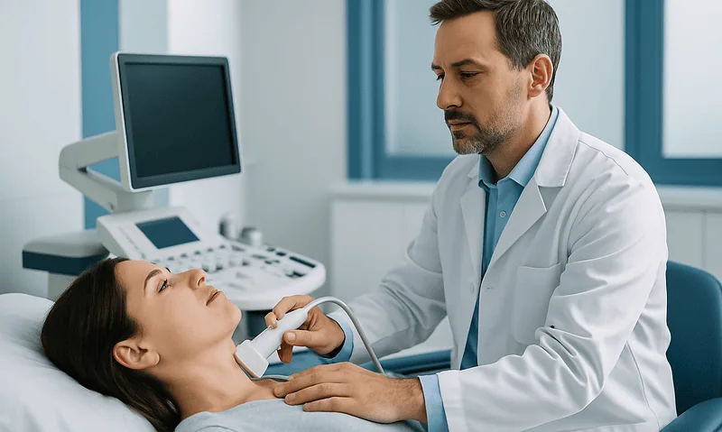
You’ll lie on your back, usually with a small pillow tucked under your shoulders so your neck extends gently backward: the least glamorous yoga pose in medicine, and the one part some people find mildly uncomfortable, especially with neck arthritis. Say so; the sonographer can adjust.
Warm-ish gel goes on your throat. Then the transducer, a smooth wand about the size of an electric razor head, glides across your neck in slow, overlapping passes. The sonographer images each lobe of the thyroid in two planes, measures the gland, and documents any nodule in three dimensions. You may be asked to swallow (the thyroid slides visibly when you do, which helps distinguish it from neighboring structures) or to turn your head to open up views of the lymph node chains.
Expect 15 to 30 minutes. Longer doesn’t mean worse; a thorough sonographer measuring several small, boring nodules takes more time than one confirming a single simple cyst. You may notice the machine switch to a mode with orange-and-blue flashes: that’s Doppler, mapping blood flow, a routine part of characterization rather than a red flag.
Two things worth knowing in advance. First, sonographers generally can’t discuss findings; interpretation is the radiologist’s job, and a neutral poker face is professional standard, not a bad omen. Second, your report may reach your portal before your doctor has reviewed it. If it does, resist the urge to sentence yourself based on unfamiliar vocabulary. The next sections are that vocabulary, decoded.
Thyroid Nodules Are Astonishingly Common: Here's the Math
This is the single most important piece of context for reading any thyroid ultrasound report, so it deserves its own numbers.
By touch alone, thyroid nodules seem uncommon: a clinician can feel one in roughly 5% of women and 1% of men. Put those same people under a high-resolution ultrasound, and the picture flips, studies have found nodules in anywhere from about 20% to as many as 68% of randomly selected adults, with prevalence climbing steadily with age. In other words, if you’re past middle age, having at least one thyroid nodule is closer to the rule than the exception.
Now the second number, the one that should sit next to the first in your mind: the overwhelming majority of these nodules, more than 90% by most estimates cited by Mayo Clinic and Cleveland Clinic, are benign. Colloid nodules, simple cysts, spongiform nodules, little zones of thyroid tissue that grew unevenly: harmless variations, not diseases in waiting.
Hold both facts at once and the whole enterprise of thyroid imaging makes sense. Radiologists aren’t trying to find nodules; nodules find themselves. The real job is triage, separating the small minority of nodules with worrisome features from an ocean of benign ones, without biopsying half the adult population. That’s precisely the problem TI-RADS was built to solve, and it’s why an ultrasound report that mentions a nodule, even several, is usually a description of normal human variation rather than a warning.
The Five Features Radiologists Actually Grade
Before any score exists, the radiologist assesses five specific characteristics of each significant nodule. These aren’t arbitrary; each one reflects something about how benign and malignant tissue tend to grow differently.
- Composition. Is the nodule fluid (cystic), solid, or mixed? Pure fluid is essentially always benign. A spongiform nodule, one that looks like a sponge, riddled with tiny fluid pockets, is nearly as reassuring. Solid tissue carries more uncertainty.
- Echogenicity. How bright or dark does the tissue appear compared with normal thyroid? Darker (hypoechoic) tissue reflects densely packed cells, which is more typical of malignancy, though plenty of benign nodules are hypoechoic too.
- Shape. Benign nodules tend to grow along the natural horizontal planes of the neck, ending up wider than tall. A nodule that’s taller than wide on a cross-sectional view has grown against those planes: a behavior more characteristic of invasive tissue, and one of the strongest single predictors in the system.
- Margins. Smooth, well-defined borders suggest a nodule politely displacing its neighbors. Irregular, lobulated, or spiky margins suggest infiltration. Evidence of extension beyond the thyroid capsule is the most serious margin finding.
- Echogenic foci. Bright specks within the nodule. Tiny punctate ones can represent microcalcifications, which are associated with papillary thyroid cancer. Larger, coarse calcifications and “comet-tail” artifacts (a benign fluid signature) carry different, generally lower, weight.
No single feature is a diagnosis. Each is a probability nudge, and TI-RADS turns those nudges into arithmetic.
What Is TI-RADS, and Why Did It Replace Gut Feeling?
TI-RADS stands for Thyroid Imaging Reporting and Data System. The version most widely used in the United States, ACR TI-RADS, was published in 2017 by a committee of the American College of Radiology, and the underlying white paper is one of the most cited documents in modern radiology (you can find it on PubMed).
The problem it addressed was inconsistency. Before standardized scoring, two skilled radiologists could look at the same nodule and write meaningfully different reports, one recommending biopsy, the other reassurance, because each was weighing suspicious features by experience and instinct. With nodules present in over half of older adults, that variability translated into enormous numbers of biopsies performed on lesions that were almost certainly benign. Fine-needle biopsies are safe, but they aren’t free of cost, anxiety, or ambiguous results that snowball into surgery.
TI-RADS borrowed a strategy that had already worked in breast imaging (BI-RADS): define the features precisely, assign each a point value proportional to its statistical association with cancer, add up the points, and let the total, together with the nodule’s size, dictate a recommendation. The design goal was explicit and, to my mind, the right one: reduce unnecessary biopsies of clearly low-risk nodules while keeping a tight net around genuinely suspicious ones.
Validation studies since 2017 have generally borne this out, showing that ACR TI-RADS recommends fewer biopsies than older guideline systems while missing few significant cancers. It isn’t perfect, no risk model is, but it replaced adjectives with arithmetic, and that alone made thyroid reports dramatically more consistent from one hospital to the next.
How TI-RADS Points Add Up
Here’s the actual math your radiologist performs, feature by feature. Each of the five graded characteristics contributes points; the totals run from 0 to a theoretical maximum well into double digits.
- Composition: purely cystic or spongiform, 0 points; mixed cystic and solid, 1 point; solid, 2 points.
- Echogenicity: anechoic (pure fluid), 0; hyperechoic or isoechoic (as bright as or brighter than normal thyroid), 1; hypoechoic (darker), 2; very hypoechoic (darker than neck muscle), 3.
- Shape: wider than tall, 0; taller than wide, 3. Note the jump: there’s no middle ground, because this single feature carries so much predictive weight.
- Margins: smooth or ill-defined, 0; lobulated or irregular, 2; extension beyond the thyroid, 3.
- Echogenic foci: none or benign comet-tail artifacts, 0; macrocalcifications, 1; peripheral rim calcification, 2; punctate echogenic foci (possible microcalcifications), 3.
A worked example makes it concrete. Say your report describes a solid (2), hypoechoic (2), wider-than-tall (0) nodule with smooth margins (0) and no echogenic foci (0). Total: 4 points. That’s a common, middle-of-the-road profile, moderately suspicious on paper, yet the large majority of such nodules prove benign.
Contrast that with a solid (2), very hypoechoic (3), taller-than-wide (3) nodule with irregular margins (2) and punctate foci (3): 13 points, a profile that concentrates every high-risk feature at once and would earn the most urgent category regardless of size thresholds for follow-up.
The total then maps to a level, TR1 through TR5, which is where size enters the equation.
TR1 to TR5: What Each Level Means for You
Points alone don’t trigger a biopsy. TI-RADS pairs the risk level with the nodule’s largest dimension, on sensible logic: a suspicious-looking 4-millimeter speck is far less consequential than a suspicious 2-centimeter mass, and even proven small papillary cancers often behave indolently. Here’s how the levels, approximate malignancy risks, and size thresholds fit together under ACR TI-RADS:
| Level | Points | Meaning | Approx. cancer risk | Typical action |
|---|---|---|---|---|
| TR1 | 0 | Benign | Under 2% | No follow-up needed |
| TR2 | 2 | Not suspicious | Under 2% | No follow-up needed |
| TR3 | 3 | Mildly suspicious | Roughly 5% | Biopsy if ≥2.5 cm; follow-up scans if ≥1.5 cm |
| TR4 | 4–6 | Moderately suspicious | Roughly 5–20% | Biopsy if ≥1.5 cm; follow-up scans if ≥1 cm |
| TR5 | 7+ | Highly suspicious | Over 20% | Biopsy if ≥1 cm; follow-up scans if ≥0.5 cm |
Read that risk column carefully, because it’s the emotional center of the whole system. Even TR4, the level that most often rattles people, means the odds still favor benign, often heavily. And TR5, the top category, means elevated risk that warrants a biopsy, not a diagnosis of cancer.
Notice, too, what falls below every threshold: a small TR3 or TR4 nodule under a centimeter typically needs nothing but time and perhaps a repeat scan. If your report ends with “no FNA recommended,” that isn’t the radiologist shrugging: it’s the system working exactly as designed.
What Are the Red Flags on a Thyroid Ultrasound?
People search this phrase constantly, so let’s answer it plainly. A handful of findings carry genuinely more weight than the rest, and they’re worth knowing, with the standing caveat that every one of them also appears in nodules that turn out benign.
- A taller-than-wide shape. Growth against the neck’s natural tissue planes is among the most specific single warning signs in the system, which is why it earns 3 points on its own.
- Punctate echogenic foci in a solid nodule. These tiny bright dots may represent microcalcifications, a feature statistically linked with papillary thyroid cancer.
- Irregular, lobulated, or infiltrative marginsand above all, any suggestion the nodule extends beyond the thyroid’s capsule into surrounding tissue.
- Markedly hypoechoic solid tissue, darker than the neighboring strap muscles of the neck.
- Abnormal lymph nodes. Rounded shape, loss of the normal fatty center, calcifications, or cystic change in a neck node can matter as much as anything inside the gland itself.
- Meaningful growth over time. A nodule that has clearly enlarged between scans draws more scrutiny than one that has sat unchanged for five years.
Just as useful is the anti-list, findings that are reassuring by design: purely cystic nodules, spongiform texture, comet-tail artifacts, smooth margins, stability across years of imaging. A report can contain the word “nodule” a dozen times and still describe a completely quiet neck.
The features cluster deliberately in TI-RADS: one red flag rarely stands alone in a cancer, which is why the score rewards accumulation rather than panicking over any single trait.
Can You See Cancer on a Thyroid Ultrasound?
The honest answer: ultrasound can strongly suspect cancer, but it cannot diagnose it. No pattern of gray pixels, however ominous, is definitive, and no pattern, however tranquil, is an absolute guarantee. What the scan provides is probability, and it’s genuinely good at that job.
At the reassuring end, ultrasound performs superbly. A purely cystic nodule or a classic spongiform nodule carries a malignancy risk low enough, well under 2%, that guidelines say no follow-up is needed at all. When radiologists call something TR1 or TR2, the evidence behind that confidence is deep.
At the suspicious end, the picture is a strong hint rather than a verdict. Even TR5 nodules, the highest category, prove benign more often than not in many validation series, elevated risk means roughly one in three to one in two in some studies, not certainty. That’s why the pathway always runs through a fine-needle aspiration (FNA) biopsy: a thin needle, guided by the same ultrasound, samples cells for a pathologist to examine under a microscope. Tissue, not imaging, makes the diagnosis.
Two more honest limits. Ultrasound is better at flagging papillary thyroid cancer, the most common and generally most treatable type, than follicular lesions, which can look deceptively bland on imaging and often require biopsy or even surgical pathology to sort out. And a normal-feeling report doesn’t extend to structures ultrasound can’t reach, such as thyroid tissue tucked behind the breastbone, which sometimes needs other imaging.
So: can it see cancer? It can see the shadow cancer tends to cast. Confirming the substance is the biopsy’s job.
What a Thyroid Ultrasound Can't Tell You
Every test has a blind side, and knowing ultrasound’s keeps expectations calibrated.
It cannot measure function. This is the misunderstanding I encounter most often: people expect the scan to explain their fatigue, weight change, or heart palpitations. Hormone output, whether the gland is overactive or underactive, is assessed with blood tests such as TSH, not images. A thyroid can look textbook-normal on ultrasound while its hormone levels are significantly off, and a gland studded with benign nodules can hum along producing perfectly normal hormones. Structure and function are separate questions requiring separate tests.
It cannot classify inflammation on its own. Ultrasound may show the heterogeneous, patchy texture typical of autoimmune thyroiditis, but the diagnosis rests on bloodwork and clinical context. The image supports the story; it doesn’t write it.
It cannot see everything anatomically. Sound waves don’t penetrate bone or air, so thyroid tissue extending down behind the sternum falls off the map, and very deep neck structures are evaluated less reliably. Body habitus and prior surgical changes can also degrade image quality.
It cannot escape its operators or its own success. Ultrasound is operator-dependent, measurements can vary a few millimeters between sonographers, which is why “growth” is only called when change exceeds defined thresholds. And its exquisite sensitivity is a double-edged sword: it finds tiny, clinically meaningless nodules in most older adults, which is exactly why experts advise against screening symptom-free necks.
None of this diminishes the test. It just means an ultrasound answers the question it was designed forwhat is in this gland?and defers what is this gland doing? to the lab.
What Happens After a Suspicious Result: Biopsy and Surveillance
Suppose your report lands at TR4 with a 1.8-centimeter nodule, squarely in biopsy territory. Here’s the realistic road ahead.
The fine-needle aspiration itself is a modest event, usually 20 to 30 minutes door to door. Under local numbing and continuous ultrasound guidance, a clinician passes a needle thinner than the one used for most blood draws into the nodule several times, collecting cells. Most people describe pressure more than pain, and typical aftercare amounts to a small bandage and taking it easy for a day. Complications are uncommon, with minor bruising the most frequent.
Results generally come back within about a week, classified under a standardized pathology framework (the Bethesda system). Roughly speaking, results fall into three buckets: clearly benign: the most common outcome, usually followed by periodic ultrasound rather than any treatment; clearly malignant or suspicious, which opens a conversation with a specialist about options; and indeterminate: the frustrating middle, where cells look neither clearly benign nor clearly malignant. Indeterminate results often lead to molecular testing of the sample or a repeat biopsy before any bigger decision, and it’s worth knowing in advance that this ambiguity happens in a meaningful minority of biopsies. It reflects biology, not a botched procedure.
For nodules below biopsy thresholds, the plan is surveillance: repeat ultrasounds at intervals that stretch out, commonly one to two years initially, then longer, as stability accumulates. Radiologists look for defined growth (typically a 20% increase in two dimensions or a volume increase of 50%), not millimeter jitter. Years of unchanged scans are, in themselves, evidence of benignity, and many surveillance plans eventually end with a clean discharge.
When to See a Doctor About Your Thyroid
Imaging aside, certain symptoms deserve a clinical evaluation on their own timetable, some promptly, a few urgently.
Make an appointment soon if you notice: a new lump or visible swelling in the front of your neck; a lump anywhere in the neck that persists beyond two to three weeks; hoarseness or a voice change lasting more than a few weeks without a cold to explain it; a sensation of pressure or difficulty when swallowing; or a known nodule that seems to be getting larger. None of these reliably means cancer, benign nodules and goiters cause most of them, but each warrants examination rather than watchful worrying at home.
Seek care promptly, the same day, if a neck mass is enlarging rapidly over days, is painful with fever (possible infection or acute inflammation), or is accompanied by any difficulty breathing. Trouble breathing with neck swelling is an emergency.
Bring symptoms of hormone imbalance to your doctor as well, since they call for blood tests rather than imaging first: unexplained fatigue, feeling persistently cold or hot when others don’t, unintentional weight change, a racing or irregular heartbeat, tremor, hair thinning, or changes in mood and concentration. Mayo Clinic and MedlinePlus both outline these as reasons for thyroid evaluation.
And a quieter scenario worth naming: if an old imaging report mentioned a thyroid nodule and nobody ever followed up, a genuinely common loose end, raise it at your next visit. Most such findings need nothing, but that conclusion should be reached deliberately, with the report in hand, rather than by default.
How to Read Your Own Report Without Spiraling
A short field guide for the moment the portal notification arrives before your doctor’s call does.
Start at the bottom. The “Impression” section is the radiologist’s actual conclusion; everything above it is methodical description. “Findings” sections are contractually thorough, every 3-millimeter cyst gets its measurements, and thoroughness reads as alarm to the uninitiated. It isn’t.
Then decode the vocabulary that most often frightens people unnecessarily. Heterogeneous means uneven texture, common in inflammation and in perfectly ordinary aging glands. Hypoechoic means darker than surrounding tissue, one point in the scoring system, present in legions of benign nodules. Vascularity on Doppler describes blood flow and, notably, was dropped from ACR TI-RADS scoring because evidence showed it didn’t reliably distinguish benign from malignant. Colloid and comet-tail artifact are actively reassuring words. Unchanged from prior may be the single most comforting phrase in radiology.
Look for the pairing that actually drives decisions: the TR level plus the size. A TR4 designation on a 7-millimeter nodule typically means follow-up imaging at most; the same TR4 on a 2-centimeter nodule means a biopsy conversation. The level without the size is half a sentence.
Finally, hold on to the base rates one more time: nodules in up to two-thirds of adults, more than 90% benign, and a scoring system engineered specifically to leave low-risk findings alone. Your report is a probability document. Let your doctor put it in the context of your labs, your history, and your neck: that synthesis, not any single line of the report, is the real result.
Frequently asked questions
What are the red flags on a thyroid ultrasound?
The most significant findings are a taller-than-wide shape, punctate echogenic foci (possible microcalcifications) in solid tissue, irregular or infiltrative margins, markedly dark (very hypoechoic) solid tissue, extension beyond the thyroid capsule, and abnormal-looking neck lymph nodes. Each raises statistical suspicion without proving anything, every one of these features also occurs in benign nodules. TI-RADS scoring adds them up precisely because clusters of features matter far more than any single one.
Can you see cancer on a thyroid ultrasound?
Ultrasound can identify features that make cancer more or less likely, but it cannot diagnose cancer by itself. Reassuring patterns like purely cystic or spongiform nodules carry under a 2% malignancy risk, while highly suspicious TR5 nodules carry an elevated risk, yet even many of those prove benign. Confirming or ruling out cancer requires a fine-needle biopsy, in which cells are sampled under ultrasound guidance and examined by a pathologist.
What should you not do before a thyroid ultrasound?
Very little preparation is needed, no fasting, no medication changes, no contrast dye. Practical tips: don’t wear necklaces or high, tight collars, since you’ll need your neck exposed; skip heavy lotion or makeup on the neck; and don’t forget prior imaging reports if you were scanned elsewhere, because comparison with old studies is one of the most valuable pieces of information a radiologist can have. There’s no reason to cancel for a cold or a full stomach.
Does a thyroid ultrasound hurt?
No. The scan involves gel and a smooth handheld probe gliding over the front of your neck, most people feel only light pressure. The one common discomfort is positional: lying with your neck gently extended over a pillow can strain people with neck or back arthritis. Tell the sonographer if the position is uncomfortable; it can usually be adjusted without compromising the images. There’s no radiation, no needles, and no recovery time.
How long does a thyroid ultrasound take?
Typically 15 to 30 minutes. The sonographer images both lobes of the thyroid in two planes, measures the gland and any nodules in three dimensions, checks blood flow with Doppler, and surveys the neck’s lymph nodes. A longer scan usually reflects thoroughness, several small nodules each need careful measurement, rather than anything alarming. Results are interpreted afterward by a radiologist, so you generally won’t get answers in the room.
What does TI-RADS 3 mean on a thyroid ultrasound?
TR3 means “mildly suspicious”: the nodule scored 3 points across the five graded features, corresponding to a malignancy risk of roughly 5%. Under ACR TI-RADS, a TR3 nodule is biopsied only if it measures 2.5 centimeters or larger, and followed with periodic ultrasound if it’s 1.5 centimeters or larger. Smaller TR3 nodules typically need no follow-up at all. The large majority of TR3 nodules are benign.
What is a hypoechoic thyroid nodule?
Hypoechoic simply means the nodule appears darker than the surrounding thyroid tissue on ultrasound, usually because its cells are more densely packed. It contributes 2 points in TI-RADS scoring because malignant nodules are more often hypoechoic, but the finding is extremely common in benign nodules too, so by itself it proves nothing. What matters is the complete profile: a hypoechoic nodule with smooth margins, a wider-than-tall shape, and no calcifications is usually low risk.
Do all thyroid nodules need a biopsy?
No, most don’t. TI-RADS was designed specifically to spare low-risk nodules from unnecessary needles. TR1 and TR2 nodules need no biopsy or follow-up regardless of size, and even moderately or highly suspicious nodules are only biopsied above defined size thresholds (1.5 cm for TR4, 1 cm for TR5). Nodules below those cutoffs are typically watched with periodic ultrasound, and years of stability are themselves strong evidence of benign behavior.
Can a thyroid ultrasound show hypothyroidism or hyperthyroidism?
Not directly. Ultrasound shows the gland’s structure, size, texture, nodules, blood flow, but hormone output is measured with blood tests such as TSH. A thyroid can look completely normal on ultrasound while producing too much or too little hormone, and a nodular-looking gland can function perfectly. Ultrasound can support a diagnosis (inflammation often produces a patchy, heterogeneous texture), but the functional diagnosis itself always comes from the lab.
How often should a thyroid nodule be rechecked with ultrasound?
It depends on the TI-RADS level and size. Common ACR TI-RADS surveillance intervals start at one to two years, stretching to five years as stability accumulates; TR5 nodules under biopsy size are typically rechecked annually. Radiologists define real growth as roughly a 20% increase in two dimensions or a 50% volume increase, small millimeter fluctuations between scans reflect measurement variation, not change. Many surveillance plans end entirely after about five years of stability.
References
- MedlinePlus Medical Encyclopedia: Thyroid Ultrasound
- Cleveland Clinic: Thyroid Nodule
- NHS: Ultrasound Scan
This article is for general information only and is not a substitute for professional medical advice. Please consult a qualified doctor about your individual situation.

More from the Blog

How Long to Fast Before Blood Work, and What Breaks a Fast
Most blood tests that require fasting call for 8 to 12 hours without food or calorie-containing drinks; plain water is fine and usually encouraged.…
Thyroid Antibody Tests: TPO, Thyroglobulin and What Positivity Means to Discuss
Positive thyroid antibodies, most often against thyroid peroxidase (TPO) or thyroglobulin, mean the immune system is reacting to thyroid tissue. That raises the likelihood…

Bruising After a Blood Test: Why It Happens and When It Matters
Bruising after a blood test is common and almost always harmless. It happens when a small amount of blood leaks from the tiny puncture…

Is Blood Blue? The Vein Illusion, Explained
No, human blood is never blue. It ranges from bright cherry red when carrying oxygen to a dark brick red when oxygen-poor, but it…

RDW: The Variability Number That Flags Early Deficiency
RDW (red cell distribution width) is a routine part of a complete blood count that measures how much your red blood cells vary in…

The Homocysteine Test: What It Measures and Its B-Vitamin Connection
A homocysteine test measures the level of homocysteine, an amino acid, in your blood. Levels above roughly 15 micromoles per liter most often point…